
Give me the overall, all the differences with diagram, uh, I mean, picture of central and leteral incisor teeth with principal identification Nur Sly drawing level the level lingual missile lateral all the views that is needed so give me a compact all the things so that all the relative all the related information can be implemented in this
central incisor lateral incisor teeth anatomy labial lingual mesial distal views
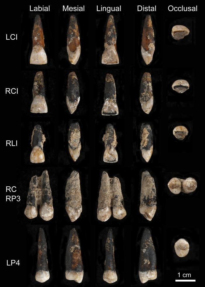
A clinical photograph of six permanent maxillary hominin teeth from the Magubike archaeological site, presented in multiple orientations: Labial, Mesial, Lingual, Distal, and Occlusal views. The specimen set includes the left central incisor (LCI), right central incisor (RCI), right lateral incisor (RLI), right canine (RC) cemented with the right third premolar (RP3), and the left fourth premolar (LP4). The teeth exhibit significant taphonomic erosive damage, characterized by partial dissolution of the enamel and rough, granular surface textures across the crowns and roots. Coloration varies from light enamel tones to dark, mottled brown/black staining, particularly on the roots and proximal surfaces. The occlusal views demonstrate diverse wear patterns, with the incisors showing irregular shapes and dark bands across biting surfaces, and the canine/premolar complex revealing distinct cusp morphology (single cusp on RC, bicuspid on RP3). This display serves as an educational resource for studying dental anthropology, paleopathology, and taphonomic degradation of human dental tissues.
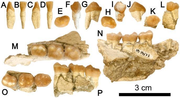
This composite educational image displays dentognathic remains from a Late Miocene hominoid (Hispanopithecus laietanus), providing key morphological details for dental anatomy and paleontological study. The visual includes multiple views of individual teeth and mandibular fragments. Panels A-D show a lower central incisor (i1) in mesial, lingual, distal, and labial views, highlighting a spatulate and waisted crown. Panels E-J depict left and right lower third premolars (p3) in occlusal, mesial, and buccal views, characterized by a sectorial shape and prominent mesiobuccal honing facets. Panel K-L shows a lower fourth premolar (p4) with a suboval occlusal profile. Mandibular fragments (M-P) contain the lower molar series (m1-m3). The occlusal views (M, O) reveal subrectangular crowns featuring a Y5 cusp pattern, short mesial foveae, and extensive talonid basins. Buccal views (N, P) show the root structures and relative height of the molar crowns. This anatomical collection demonstrates hominoid-specific dental traits used for taxonomic classification and dietary reconstruction.
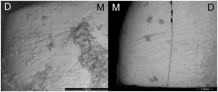
This diagnostic microscopy image displays two high-magnification views of human dental specimens (LI2 and LI1) highlighting instrumental striations from an archaeological context. The left panel shows an upper lateral incisor (LI2) with clear, fine linear grooves etched into the labial surface, predominantly in oblique and horizontal orientations. Clusters of granular sediment deposits are visible on the mesial (M) aspect, partially obscuring the enamel surface. The right panel displays an upper central incisor (LI1) with similar linear striations across the labial face, including a prominent vertical crack or post-depositional feature near the midline. Both specimens are marked with 'M' (mesial) and 'D' (distal) to indicate anatomical orientation. The image demonstrates cultural dental wear patterns, specifically 'stuff-and-cut' striations associated with non-masticatory use of teeth in prehistoric populations. The well-preserved surface morphology facilitates the study of manual laterality and dental biomechanics in paleoanthropology and forensic odontology. Scale bars of 1.0mm are provided for both frames.
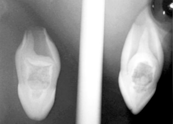
This diagnostic periapical radiograph displays labial and mesial views of a maxillary left lateral incisor (tooth 22) demonstrating complex developmental anomalies. The primary finding is a Oehlers Type III dens invaginatus, characterized by a radiopaque, pouch-like invagination originating from the lingual/palatal surface and extending apically beyond the cemento-enamel junction. This invagination creates a 'tooth within a tooth' appearance with a central radiolucent core. Additionally, the tooth exhibits an abnormally wide pulp chamber containing a large, irregular radiopacity consistent with a pulp stone (denticle), which occupies a significant portion of the internal coronal space. The radiographic features illustrate developmental disturbances in dental morphodifferentiation, specifically invaginated teeth and calcifications within the pulp system, which are clinically significant for endodontic planning and potential periapical pathology.
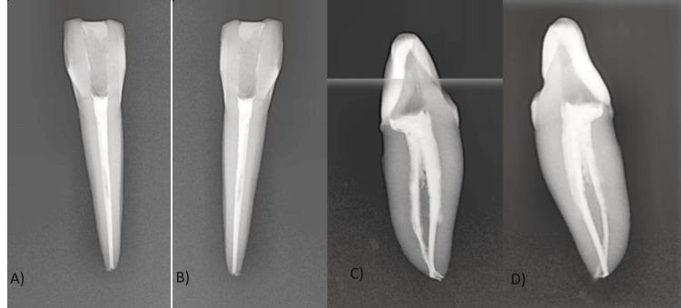
This set of four digital periapical radiographs (labeled A-D) displays an endodontically treated mandibular lateral incisor from multiple projections: A) labial, B) lingual, C) mesial, and D) distal. The images illustrate a complex Vertucci type VII root canal configuration (1-2-1-2). In the labial and lingual views, the obturation appears as a single, uniform radiopaque mass tapering toward the apex. However, the proximal mesial and distal projections reveal a bifurcation into two distinct buccal and lingual canals in the middle third of the root. These canals follow a parallel path before converging and then diverging again in the apical third, forming an apical delta and a lateral canal as visible in D. The obturation material (gutta-percha and resin sealer) shows high radiopacity and adequate apical extent, though slightly shorter in the lingual canal. This visual is used to teach dental students about internal anatomical variations and the importance of multi-angled radiography in identifying complex pulp chamber and canal morphology.
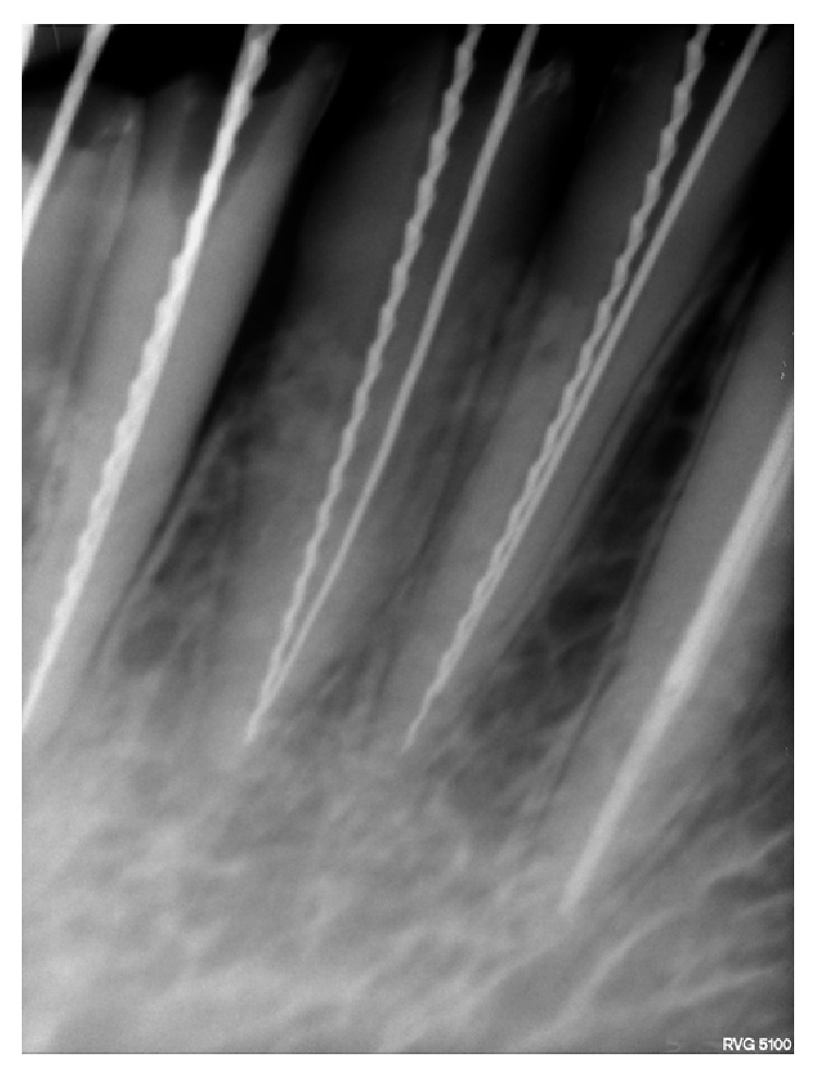
**Imaging Modality:** Intraoral periapical radiograph (digital). **Anatomical Region:** Mandibular anterior segment, specifically visualizing the mandibular right permanent central incisor (FDI 41) and lateral incisor (FDI 42). **Observed Findings:** The image depicts an endodontic working length determination or instrumentation phase. Both teeth 41 and 42 exhibit anatomical variations in the form of two distinct root canals per tooth (labial and lingual/palatal). **Characteristic Visual Features:** Four radiopaque endodontic instruments are visible within the root canal systems: * **Hedström (H-type) Files:** Identified by their characteristic teardrop-shaped, fluted profile with sharp, stacked cones, positioned in the distal/lingual canals. * **Reamers:** Identified by a more slender, twisted triangular or square cross-sectional appearance with a coarser pitch compared to the H-files, positioned in the mesial/labial canals. **Clinical Context:** The radiograph confirms the presence of dual canals in mandibular incisors, a critical morphological finding for successful endodontic therapy. The use of different instrument types (H-file vs. Reamer) serves as a visual marker to differentiate between the buccal and lingual canals on a two-dimensional radiographic plane. The surrounding alveolar bone shows a normal trabecular pattern.
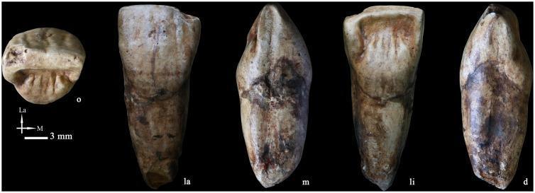
A clinical specimen image showing multiple views of a high-quality replica of a right upper central incisor (PA835). The tooth is displayed from five different anatomical perspectives: occlusal (o), labial (la), mesial (m), lingual (li), and distal (d). The occlusal view illustrates the biting surface morphology with visible ridges and grooves. The lingual aspect highlights a prominent basal eminence and several finger-like vertical projections near the incisal edge. The labial surface demonstrates a distinctive wrinkled texture with longitudinal ridges, sometimes associated with primitive cingulum-like structures. Both the crown and root portions show complex surface characteristics including beige-to-brown staining and textural variations. The root is robust, maintaining its breadth significantly before narrowing towards the apex. This specimen is used in dental anthropology and clinical education to study morphological variations in human dentition, specifically comparing modern and paleoanthropological features such as shovel-shaping and surface crenulation. A 3 mm scale bar is provided for reference.
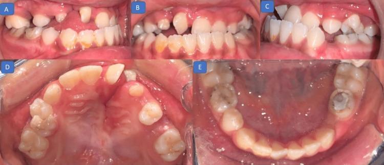
A composite of five intraoral clinical photographs documenting pretreatment dental status in a pediatric patient with a history of cleft lip and palate. (A, C) Right lateral views demonstrate an Angle Class III molar relationship, characterized by the mandibular teeth being mesial to the maxillary teeth, and a missing deciduous first molar. (B) Frontal view reveals a negative overjet (anterior crossbite), with maxillary incisors positioned lingual to the mandibular incisors. (D) Maxillary occlusal view shows a rotated right central incisor and a significant discontinuity of the alveolar ridge on the right side, consistent with a cleft defect. (E) Mandibular occlusal view shows a well-aligned arch with a large restoration and discoloration on the distal-most lower right tooth. The images illustrate malocclusion and anatomical discrepancies typical of orofacial cleft sequelae, including dental rotations, spacing, and skeletal Class III disharmony.
maxillary mandibular central lateral incisor crown morphology diagram dental anatomy
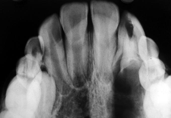
This diagnostic image is an occlusal radiograph showing the maxillary or mandibular anterior dentition. The central and lateral incisors are prominently visible as radiopaque structures, with their respective pulp chambers and root canals discernible. A notable finding is a well-defined area of radiolucency in the crown of a lateral incisor, characteristic of dental caries or a pathological lesion. The surrounding alveolar bone demonstrates typical trabecular patterns with varying radiopacity. The periodontal ligament space is visible as a thin, continuous radiolucent line encompassing the roots, and the lamina dura appears as a thin radiopaque border. The radiograph illustrates key dental anatomy, including the relationship between crown density, root morphology, and supporting periodontal structures, making it an essential educational resource for studying dental pathology and intraoral imaging techniques.
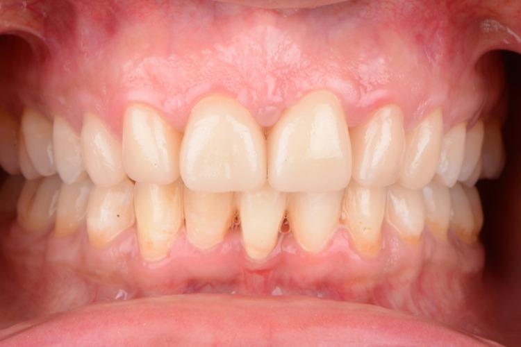
Frontal intraoral clinical photograph showing a five-year follow-up of a patient with congenitally missing maxillary central incisors, now restored with monolithic lithium disilicate crowns. The image demonstrates the esthetic result of orthodontic tooth repositioning, where lateral incisors were moved to the central positions and canines to lateral positions. The maxillary anterior teeth exhibit a uniform off-white shade with a morphology following the 'golden ratio' (central incisor width approximately 1.6 times that of lateral incisors). The gingival tissue shows a scalloped contour, post-esthetic crown lengthening, with slight blunting of the interdental papillae between the maxillary central incisors. The mandibular teeth show signs of mild gingival inflammation and visible dental plaque at the cervical margins. This clinical image illustrates complex multidisciplinary management involving orthodontics, periodontal surgery, and fixed prosthodontics in a patient with a history of cleft palate.
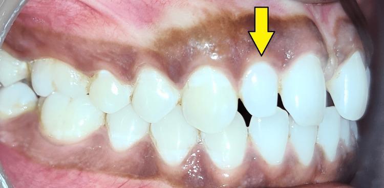
This clinical photograph is an intraoral, right lateral view of the maxillary and mandibular dentition. The primary focus is a peg-shaped lateral incisor (indicated by a yellow arrow), which demonstrates a characteristic microdontia characterized by a conical or tapered crown morphology that is significantly smaller than the adjacent central incisor. This dental anomaly has resulted in a localized diastema (gap) between the lateral and central incisors. The dentition displays a Class I molar and canine relationship. The surrounding gingival tissue and alveolar mucosa exhibit physiological hyperpigmentation, appearing as a diffuse dark brownish-purple hue across the keratinized and non-keratinized tissue. This image is an educational example of common dental developmental anomalies and their aesthetic impact on orthodontic alignment, typically addressed through restorative build-up or orthodontic space management.
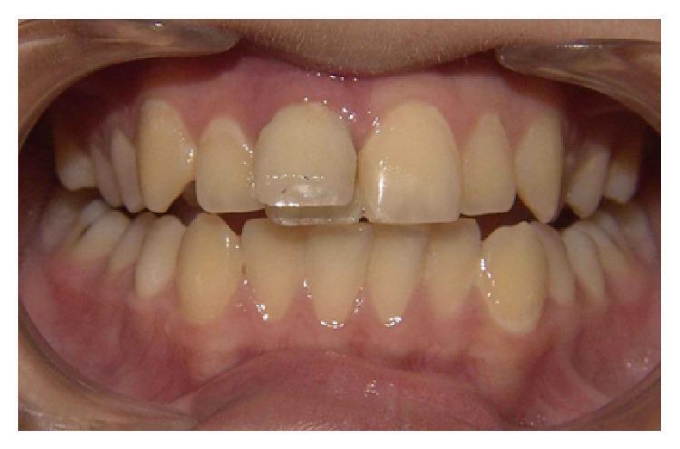
This clinical intraoral photograph demonstrates an anterior view of the maxillary and mandibular dentition with a primary focus on dental trauma. A significant crown fracture is visible on the maxillary right central incisor (tooth #8 or 11). The fracture line appears complex, with a vertical component and a transverse mesiodistal separation across the middle third of the crown. The incisal fragment is partially detached or displaced, showing a clear discrepancy in the incisal edge alignment compared to the intact maxillary left central incisor. The fracture extends cervically toward the gingival margin, and the clinical context indicates pulp exposure, making this a complicated crown fracture. The surrounding gingival tissue appears healthy with no immediate signs of acute swelling or laceration. The adjacent lateral incisors and mandibular anterior teeth remain intact, though they are visible for comparative assessment of tooth morphology and alignment. This image serves as an educational example of traumatic dental injury, specifically crown fractures in permanent dentition that necessitate endodontic evaluation and restorative management.
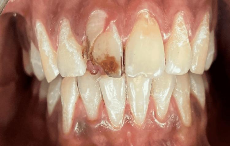
This clinical photograph displays a front-on intraoral view of the maxillary and mandibular dentition, focusing on a significant dental trauma. The maxillary right central incisor (tooth 11) presents a complicated crown fracture. The fracture line is oblique, extending from the mesial aspect toward the cervical area, resulting in a mobile coronal fragment that appears partially displaced. Notable brownish discoloration and exposed dentin are visible within the fractured area, suggesting possible pulpal involvement. The adjacent gingival tissues, particularly the interdental papilla and marginal gingiva surrounding tooth 11, exhibit signs of acute inflammation, including erythema and swelling. The surrounding teeth, including the maxillary left central incisor and lateral incisors, appear intact with normal morphology and alignment, though light plaque accumulation is visible at the gingival margins. This image serves as a clinical example of acute dental trauma requiring endodontic evaluation and restorative intervention, such as fragment reattachment or post-and-core rehabilitation.
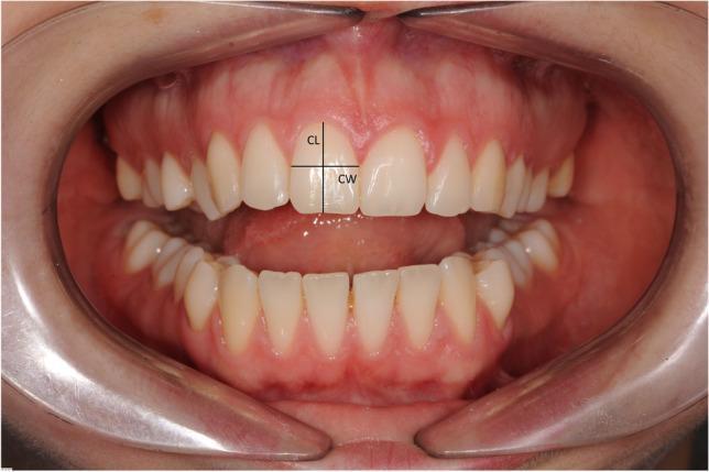
A frontal intraoral clinical photograph showing the maxillary and mandibular dental arches in a slightly open position, maintained by clear cheek retractors. The focus is on the crown morphology of the maxillary central incisors. The image displays linear measurement annotations on the right maxillary central incisor: Crown Length (CL), represented by a vertical line from the incisal edge to the gingival margin, and Crown Width (CW), represented by a horizontal line across the widest part of the tooth between the approximal surfaces. The gingival tissue appears healthy and pink with intact interdental papillae. The primary educational objective is to illustrate the parameters used to calculate the width-to-length ratio (W/L), a key metric in esthetic dentistry and orthodontics for assessing tooth proportions and the impact of crown morphology on periodontal outcomes, such as papilla recession following maxillary expansion.
maxillary mandibular central lateral incisor teeth principal identification features all views labial lingual mesial distal incisal dental anatomy
https://pocketdentistry.com/type-traits-that-differentiate-m…
maxillary central incisor labial view crown root morphology dental
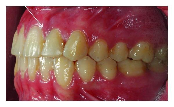
This intraoral clinical photograph provides a left labial view of the permanent maxillary dentition in a 14-year-old female. The primary focus is the maxillary left lateral incisor (tooth 22), which exhibits an abnormal crown morphology characterized by increased mesiodistal width. A vertical developmental groove is visible on the labial surface of the clinical crown, a hallmark feature indicative of dental fusion between the lateral incisor and a supernumerary tooth. The adjacent teeth, including the central incisor and canine, show signs of anterior crowding. The surrounding gingival tissues in the upper left quadrant appear erythematous and glossy with irregular contours, suggesting localized gingival inflammation or gingivitis. This image serves as a clinical example of a rare dental anomaly often associated with conditions like unilateral cleft lip, illustrating the aesthetic and functional challenges that necessitate combined orthodontic and restorative interventions.
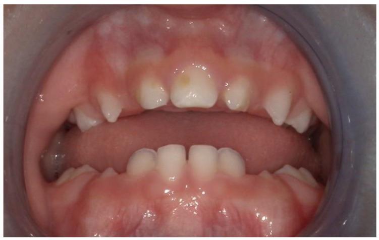
This clinical photograph displays an intraoral view of a pediatric patient's dentition, highlighting a solitary median maxillary central incisor (SMMCI). A single, symmetrically positioned, broad rectangular incisor is visible at the maxillary midline, replacing the normal pair of central incisors. The tooth crown shows a subtle, small yellow focal discoloration on the labial surface. The surrounding gingiva and oral mucosa appear healthy, moist, and pink without signs of inflammation. The palate exhibits a high-arched, ogival morphology with a prominent anterior vertex. This finding is a key clinical marker often associated with midline developmental anomalies such as congenital nasal pyriform aperture stenosis (CNPAS) or holoprosencephaly (HPE) spectrum. The mandibular dentition shows typical primary incisors with normal spacing. The image serves as an educational example of dental dysmorphology related to genetic syndromes involving midline facial development.
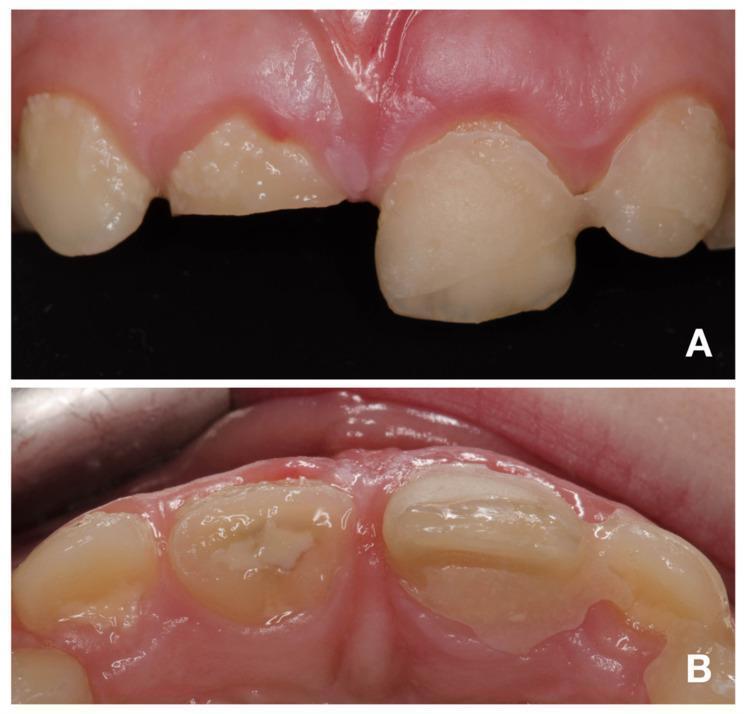
Two intraoral clinical photographs showing a traumatic dental injury in the maxillary anterior region (teeth 12–22). View A (buccal/labial) demonstrates a complicated crown-root fracture of the maxillary right central incisor (tooth 11). The fracture is oblique, extending apically from the labial toward the palatal surface. Stabilization is provided by a dental splint connecting tooth 21 to the adjacent primary canine (63). View B (occlusal) provides a superior perspective of the injury, highlighting the palatal margin of the fracture on tooth 11, located approximately 2 mm above the marginal gingiva. The image illustrates severe dental trauma, likely classified as Ellis Class III, involving the enamel, dentin, and pulp. The surrounding gingival tissue shows slight erythema consistent with recent trauma. This material is used in pediatric dentistry and endodontics to teach the visual assessment of crown-root fractures and the clinical application of emergency stabilization splints in mixed dentition.
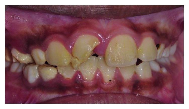
Frontal view intraoral clinical photograph showing the maxillary and mandibular anterior teeth. The primary pathology is a complicated crown-root fracture of the maxillary right central incisor (tooth 11). The fracture line is oblique, originating on the labial surface and extending palatally and subgingivally. A significant portion of the distal-incisal crown structure is missing, and the remaining coronal fragment appears slightly displaced. The surrounding gingival tissue at the site of injury shows signs of acute inflammation, including erythema and swelling, specifically in the interdental papilla area. The dentition exhibits generalized yellowish discoloration and extrinsic staining, with visible plaque accumulation along the gingival margins. The alignment of the maxillary and mandibular incisors is slightly irregular, characterized by minor crowding. The clinical presentation is consistent with traumatic dental injury requiring multidisciplinary management, including endodontic treatment, orthodontic extrusion, and restorative rehabilitation.
A clean dental anatomy educational diagram showing side-by-side comparison of four teeth in multiple views: Maxillary Central Incisor, Maxillary Lateral Incisor, Mandibular Central Incisor, and Mandibular Lateral Incisor. Each tooth shown from 5 views: Labial, Lingual, Mesial, Distal, and Incisal. Label all key features: mesioincisal angle, distoincisal angle, cingulum, marginal ridges, lingual fossa, contact areas, root shape, crown outline. Use clear black outlines on white background with neat labels and a title. Educational dental morphology chart style.
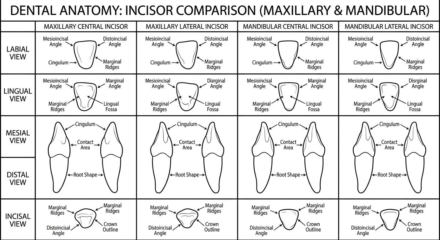
Central vs. Lateral Incisors - Complete Dental Anatomy Reference
1. OVERVIEW - At a Glance
| Feature | Maxillary Central | Maxillary Lateral | Mandibular Central | Mandibular Lateral |
|---|---|---|---|---|
| FDI Notation | 11, 21 | 12, 22 | 41, 31 | 42, 32 |
| Crown Size | Largest anterior | Smaller than central | Narrowest crown in mouth | Slightly larger than mandib. central |
| Eruption Age | 7-8 yrs | 8-9 yrs | 6-7 yrs | 7-8 yrs |
| Root | Shortest root/crown ratio of any tooth | Longer root than central | Single, narrow, flat | Slightly longer & wider than central |
| Symmetry | Asymmetric (distal more rounded) | More asymmetric | Nearly perfectly symmetric | Less symmetric |
2. MAXILLARY INCISORS - All Views
LABIAL VIEW
- Largest and most prominent anterior tooth
- Crown is wider mesiodistally than the lateral
- Mesioincisal angle: nearly a right angle (sharp, 90°)
- Distoincisal angle: rounded/obtuse
- Three developmental lobes visible as faint ridges (mamelons in young teeth)
- Cervical line curves toward root (convex toward incisal)
- Root: conical, tapers evenly, relatively short compared to crown (smallest root-to-crown ratio of any permanent tooth)
- Crown is smaller, narrower, and more rounded than the central
- More variation in shape (peg lateral is a common anomaly)
- Both mesioincisal and distoincisal angles are more rounded than the central
- Distoincisal angle is noticeably more rounded than on the central
- Root: longer in proportion to the crown compared to the central
LINGUAL VIEW
- Prominent cingulum slightly off-center to the distal
- Mesial marginal ridge is longer than distal marginal ridge
- Shallow-to-moderate lingual fossa
- Incisal ridge, mesial and distal marginal ridges, and cingulum form the boundaries of the lingual fossa
- Cingulum is more prominent and centered
- Deeper lingual fossa with a caries-prone pit adjacent to the cingulum (more susceptible to decay than the central)
- Marginal ridges are more prominent
- Deep pit at the fossa near the cingulum is a hallmark identification feature of the lateral
MESIAL VIEW
- Crown appears triangular
- Labial surface is slightly convex
- Incisal edge lies just labial to the root axis line or on the root axis line
- Slight distolingual twist of the incisal ridge (distal portion is slightly more lingual)
- Cervical line: greatest curvature of any tooth (deepest CEJ curve mesially)
- Similar triangular profile
- Cervical line curvature is less than the central
- Crown is narrower labiolingually
DISTAL VIEW
- Less cervical curvature than mesial view
- Incisal edge appears slightly more lingual due to the distolingual twist
- Cervical line curvature is less than mesial side
INCISAL (OCCLUSAL) VIEW
- Crown outline is wider mesiodistally than labiolingually (roughly triangular)
- Labial surface forms the broad curved base of the triangle
- Cingulum is off-center toward the distal
- Mesial marginal ridge appears longer than distal marginal ridge
- Crown outline is more oval/round - mesiodistal and labiolingual dimensions are more equal
- Less wide mesiodistally compared to central
- Cingulum appears more centered
PROXIMAL CONTACT AREAS - MAXILLARY (Key Identification Feature)
| Mesial Contact | Distal Contact | |
|---|---|---|
| Central | Incisal third (near incisal edge) | Near incisal/middle third junction |
| Lateral | Incisal third (slightly more cervical than central) | Middle third (most cervical of all incisors) |
3. MANDIBULAR INCISORS - All Views
LABIAL VIEW
- Narrowest crown in the entire mouth
- Nearly bilaterally symmetric (hardest to distinguish left from right)
- Mesioincisal angle = distoincisal angle (both form nearly right angles)
- Smooth labial surface (may have 2 shallow developmental depressions in incisal third)
- Incisal edge is essentially straight and horizontal
- Crown is slightly larger in all dimensions than the mandibular central (OPPOSITE of maxillary - lateral is bigger than central in mandible)
- Less symmetric than the central
- Distoincisal angle is noticeably more rounded than the mesioincisal angle
- Incisal edge slopes toward the distal
- This asymmetry helps identify right vs. left
LINGUAL VIEW
- Small cingulum
- Shallow lingual fossa
- Marginal ridges are barely visible (less developed)
- Symmetric appearance
- Cingulum is slightly more developed
- Lingual fossa is deeper and more concave
- Marginal ridges are more prominent
- Asymmetry visible (distal side more developed/prominent)
MESIAL VIEW
- Crown is narrow labiolingually
- Incisal edge is nearly centered over the root axis
- Cervical line curves toward the incisal (mesial curvature greater than distal)
- Similar narrow profile
- Root may show slight distal inclination
- Incisal edge may be slightly lingual to root axis
DISTAL VIEW
- Less curvature of cervical line than mesial
- Similar appearance; crown appears twisted slightly distally (distolingual rotation is common in mandibular laterals)
INCISAL VIEW
- Crown outline is very narrow and nearly symmetric
- Appears like a thin rectangle or narrow oval
- Crown outline shows slight asymmetry
- Distal half appears slightly more developed/fuller
- May show slight distolingual rotation
PROXIMAL CONTACT AREAS - MANDIBULAR (Key Identification Feature)
| Mesial Contact | Distal Contact | |
|---|---|---|
| Central | Incisal third | Incisal third (SAME level as mesial - unique!) |
| Lateral | Incisal third | Incisal third but noticeably more cervical than mesial |
Rule to remember: The mandibular central is the ONLY incisor where mesial and distal contacts are at the same height.
4. MASTER COMPARISON TABLE - Central vs. Lateral (All Features)
| Feature | Central Incisor | Lateral Incisor |
|---|---|---|
| Crown width | Maxillary: widest; Mandibular: narrowest | Max: smaller; Mand: slightly larger than central |
| Mesioincisal angle | Sharp/right angle | More rounded |
| Distoincisal angle | Rounded (less than mesio) | More rounded (especially mandibular) |
| Symmetry | Max: asymmetric; Mand: nearly symmetric | Both: more asymmetric |
| Root length | Maxillary: shortest R:C ratio | Longer root proportionally |
| Lingual fossa | Shallow | Deeper (especially maxillary - with pit) |
| Cingulum | Maxillary: off-center distal | More centered/prominent |
| Distal contact level | Near incisal/middle junction (max) | Middle third (max) - most cervical of incisors |
| Incisal edge | Straight/horizontal (mand central) | Slopes toward distal (mand lateral) |
| Root shape (proximal) | Single, flattened | Similar, may have distal concavity |
| Anomaly tendency | Less variation | More (peg lateral, dens invaginatus, pit) |
5. PRINCIPAL IDENTIFICATION FEATURES (Summary Cards)
MAXILLARY CENTRAL - "Identify by:"
- Largest anterior crown
- Sharp mesioincisal angle
- Short root relative to crown
- Widest mesiodistally among incisors
- Off-center distal cingulum
MAXILLARY LATERAL - "Identify by:"
- Deep lingual pit/fossa (caries-prone)
- Distal contact in the middle third (most cervical)
- Longer root relative to crown than central
- More rounded corners
- More oval incisal outline
MANDIBULAR CENTRAL - "Identify by:"
- Narrowest crown in the mouth
- Nearly perfect bilateral symmetry
- Mesial and distal contacts at same height
- Straight horizontal incisal edge
- Very shallow lingual fossa
MANDIBULAR LATERAL - "Identify by:"
- Slightly larger than mandibular central (reversed from maxillary)
- Rounded distoincisal angle (sloping incisal edge)
- Distal contact more cervical than mesial
- Often shows distolingual rotation
- Deeper lingual fossa than central
6. Visual Reference - Multi-View Specimen