
Everything about urinary bladder anatomy physiology and carcinoma in very detail with reference to standard textbooks
Reading File
Finding Sources
Finding Sources
Searching PubMed
"bladder cancer"[MeSH Terms] AND "urothelial carcinoma"[MeSH Terms]
Now let me read the key content blocks in parallel from the most comprehensive sources:
Reading File
Reading File
Reading File
Reading File
Reading File
Reading File
Reading File
Searching Images
urinary bladder anatomy layers wall structure
Reading File
Searching Images
bladder carcinoma urothelial transitional cell carcinoma cystoscopy histology
Searching PubMed
"bladder cancer"[MeSH Terms] AND "treatment"[MeSH Terms]
Reading File
Finding Sources
Reading File
Finding Sources
Reading File
Reading File
I now have comprehensive content from multiple major textbooks. Let me compile the complete, detailed answer.# Urinary Bladder: Anatomy, Physiology, and Carcinoma
PART I: ANATOMY
1. Location and General Description
The urinary bladder is a hollow, distensible muscular reservoir for urine situated in the lesser pelvis, posterior to the pubic symphysis, and embedded in subperitoneal connective tissue. Its size and shape change dynamically with filling: when empty it is bowl-shaped and the apex descends; as it fills, the apex and wall are drawn forward and upward between the peritoneum and abdominal wall, forming an ovoid shape. A full bladder can extend as far as the superior border of the pubic symphysis — this anatomical fact permits suprapubic aspiration without entering the peritoneal cavity.
- Normal capacity: approximately 500 mL
- Urge to void: typically sensed at ~300 mL
— Color Atlas of Human Anatomy Vol. 2, Internal Organs
2. Parts of the Bladder
| Part | Description |
|---|---|
| Body | Largest part; constitutes most of the organ |
| Apex (vertex) | Anterosuperior; gives attachment to the obliterated urachus (median umbilical ligament) running to the umbilicus |
| Fundus (base) | Posterior and inferior; the two ureters open into its lateral aspects |
| Neck | Inferior; continuous anteriorly with the urethra |
— Color Atlas of Human Anatomy Vol. 2, Internal Organs
3. Internal Surface and Trigone
The inner surface of the bladder is pale red. It has two distinct zones:
Rugose mucosa: Throughout most of the bladder, the mucosa is thrown into folds due to its mobility against the underlying muscular layer. When the bladder is distended these folds completely flatten.
Trigone of the bladder: The triangular region on the fundus bounded by:
- The two ureteric orifices (posterolateral angles)
- The internal urethral orifice (anterior angle)
The trigone mucosa is flat and firmly attached to the underlying muscle — it does not form folds at any degree of filling. In the male, the uvula of the bladder (a conical elevation produced by the prostate) projects into the internal urethral orifice.
Embryological basis: The trigone is derived from the mesonephric (Wolffian) ducts and therefore lacks a muscularis mucosae and submucosal layer — the remainder of the bladder wall originates from the cloaca (hindgut) and has a fully developed layered wall analogous to the gastrointestinal tract.
— Histology: A Text and Atlas with Correlated Cell and Molecular Biology; Color Atlas of Human Anatomy Vol. 2
4. Wall Layers (Microanatomy)
The bladder wall is composed of three fundamental layers:
a) Mucosa (Urothelium + Lamina Propria)
- Lined by transitional epithelium (urothelium) — a specialized stratified epithelium capable of stretching
- The urothelium consists of basal cells, intermediate cells, and superficial umbrella cells (large, dome-shaped cells that expand during distension)
- Beneath lies the lamina propria (loose connective tissue); this layer is absent at the trigone
b) Muscularis Propria (Detrusor Muscle)
- The smooth muscle bundles are randomly mixed with collagen — less regularly arranged than in tubular portions of the urinary tract
- Toward the urethral opening, the muscle fibers form the involuntary internal urethral sphincter (a ring-like arrangement of smooth muscle)
- At the trigone, the muscular layer is a direct continuation of the ureteral smooth muscle (only two layers instead of three)
- Around the ureteric openings, smooth muscle is organized in a complex circular arrangement
c) Adventitia / Serosa
- The serosa (peritoneum + subserosa connective tissue) covers only the superior surface and the portion of the posterior surface above the trigone
- The remainder is covered by adventitia only
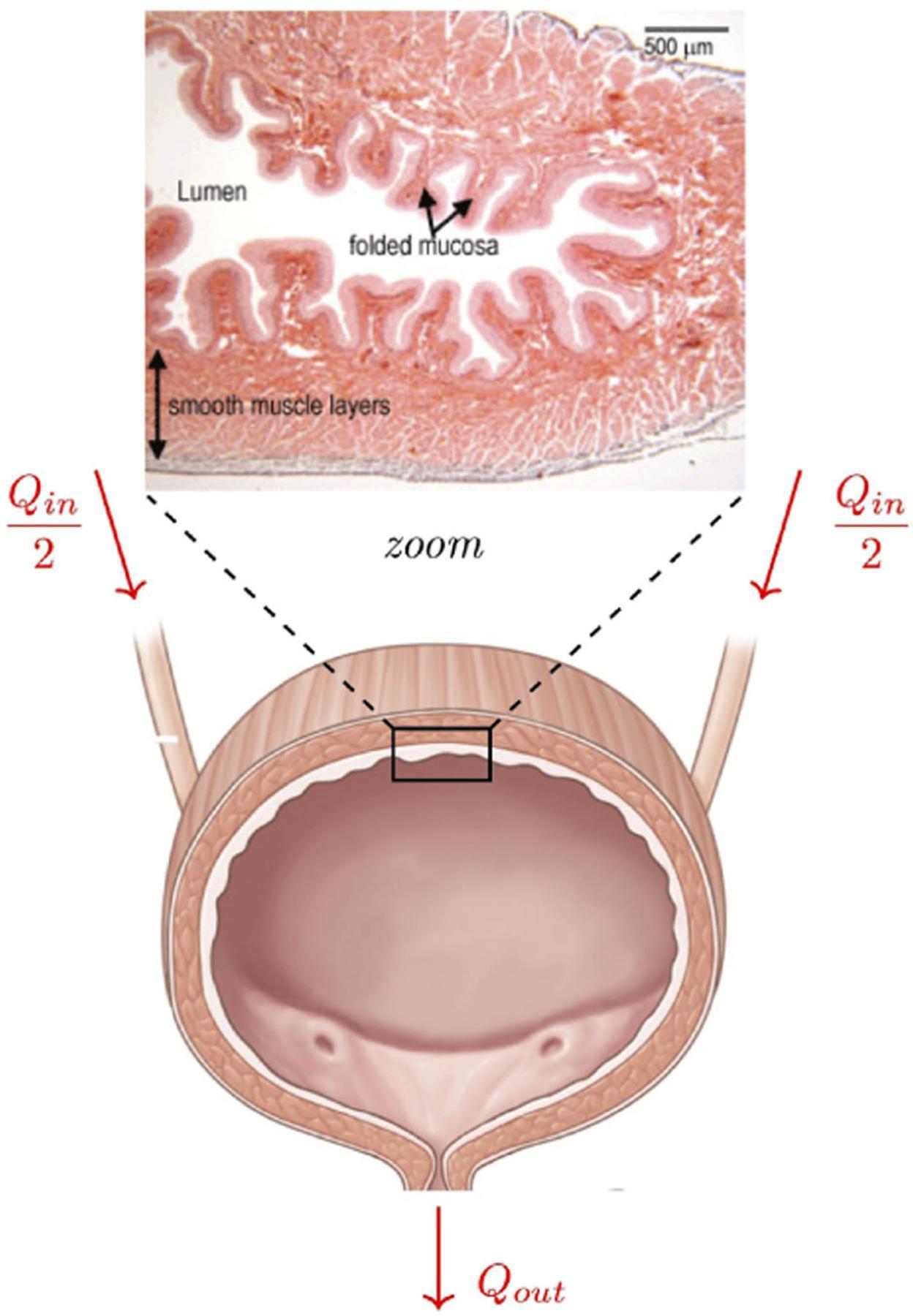
Histomicrograph of bladder wall: folded urothelial mucosa (rugae) and underlying smooth muscle (detrusor). The folded geometry enables compliance during filling.
— Histology: A Text and Atlas; Color Atlas of Human Anatomy Vol. 2
5. Blood Supply
- Arteries: Branches of the bilateral internal iliac arteries:
- Superior vesical artery (from the umbilical artery) — supplies the upper bladder and trigone
- Inferior vesical artery — supplies the fundus and neck
- Veins: The vesical venous plexus surrounds the fundus and drains directly into the internal iliac veins
6. Lymphatic Drainage
Lymphatics drain to:
- External iliac nodes (primary)
- Internal iliac (obturator) nodes
- Common iliac nodes (advanced spread)
7. Nerve Supply
Innervation is divided into extrinsic and intrinsic systems:
| System | Origin | Action |
|---|---|---|
| Parasympathetic | S2–S4 (pelvic splanchnic nerves → terminal ganglia in bladder wall) | Contracts detrusor (via M3 muscarinic receptors); relaxes internal sphincter (via nitric oxide) |
| Sympathetic | Hypogastric plexus (L1–L3) | Relaxes detrusor (via β₃ adrenoceptors); contracts internal urethral sphincter (via α₁ adrenoceptors) |
| Somatic | Pudendal nerve (S2–S4) | Maintains tonic contraction of the external urethral sphincter (skeletal muscle, via nicotinic ACh receptors) |
— Histology: A Text and Atlas; Costanzo Physiology 7th Edition; Color Atlas of Human Anatomy Vol. 2
8. Embryology
The urinary bladder develops from the urogenital sinus, which is partitioned from the cloaca by the urorectal septum around the 4th–7th week. The urogenital sinus is divided into three parts:
- Vesical part → forms the main body and dome of the bladder
- The ureters are absorbed into the posterior wall → form the trigone
- The urachus (allantois remnant) connects the fetal bladder to the umbilicus; normally it obliterates at birth to form the median umbilical ligament
— The Developing Human: Clinically Oriented Embryology
PART II: PHYSIOLOGY — MICTURITION
1. Overview
Micturition (voiding) is controlled by the autonomic nervous system but subject to voluntary modulation via the somatic nervous system. The two phases are filling/storage and voiding.
2. Filling Phase (Sympathetic Predominance)
When the bladder fills with urine:
- Sympathetic activity (L1–L3) predominates
- Noradrenaline (NA) stimulates β₂/β₃ receptors on the detrusor → relaxes the bladder wall (increases compliance, reduces intravesical pressure)
- NA stimulates α₁ receptors on the internal sphincter → contracts the sphincter
- The external urethral sphincter is maintained closed by voluntary somatic (pudendal nerve) action
3. Voiding Phase (Parasympathetic Predominance)
When the bladder is full (~300 mL):
- Mechanoreceptors in the bladder wall sense stretch and transmit afferent signals via sacral spinal cord → brain stem
- The micturition reflex is coordinated in the midbrain (pontine micturition center)
- Parasympathetic activity (S2–S4) predominates:
- ACh stimulates M3 muscarinic receptors on the detrusor → contraction → increased intravesical pressure
- Nitric oxide released → relaxes the internal sphincter
- Simultaneously, voluntary inhibition of pudendal nerve firing → relaxation of the external sphincter
- Urine is expelled
Key concept: Sympathetic and parasympathetic actions are precisely reciprocal — sympathetic dominates filling (bladder relaxes, sphincter contracts); parasympathetic dominates voiding (bladder contracts, sphincter relaxes). — Costanzo Physiology 7th Edition
PART III: BLADDER CARCINOMA
1. Epidemiology
- Bladder cancer is the 9th most common cancer worldwide and carries significant morbidity and mortality
- More common in men (M:F ≈ 3–4:1), in higher-resource countries, and in urban populations
- ~80% of patients are 50–80 years of age
- ~90% of bladder cancers in the United States are urothelial carcinomas (UC)
- Squamous cell carcinoma (SCC): 2–5% in the US, but much more prevalent in regions where urinary schistosomiasis is endemic (East Africa, Middle East)
- Adenocarcinoma: rare (~1%)
— Robbins & Kumar Basic Pathology (Robbins Pathology); Robbins, Cotran & Kumar Pathologic Basis of Disease
2. Etiology and Risk Factors
| Risk Factor | Notes |
|---|---|
| Cigarette smoking | Single most important risk factor; 2–3× increased risk; responsible for ~50% of cases |
| Occupational carcinogens | Aromatic amines (2-naphthylamine, benzidine) — workers in aniline dye, rubber, leather, paint industries |
| Schistosoma haematobium | Parasite in endemic regions → chronic bladder irritation → squamous metaplasia → SCC |
| Radiation therapy | Prior pelvic irradiation (prostate, uterine cancer) increases UC risk years later |
| Cyclophosphamide | Hemorrhagic cystitis + increased bladder cancer risk with prolonged exposure |
| Analgesic abuse | Phenacetin-containing analgesics implicated (as in analgesic nephropathy) |
| Family history | Known risk factor |
| Chronic cystitis / bladder stones | Chronic irritation predisposes to squamous metaplasia and SCC |
— Robbins, Cotran & Kumar Pathologic Basis of Disease
3. Pathogenesis and Molecular Biology
There are two largely distinct molecular pathways of tumor progression:
Pathway 1: Superficial Papillary (Non-Muscle-Invasive) Tumors
Normal urothelium → Papilloma / PUNLMP → Low-grade papillary UC → (rarely) High-grade UC → Muscle invasion
- Gain-of-function mutations activating growth factor receptor pathways:
- FGFR3 tyrosine kinase receptor amplifications/mutations (most common, ~70% of low-grade papillary UC)
- RAS activating mutations
- PI3-kinase activating mutations
- These tumors frequently recur but progress to muscle invasion in only ~20% of cases
- TP53 mutations occur later in progression if muscle invasion does occur
Pathway 2: Carcinoma in Situ (CIS) → Muscle-Invasive Cancer
Normal urothelium → Urothelial dysplasia → Flat CIS → Muscle-invasive UC
- Driven by loss-of-function mutations in tumor suppressor genes:
- TP53 mutations (early in CIS development)
- RB (retinoblastoma) gene inactivation
- Chromatin-remodeling gene mutations (e.g., KDM6A, ARID1A, KMT2D)
- DNA repair pathway defects
- Bladder cancers have a high burden of somatic mutations (comparable to lung cancer and melanoma — consistent with carcinogen-driven mutagenesis)
RNA expression analysis suggests several relatively distinct molecular subtypes of bladder cancer (luminal vs. basal subtypes), with implications for prognosis and treatment.
— Robbins, Cotran & Kumar Pathologic Basis of Disease; Robbins & Kumar Basic Pathology

4. WHO Classification (2022) of Urothelial Tumors
Flat (Non-Papillary) Lesions:
- Urothelial dysplasia
- Urothelial carcinoma in situ (CIS) — flat, high-grade lesion
Exophytic Papillary Lesions (in order of increasing malignancy):
| Category | Features |
|---|---|
| Papilloma | 1% of bladder tumors; histologically identical to normal urothelium; rare recurrence |
| Inverted papilloma | Benign; anastomosing cords of urothelium growing into lamina propria |
| PUNLMP (Papillary urothelial neoplasm of low malignant potential) | Thicker epithelium than papilloma; rare progression |
| Low-grade papillary UC | Orderly architecture, low-grade cytologic atypia |
| High-grade papillary UC | Disordered architecture, significant cytologic atypia |
| Invasive UC | Lamina propria or beyond |
— Robbins, Cotran & Kumar Pathologic Basis of Disease (Table 21.3)
5. Morphology
Macroscopic Appearances:
- Papillary tumors: Red, elevated excrescences ranging from <1 cm to 5 cm in diameter; multiple tumors often present; frond-like (cauliflower) gross appearance
- Flat/CIS: Flat, velvety, red mucosal areas; may be difficult to see on cystoscopy
- Invasive/high-grade tumors: Nodular, ulcerated, fungating masses; yellow areas of necrosis; may spread over wide areas
Microscopic Features:
- Low-grade UC: Uniform urothelial cells with preserved polarity, evenly spaced nuclei, rare mitoses
- High-grade UC: Marked nuclear pleomorphism, loss of polarity, numerous mitoses, necrosis
- CIS: Full-thickness atypia of urothelium without invasion; discohesive cells, hyperchromatic pleomorphic nuclei, increased mitoses, intact basement membrane
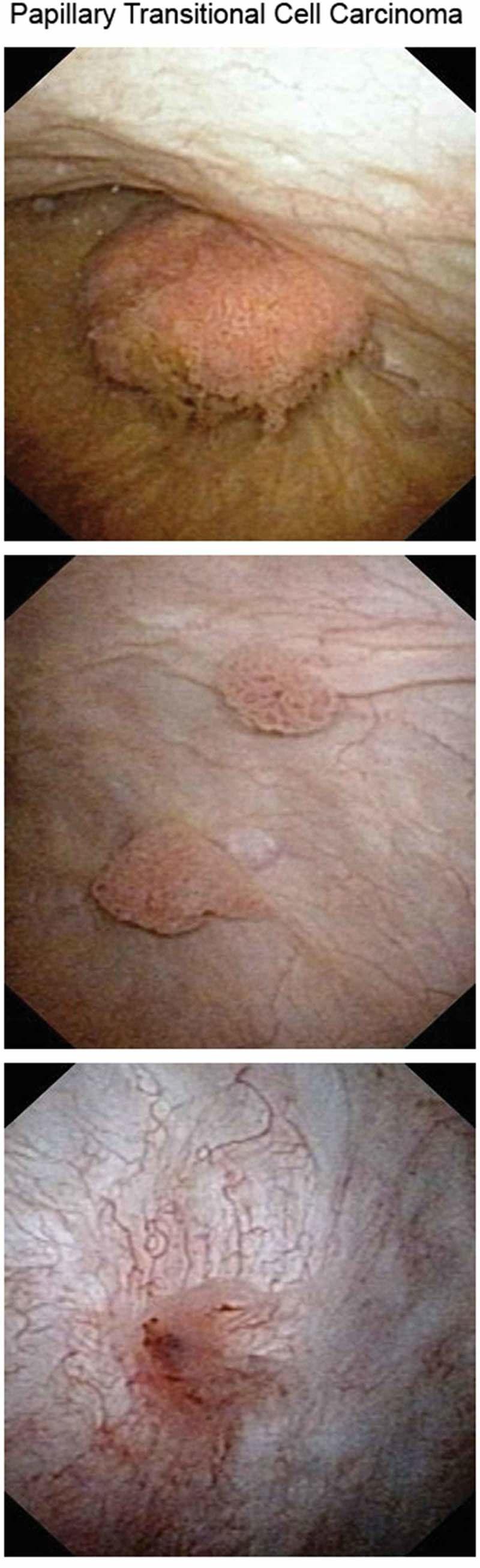
Cystoscopic appearance: papillary urothelial carcinoma with frond-like projections (top/middle frames) and irregular tumor vascularity (bottom frame).
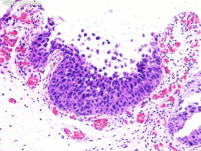
Urothelial carcinoma in situ: discohesive, pleomorphic cells with high N:C ratio and mucosal denudation.
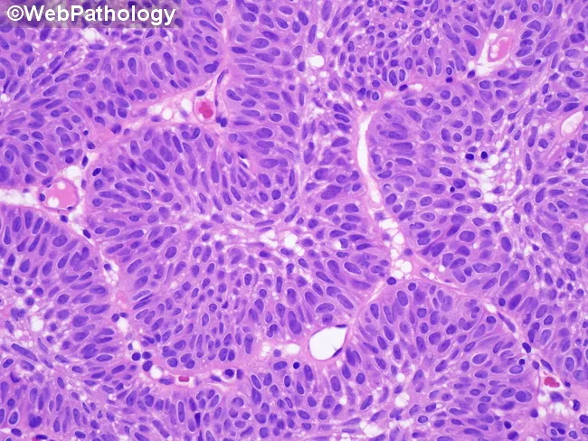
Inverted papilloma: endophytic growth with bland cytology — contrasts with CIS and invasive carcinoma.
6. Pathologic Staging — AJCC 8th Edition (pTNM)
| Stage | Definition |
|---|---|
| pTa | Noninvasive papillary carcinoma (confined to urothelium) |
| pTis | Flat urothelial CIS |
| pT1 | Invades lamina propria (subepithelial connective tissue) |
| pT2a | Invades superficial muscularis propria (inner half) |
| pT2b | Invades deep muscularis propria (outer half) |
| pT3a | Invades perivesical soft tissue microscopically |
| pT3b | Invades perivesical soft tissue macroscopically |
| pT4a | Invades prostatic stroma, seminal vesicles, uterus, or vagina |
| pT4b | Invades pelvic wall or abdominal wall |
| N1 | Single regional node (true pelvis: perivesical, obturator, internal/external iliac, sacral) |
| N2 | Multiple regional nodes in true pelvis |
| N3 | Common iliac lymph node metastasis |
| M1a | Distant lymph nodes beyond common iliac arteries |
| M1b | Non-lymph node distant metastases |
pTa, Tis, T1 = Non-Muscle-Invasive Bladder Cancer (NMIBC) (~70% at presentation) T2 and above = Muscle-Invasive Bladder Cancer (MIBC) (~20–30%)
— Robbins, Cotran & Kumar Pathologic Basis of Disease (Table 21.4); Sabiston Textbook of Surgery
7. Clinical Features
- Painless hematuria (gross or microscopic) — hallmark presenting symptom; even a single episode on urinalysis warrants full workup in at-risk patients
- Irritative voiding symptoms (frequency, urgency, dysuria) in the absence of infection — especially common with CIS
- Advanced disease: flank pain (ureteric obstruction), pelvic pain, lower extremity edema (lymph node involvement)
8. Diagnosis
- Urine cytology — sensitive for high-grade tumors and CIS; can be equivocal in low-grade tumors
- Cystoscopy — gold standard; direct visualization of the urothelial surface
- TURBT (Transurethral Resection of Bladder Tumor) — both diagnostic (histology + staging) and therapeutic
- Upper tract imaging — contrast-enhanced CT urography to evaluate upper urothelial tract
- Bimanual examination under anesthesia — palpable mass after TURBT = extravesical extension (T3); fixed mass = possible T4
- Urine biomarkers — not routinely recommended due to cost and low specificity
9. Treatment
Non-Muscle-Invasive Bladder Cancer (NMIBC)
| Risk | Management |
|---|---|
| Low-risk (small, low-grade, solitary, Ta) | TURBT alone; single immediate intravesical mitomycin C (or gemcitabine) within 24 hrs |
| Intermediate-risk | TURBT + intravesical chemotherapy |
| High-risk (high-grade, T1, CIS, multifocal, recurrent) | TURBT + intravesical BCG (Bacillus Calmette-Guérin) — induction 6 weeks + maintenance; BCG elicits local inflammatory destruction of tumor |
| BCG-unresponsive | Consider radical cystectomy or clinical trial |
- NMIBC has up to 70% recurrence rate — requires lifelong surveillance cystoscopy
- Most recurrences are clonally related (shed and implanted from original tumor at distant sites)
- Low-grade papillary UC, PUNLMP, and papilloma: 98% 10-year survival regardless of recurrences
Muscle-Invasive Bladder Cancer (MIBC)
- Neoadjuvant cisplatin-based chemotherapy followed by radical cystectomy (gold standard for resectable MIBC)
- Radical cystectomy includes: cystoprostatectomy in males, anterior exenteration in females, plus pelvic lymph node dissection and urinary diversion
- Bladder-sparing trimodality therapy: maximal TURBT + concurrent chemoradiation — for selected patients unfit for or refusing cystectomy
- Adjuvant immunotherapy (e.g., nivolumab) for high-risk pathologic stage after cystectomy
Metastatic Bladder Cancer
- First-line: Platinum-based chemotherapy (cisplatin + gemcitabine or MVAC — methotrexate, vinblastine, doxorubicin, cisplatin)
- Immune checkpoint inhibitors: Pembrolizumab, atezolizumab — for platinum-ineligible patients or after platinum failure
- Enfortumab vedotin (antibody-drug conjugate targeting Nectin-4) ± pembrolizumab — emerging standard for advanced UC
- Erdafitinib — FGFR3 inhibitor for tumors with FGFR3/FGFR2 alterations
— Robbins & Kumar Basic Pathology; Sabiston Textbook of Surgery
10. Special Types of Bladder Carcinoma
Squamous Cell Carcinoma (SCC)
- 3–7% of US bladder cancers; much more common in endemic schistosomiasis regions
- Arises from atypical keratinizing mucosa (squamous dysplasia and CIS)
- Nearly always invasive, fungating, or ulcerative at presentation
- Chronic irritation/infection-driven (bladder stones, indwelling catheters, chronic cystitis)
Adenocarcinoma
- Rare (<2%)
- May arise from: cystitis glandularis/cystica with intestinal metaplasia, urachal remnants (20–40% of bladder adenocarcinomas), or bladder exstrophy
- Urachal adenocarcinoma: located at the dome/anterior wall; difficult to stage by TNM; arises from patent urachus
- Histologically identical to GI adenocarcinomas
Small Cell Carcinoma
- Rare; indistinguishable from small-cell carcinoma of the lung
- Highly aggressive; treated with chemotherapy analogous to lung small-cell protocols
11. Congenital Conditions with Neoplastic Risk
| Condition | Risk |
|---|---|
| Bladder exstrophy | Exposed mucosa undergoes intestinal glandular metaplasia + chronic infection → increased risk of adenocarcinoma |
| Urachal cyst/anomalies | Lined by urothelial or metaplastic glandular epithelium → adenocarcinoma (0.1–0.3% of all bladder cancers, 20–40% of bladder adenocarcinomas) |
| Bladder diverticula | Carcinoma arising in diverticula is more advanced at diagnosis due to thin/absent muscularis propria layer |
| Vesicoureteral reflux | Predisposes to ascending pyelonephritis; not directly neoplastic |
— Robbins, Cotran & Kumar Pathologic Basis of Disease
12. Prognosis
| Stage | 5-Year Survival |
|---|---|
| pTa / PUNLMP / Papilloma | ~98% (10-year) |
| pT1 (lamina propria invasion) | ~70–80% |
| pT2 (muscle invasion) | ~50–60% |
| pT3–T4 | ~20–30% |
| Metastatic (M1) | <10% |
- CIS alone (without invasive carcinoma): 28% progress to muscle-invasive; 7% die of disease
- CIS + infiltrating UC: 59% progress; 45% die of disease
- High-grade papillary UC: invasion and cancer death in ~25%
KEY CONCEPTS SUMMARY
Anatomy: The bladder is a pelvic hollow muscular organ; its wall comprises urothelium, lamina propria, and detrusor (three smooth-muscle layers). The trigone is embryologically distinct (mesonephric duct origin), lacks a muscularis mucosae, and does not fold. Blood supply is from superior and inferior vesical arteries (internal iliac); innervation is sympathetic (L1–L3), parasympathetic (S2–S4), and somatic (pudendal, S2–S4).
Physiology: Filling is sympathetically mediated (detrusor relaxation via β₂/β₃; internal sphincter contraction via α₁). Voiding is parasympathetically mediated (detrusor contraction via M3; internal sphincter relaxation via NO). The pontine micturition center coordinates the reflex; the external sphincter is under voluntary somatic control.
Carcinoma: ~90% are urothelial carcinomas. Two molecular pathways exist — (1) FGFR3/RAS/PI3K-driven non-muscle-invasive papillary tumors that recur frequently but rarely invade, and (2) TP53/RB-driven flat CIS lesions that frequently progress to muscle-invasive cancer. TNM staging (AJCC 8th) is critical: NMIBC (Ta/Tis/T1) is managed with TURBT ± intravesical BCG; MIBC (T2+) requires neoadjuvant chemotherapy + radical cystectomy; metastatic disease is treated with platinum-based chemotherapy and checkpoint immunotherapy.
References:
- Robbins, Cotran & Kumar: Pathologic Basis of Disease, 10th Ed., Chapter 21 (Urinary Bladder)
- Robbins & Kumar Basic Pathology (Robbins Pathology), Chapter 16
- Histology: A Text and Atlas with Correlated Cell and Molecular Biology, 9th Ed. (Pawlina & Ross), Chapter on Urinary System
- Costanzo Physiology, 7th Ed., Chapter 2 (Autonomic Nervous System / Micturition)
- Color Atlas of Human Anatomy, Vol. 2: Internal Organs (Thieme)
- The Developing Human: Clinically Oriented Embryology, Chapter 12 (Urogenital System)
- Campbell-Walsh-Wein Urology, 12th Ed., Chapters on Bladder Cancer and Micturition
- Sabiston Textbook of Surgery, 21st Ed., Chapter on Bladder CancerThis is a comprehensive, textbook-referenced guide covering all three domains. Here's a quick orientation:
What's Covered
Anatomy (Part I)
- Location, parts (apex, body, fundus, neck) and the clinical significance of the distensible pelvic position
- Trigone in detail — its distinct embryological origin (mesonephric duct), absence of muscularis mucosae, and why it doesn't fold
- Wall layers: urothelium with umbrella cells → lamina propria → detrusor (three smooth-muscle layers) → serosa (partial)
- Blood supply (superior/inferior vesical from internal iliac), venous drainage (vesical plexus → internal iliac), lymphatics, and complete nerve supply table
Physiology (Part II)
- Filling phase: sympathetic dominance (β₂/β₃ → detrusor relaxation; α₁ → internal sphincter contraction)
- Voiding phase: parasympathetic dominance (M3 → detrusor contraction; NO → internal sphincter relaxation) + voluntary pudendal nerve inhibition of external sphincter
- Role of the pontine micturition center
Carcinoma (Part III)
- Epidemiology and complete risk factor table (smoking, arylamines, schistosomiasis, cyclophosphamide, radiation, etc.)
- Two molecular pathways: FGFR3/RAS/PI3K (papillary, non-muscle-invasive) vs. TP53/RB (CIS → muscle-invasive)
- WHO 2022 classification of urothelial tumors
- Full AJCC 8th Edition TNM staging table
- Treatment stratified by NMIBC (TURBT ± BCG) vs. MIBC (neoadjuvant chemo + radical cystectomy) vs. metastatic (platinum/immunotherapy/enfortumab)
- Special types: SCC, adenocarcinoma, small-cell, urachal
- Prognosis by stage
All content is sourced from Robbins & Kumar, Campbell-Walsh-Wein Urology, Histology: A Text and Atlas (Pawlina), Costanzo Physiology, Sabiston Surgery, Color Atlas of Human Anatomy, and The Developing Human (Moore & Persaud).
This is a shared conversation. Sign in to Orris to start your own chat.