
What are the principles of intra articular fracture fixation
intra-articular fracture fixation ORIF articular surface
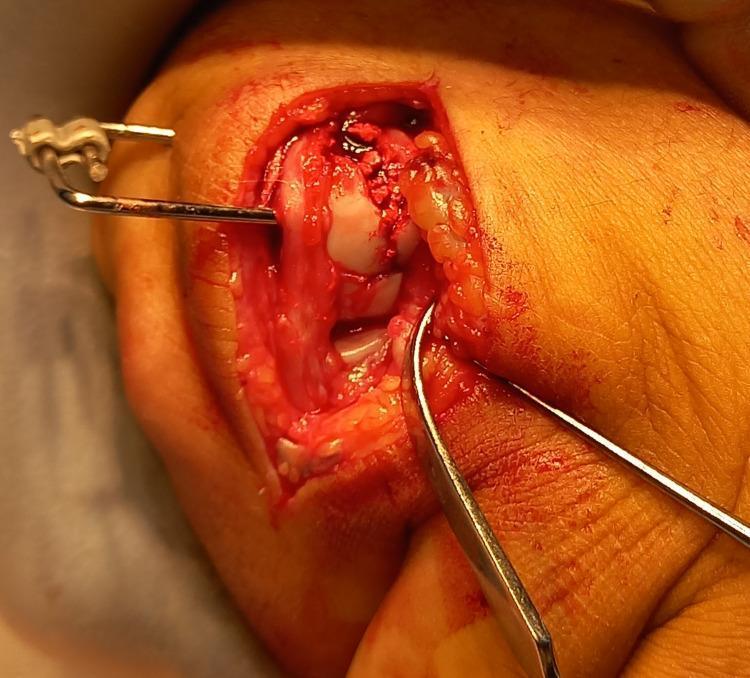
This clinical intraoperative photograph displays an open reduction and internal fixation (ORIF) procedure of a comminuted intra-articular fracture of the fifth metacarpal head in a human hand. The surgical field reveals an elliptical incision with retracted skin and subcutaneous adipose tissue, exposing the dorsal aspect of the metacarpophalangeal joint. Centrally, the articular surface of the metacarpal head is visible, showing significant impaction, depression, and bony comminution with associated hemorrhage. A metallic pointed reduction clamp is being used to manipulate and stabilize bone fragments. To the left, a K-wire is seen traversing the soft tissue and bone, connected externally to a component of an HK dynamic external fixator system. The image illustrates complex orthopedic management of hand trauma, specifically targeting the restoration of articular congruity in a joint-involved fracture where standard screw fixation may be insufficient due to bone stock loss. This material is suited for orthopedic surgical education regarding fracture stabilization and the application of external fixation devices.
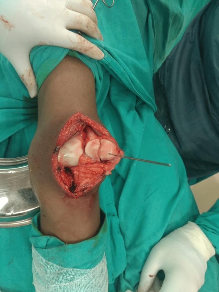
This clinical photograph shows an intraoperative view of an open reduction and internal fixation (ORIF) for a large displaced osteochondral fracture of the patella. The surgical site, accessed via a standard medial parapatellar approach on the left knee, reveals the exposed articular surface and underlying bone. The patellar joint surface is visibly fragmented, showing distinctive smooth, white cartilaginous surfaces characteristic of osteochondral fragments. A thin Kirschner wire (K-wire) or guide pin is seen traversing one of the fragments, serving as provisional fixation to align the fracture before permanent stabilization. The surgical field demonstrates typical hemorrhagic tissue and disrupted soft structures associated with acute traumatic injury. Personnel are depicted in a sterile environment, wearing white surgical gloves and utilizing green surgical drapes. This image is an educational example of orthopedic trauma management, specifically illustrating the visualization of intra-articular knee fractures and the initial steps of mechanical stabilization in a 16-year-old male patient.
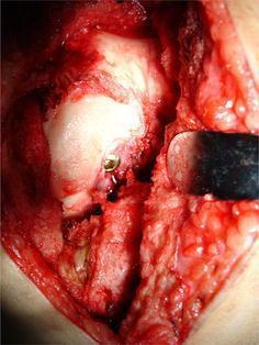
This clinical photograph captures an intraoperative view of an open reduction and internal fixation (ORIF) of a Hoffa fracture (coronal plane femoral condyle fracture). The surgical field shows the distal femur exposed through an arthrotomy, with the knee in a flexed position to visualize the articular surface. A primary focus is the femoral intercondylar notch, where a metallic cancellous screw has been inserted as part of a novel fixation technique. The screw head is visible at the entry point within the notch, directed toward the fractured condylar fragment to provide stable compression. A metallic bone repositioning clamp is visible on the right side of the frame, maintaining the reduction of the fracture during hardware placement. Surrounding structures include the articular cartilage of the femoral condyles, exposed cancellous bone, and soft tissue margins. This image serves as a clinical example of orthopedic trauma surgery, specifically demonstrating the anatomical positioning for intercondylar screw fixation in complex intra-articular knee fractures.
Principles of Intra-Articular Fracture Fixation
1. Anatomical Reduction of the Articular Surface
- Articular reduction must be confirmed - arthroscopy, fluoroscopy, or direct visualization
- In highly comminuted fractures where perfect anatomical reduction is impossible, restoration of the best achievable articular alignment remains the priority
- The joint surface is always reconstructed first, before addressing the metaphyseal or diaphyseal component
"The principle of reconstruction is to first obtain an anatomic reduction of the joint surface. This articular segment can be stabilized with independent lag screws." - Rockwood and Green's Fractures in Adults, 10th ed.
2. Rigid/Stable Fixation
- Lag screws - provide interfragmentary compression across the articular fracture; the workhorse for simple articular splits
- Plates and screws - used for metaphyseal extension and when lag screw fixation alone is insufficient; anatomically pre-contoured periarticular plates are now standard
- Locking plates - particularly important in osteoporotic bone, where standard screws risk cut-out; the screw locks into the plate, creating a fixed-angle construct with improved pull-out strength
- K-wires - used for provisional fixation and in small joints approaching skeletal maturity
- External fixation - may be used as a temporary or definitive bridge, especially with severe soft tissue injury
"AO type B and type C fractures are intra-articular and as such the principles of treating intra-articular fractures need to be respected; namely, anatomical reduction of the articular surface and rigid stabilisation to allow early joint movement and avoidance of degenerative joint disease." - Bailey and Love's Short Practice of Surgery, 28th ed.
3. Early Joint Mobilization
- Cartilage deterioration (cartilage is avascular and depends on synovial fluid diffusion through motion)
- Peri-articular fibrosis and stiffness
- Muscle wasting
4. Respect for Soft Tissues
- Use minimally invasive approaches where possible
- Avoid stripping periosteum unnecessarily
- In high-energy injuries with severe soft tissue swelling, staged surgery is frequently employed:
- Stage 1: temporary spanning external fixator to restore length and alignment
- Stage 2: definitive ORIF once soft tissues recover (typically 7-14 days)
- This staged approach is well-established for tibial pilon fractures and distal femur fractures
5. Bone Grafting / Void Filling
- Cancellous autograft - gold standard for filling impaction defects (iliac crest, local cancellous bone)
- Bone substitutes - injectable calcium phosphate or calcium sulfate bone cements may augment fixation in osteoporotic bone and fill voids
- These measures restore the subchondral bone scaffold that supports the overlying cartilage and the fixation construct
"Injectable bone substitutes may be used to fill bone voids and augment fixation." - Bailey and Love's Short Practice of Surgery, 28th ed.
6. Special Considerations in Osteoporotic Bone
- Even when anatomical reduction is achieved, fixation may fail by cut-out through soft metaphyseal bone
- Locking plate technology addresses this by converting pull-out forces into a fixed-angle construct
- If stable fixation is not achievable, options include non-operative treatment with delayed joint replacement, or primary joint replacement (e.g., arthroplasty for comminuted distal humerus or tibial plateau fractures in the elderly)
Summary Table
| Principle | Key Point |
|---|---|
| Anatomical articular reduction | Congruent joint surface to prevent post-traumatic OA |
| Stable fixation | Lag screws, plates, locking constructs - chosen by bone quality and pattern |
| Early mobilization | Cartilage nutrition, prevent stiffness |
| Soft tissue respect | Staged surgery for high-energy injuries; avoid devitalization |
| Bone void filling | Graft or substitute under elevated articular fragments |
| Address osteoporosis | Locking plates, bone substitutes, or primary arthroplasty |
- [Bailey and Love's Short Practice of Surgery, 28th ed.](p. 452-453)
- [Rockwood and Green's Fractures in Adults, 10th ed., 2025](p. 533)
- [Campbell's Operative Orthopaedics, 15th ed., 2026](p. 3337)
- [Schwartz's Principles of Surgery, 11th ed.](p. 1913)
AO principles of fracture fixation
AO fracture fixation compression plate lag screw absolute relative stability
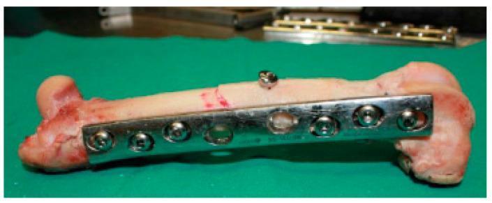
This clinical photograph displays an ex vivo femoral shaft undergoing internal fixation for a simple oblique fracture, serving as an educational example of orthopedic osteosynthesis. The primary stabilization is achieved using a shiny, metallic 8-hole Dynamic Compression Plate (DCP) contoured to the lateral aspect of the bone. The plate is secured with six bicortical screws positioned across the proximal and distal segments, acting in a neutralization function. Notably, an independent lag screw is placed perpendicularly across the fracture line to provide dynamic compression and enhance mechanical stability. The bone specimen exhibits a smooth cortical surface with visible reddish staining near the epiphyses, indicating the presence of vascularized tissue or residual soft matter. The arrangement demonstrates key principles of fracture management, including anatomical reduction and stable internal fixation to facilitate secondary bone healing. This image is relevant for surgical training and biomechanical study of plate-and-screw constructs in long bone fractures.
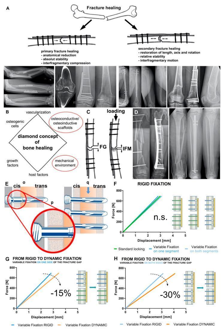
This composite educational image illustrates the mechanobiology of bone fracture healing and orthopedic fixation technologies. Section A compares primary healing (absolute stability, direct contact via compression plates/lag screws, no callus) with secondary healing (relative stability, interfragmentary motion via intramedullary nails/bridge plates, visible callus). Section B details the 'Diamond Concept' of healing, including osteogenic cells, scaffolds, growth factors, and the mechanical environment. Section C defines interfragmentary strain as the ratio of interfragmentary movement (IFM) to fracture gap (FG). Section D shows clinical radiographs of rigid versus flexible fixation in femoral and tibial shaft fractures. Sections E–H focus on Variable Fixation Locking Screw (VFLS) technology. A diagram (E) demonstrates a screw (q) within a resorbable sleeve (p) passing through the cis-cortex and trans-cortex (o). Biomechanical graphs (F, G, H) illustrate construct stiffness transitions, showing that sleeve resorption decreases stiffness by 15% to 30% depending on the configuration, thereby dynamizing the fracture site to stimulate healing.
AO Principles of Fracture Fixation
The Original Four AO Principles
| # | Principle | Core Concept |
|---|---|---|
| 1 | Anatomical reduction | Restore bone anatomy |
| 2 | Stable fixation | Appropriate to fracture type and healing mode |
| 3 | Preservation of blood supply | Atraumatic technique and soft tissue care |
| 4 | Early active mobilization | Of the patient and injured limb |
1. Anatomical Reduction
- For intra-articular fractures: anatomical reduction is mandatory (joint surface congruity to <2 mm step-off) to prevent post-traumatic arthritis
- For extra-articular fractures: functional alignment (restoring length, alignment, rotation) is the goal; perfect anatomic reduction of every fragment is not always required
- Reduction may be direct (open, with clamps, with visualization) or indirect (ligamentotaxis, using the implant as a reduction tool)
"Primary bone healing generally requires anatomic reduction, compression between fragments, and rigid fixation (absolute stability). Secondary bone healing generally requires restoration of alignment and flexible fixation (relative stability), but not necessarily anatomic reduction of individual fracture fragments." - Rockwood and Green's Fractures in Adults, 10th ed.
2. Stable Fixation - Absolute vs. Relative Stability
Absolute Stability
- Definition: No motion at the fracture site under physiologic load
- Healing mode: Direct (primary) bone healing - Haversian remodeling across the fracture without callus
- Strain: <2% (strain = ΔL/L, the change in fracture gap divided by the gap itself)
- Indications: Simple fractures (transverse, short oblique), intra-articular fractures
- Implants: Lag screws (interfragmentary compression), dynamic compression plates (DCP), tension band wiring
- Key point: Callus is absent on X-ray; healing is harder to confirm radiographically; implants must have a longer fatigue life
Relative Stability
- Definition: Controlled micromotion at the fracture site under physiologic load
- Healing mode: Indirect (secondary) bone healing via callus (endochondral ossification)
- Strain: 2-10% promotes callus; >10% prevents bone formation entirely
- Indications: Comminuted fractures, multifragmentary shaft fractures, metaphyseal fractures with biological advantages
- Implants: Intramedullary nails, bridge plates, external fixators, casts
- Key point: As callus matures and stiffens, strain decreases, creating a self-reinforcing healing cycle
"Absolute stability. Implies no displacement or movement and is achieved by accurate anatomical reduction with compression across the fracture fragments to optimise the environment for direct bone healing. Intra-articular fractures require an anatomical reduction and absolute stability." - Bailey and Love's Short Practice of Surgery, 28th ed.
Strain and Healing - Summary
| Strain (ΔL/L) | Healing Type |
|---|---|
| < 2% | Primary bone healing (no callus) |
| 2% - 10% | Secondary bone healing (callus, endochondral ossification) |
| > 10% | No bone formation possible |
3. Preservation of Blood Supply (Atraumatic Technique)
- Avoid excessive periosteal stripping
- Preserve the fracture hematoma where possible (it contains growth factors that initiate the healing cascade)
- Use minimally invasive percutaneous approaches where appropriate
- Indirect reduction techniques preserve the soft tissue envelope around comminuted fragments
- For high-energy injuries with severe soft tissue damage: staged surgery (temporary external fixation first, definitive fixation after soft tissue recovery)
"Typically, obtaining absolute stability requires removing the fracture hematoma... In contrast, the surgeon can often obtain relative stability with less trauma to soft tissues, often through indirect reduction, while preserving the fracture hematoma." - Rockwood and Green's Fractures in Adults, 10th ed.
4. Early Active Mobilization
- Patient mobilization: reduces DVT, pneumonia, deconditioning - especially in the elderly
- Joint mobilization: prevents articular cartilage degeneration (avascular cartilage depends on synovial fluid diffusion through motion), peri-articular fibrosis, and muscle atrophy
- Bone healing: physiologic loading through controlled motion stimulates callus formation (Wolff's law)
- Early mobilization is particularly emphasized around the elbow (notorious for stiffness), the ankle, and in elderly hip fracture patients
The Modern Evolution: From 4 Principles to a Biomechanical Framework
| Original (1960s) | Modern Understanding |
|---|---|
| "Rigid fixation" for all | Absolute OR relative stability - chosen based on fracture pattern |
| Open reduction preferred | Biological fixation / indirect reduction for comminuted fractures |
| Compression plates for shafts | IM nails for most diaphyseal fractures; bridge plating for comminuted patterns |
| Single doctrine | Individualized to fracture morphology, bone quality, soft tissues, patient factors |
"Constructs that mix techniques of nonanatomic reduction and relative stability often lead to failure." - Rockwood and Green's Fractures in Adults, 10th ed.
Quick Reference: Implant Choice by Stability Type
| Construct | Stability | Healing Mode |
|---|---|---|
| Lag screw alone | Absolute | Primary |
| Compression plate (DCP) | Absolute | Primary |
| Tension band wire | Absolute | Primary |
| Intramedullary nail | Relative | Secondary (callus) |
| Bridge plate | Relative | Secondary (callus) |
| External fixator | Relative | Secondary (callus) |
| Cast / splint | Relative | Secondary (callus) |
- [Rockwood and Green's Fractures in Adults, 10th ed., 2025](Chapters 1 & 12)
- [Bailey and Love's Short Practice of Surgery, 28th ed.](p. 447-453)
- [Miller's Review of Orthopaedics, 9th ed.](Basics of Fracture Healing and Fixation)
- [Pye's Surgical Handicraft, 22nd ed.](Internal Fixation Devices)