
Ogilive syndrome
Ogilvie syndrome acute colonic pseudo-obstruction
Ogilvie syndrome acute colonic pseudo-obstruction abdominal X-ray dilated colon cecum
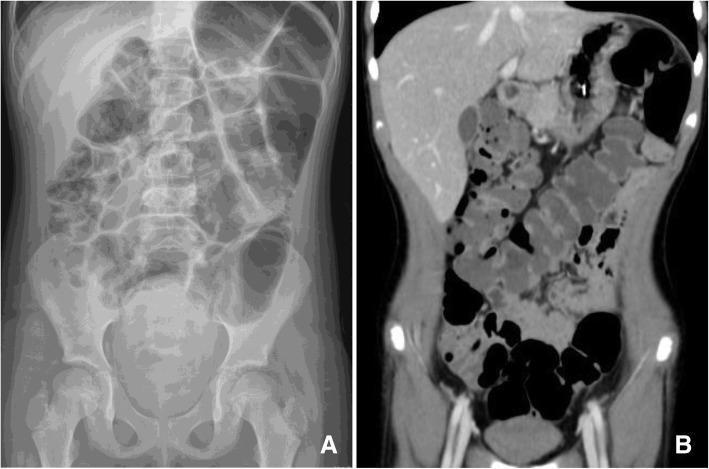
This composite diagnostic image illustrates acute colonic pseudo-obstruction, also known as Ogilvie's syndrome. Image A is an anteroposterior (AP) abdominal X-ray demonstrating marked, gaseous dilatation of the colon extending from the cecum through the splenic flexure. Notably, there is an absence of air-fluid levels or signs of pneumoperitoneum (free air). Image B is a coronal computed tomography (CT) scan of the same patient, providing a more detailed view of the massive large bowel dilatation. The CT reveals a clear transition and confirms that while the large bowel is severely distended, there is minimal involvement of the small bowel loops. Both imaging modalities highlight the lack of a mechanical obstructive lesion, pointing toward a functional motility disorder. These findings are clinically significant for diagnosing colonic pseudo-obstruction in a hospital setting, typically managed with bowel rest and pharmacological interventions.
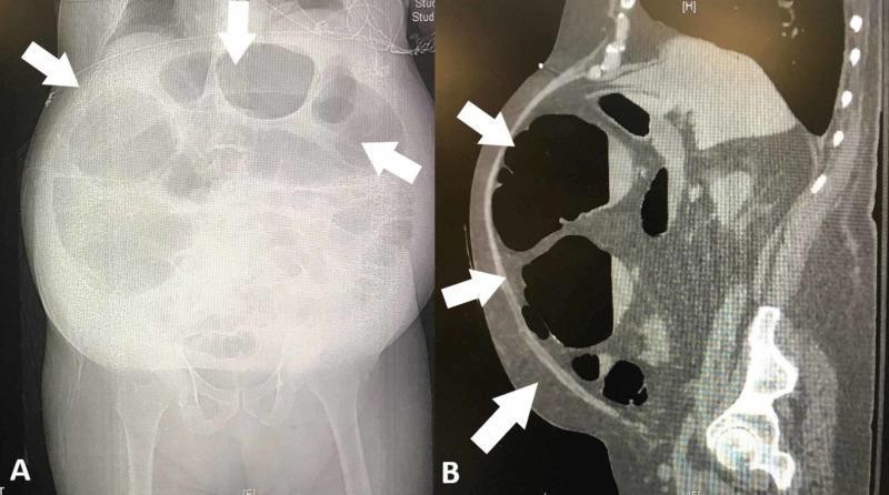
This composite diagnostic image demonstrates severe colonic dilatation in a 76-year-old male patient, consistent with Ogilvie syndrome (acute colonic pseudo-obstruction). Image A is an anteroposterior abdominal radiograph (X-ray) showing marked gaseous distention of the large bowel loops, particularly in the right and transverse colon segments. The haustral markings are visible but stretched, indicating significant intramural pressure. Image B is a sagittal view from a computed tomography (CT) scan with oral contrast, revealing a massively dilated transverse colon segment abutting the anterior abdominal wall. White arrows in both images highlight the most severely distended areas. The CT scan confirms the lack of a mechanical obstructing lesion and shows the relationship between the dilated bowel and the peritoneum. These findings illustrate typical radiological features of massive non-obstructive colonic dilatation, a critical finding in postoperative patients presenting with abdominal distension and vomiting. Educational focus includes distinguishing mechanical versus functional bowel obstruction through multi-modal imaging.
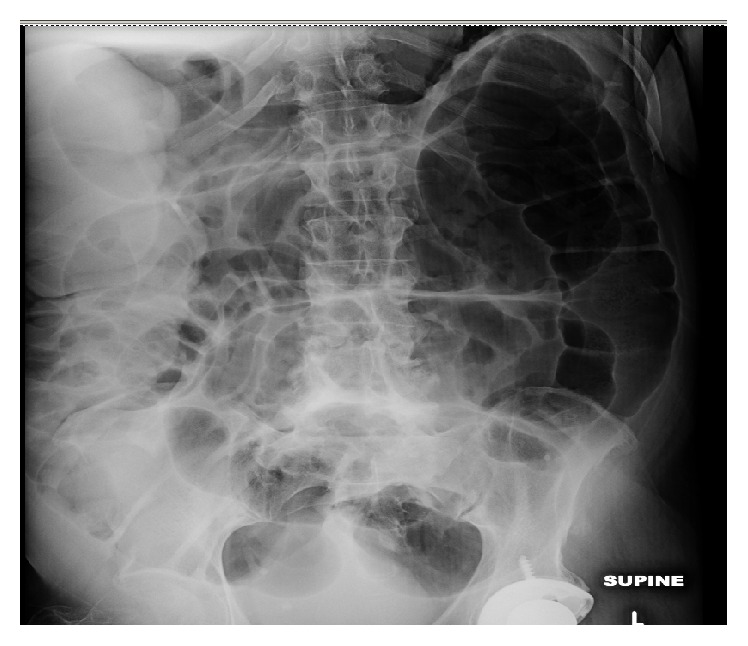
**Imaging Modality:** Abdominal X-ray (Supine view) **Anatomical Region:** Abdomen and pelvis **Observed Pathology:** Massive, generalized colonic dilatation consistent with Acute Colonic Pseudo-obstruction (Ogilvie’s Syndrome). **Characteristic Visual Features:** The radiograph demonstrates profound gaseous distension of the large bowel, most prominent in the cecum, ascending colon, and transverse colon. The haustral markings remain visible despite the severe mural stretching, a feature that helps distinguish colonic from small bowel dilatation. There is a lack of evidence of mechanical transition points or distal gas depletion, with air visualized throughout the rectum and sigmoid colon. The small bowel loops appear mildly dilated but are secondary to the primary colonic findings. **Diagnostic Features:** * **Cecal Diameter:** Significant enlargement, increasing the risk of mural ischemia or perforation. * **Gas Distribution:** Diffuse gaseous distension involving the proximal and distal colon without a definitive obstructing lesion. * **Skeletal/Metallic Findings:** Presence of a left total hip arthroplasty component in the lower field and degenerative changes in the lumbar spine. **Clinical Significance:** High-risk radiographic presentation of functional colonic obstruction requiring urgent clinical correlation to prevent cecal rupture.
Ogilvie Syndrome (Acute Colonic Pseudo-obstruction)
Definition and History
Pathophysiology
-
A relative excess of sympathetic over parasympathetic tone leads to functional inhibition of colonic motility
-
Ogilvie's original proposal: an imbalance among intrinsic, sympathetic, and parasympathetic neural control
-
The strong association with orthopedic surgery of the lower back and hips is explained by afferent stimulation of sympathetic reflexes in the thoracolumbar spinal cord, causing reflex sympathetic inhibition of colonic motility
-
Other mechanisms: disrupted colonic reflex arcs, chronic systemic illness, and medications that suppress motility
-
Yamada's Textbook of Gastroenterology, p. 1196
Risk Factors and Associated Conditions
| Category | Examples |
|---|---|
| Postsurgical | Major orthopedic/spinal surgery, solid organ transplants, cardiac procedures, C-section |
| Neurologic | Parkinson disease, Alzheimer disease, stroke, spinal cord injury |
| Cardiac | Congestive heart failure, myocardial infarction |
| Pulmonary | COPD |
| Trauma | Major trauma, shock, burns |
| Metabolic | Diabetes, renal failure, electrolyte disturbances (hypokalemia in 20-30%) |
| Infectious | CMV, varicella-zoster virus |
| Drugs | Opiates, anticholinergics, anti-Parkinson drugs, antipsychotics, clonidine, chemotherapy |
| Autoimmune | Lupus, scleroderma |
Clinical Presentation
- Abdominal distension - the dominant feature, developing over 1-7 days
- Abdominal pain - constant but usually mild
- Nausea and vomiting in >50% of patients
- Obstipation is common; paradoxically, some patients have diarrhea (from hypersecretion)
- Decreased or absent bowel sounds (or high-pitched tinkling sounds)
- Labored breathing from diaphragm elevation
- Red flags for ischemia/perforation: fever, significant tenderness, rebound, leukocytosis - these are uncommon initially but must be sought
Imaging
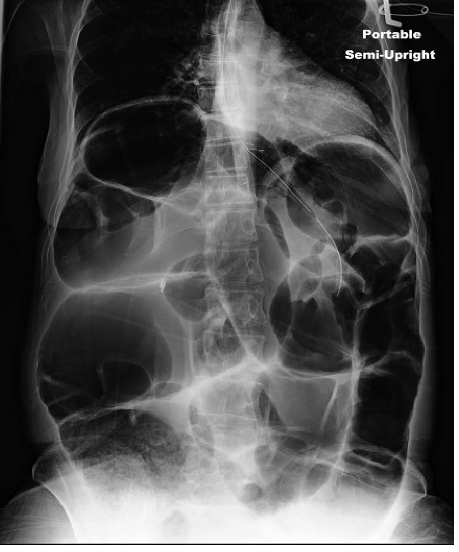
- Massive gaseous distension predominantly in the cecum and right colon (cecum typically 9-12 cm)
- Gas distributed throughout the colon including the rectum - this pattern distinguishes pseudo-obstruction from mechanical obstruction (where no gas is seen distal to the blockage)
- Small bowel is usually not dilated
Differential Diagnosis
- Mechanical large bowel obstruction (carcinoma, volvulus) - distinguished by CT/contrast enema
- Toxic megacolon - occurs in severe inflammatory bowel disease or infectious colitis (C. difficile); usually accompanied by systemic toxicity
- Ischemic colitis - shares many risk factors; female predominance; mucosal changes on CT/colonoscopy
- Colonic volvulus (sigmoid or cecal)
Management
Step 1: Supportive Care (cecal diameter < 12 cm, no ischemia/perforation)
- NPO, IV fluids, nasogastric tube
- Rectal tube placement for decompression
- Correct electrolytes (especially hypokalemia)
- Discontinue offending drugs: opiates, anticholinergics, anti-Parkinson agents, antidepressants, neuroleptics, clonidine
- Encourage ambulation, prone positioning, knee-chest position to encourage flatus
- Serial abdominal exams and X-rays
- Avoid osmotic and stimulant laxatives (worsen dilation)
- Most cases resolve with these simple measures
Step 2: Pharmacologic Decompression (cecal diameter > 12 cm OR no response to supportive care)
- Mechanism: Acetylcholinesterase inhibitor → increases muscarinic/parasympathetic activity → colonic peristalsis
- Dose: 2-2.5 mg IV over 3-5 minutes
- Response: flatus and bowel movement within 10 minutes in most responders
- Success rate: 60-94%; recurrence in up to 31% (may need repeat dosing)
- Contraindications: mechanical obstruction, ischemia, perforation, bradycardia, recent MI, asthma/COPD, renal failure, myasthenia gravis
- Monitoring: continuous cardiac monitoring; atropine 0.4-0.6 mg IV immediately available for bradycardia
- Side effects: bradycardia, bronchospasm, vomiting, cramping, excessive salivation
- Alternative: epidural anesthesia (sympathetic blockade) for selected patients
- Limited evidence also supports naloxone or methylnaltrexone when opioids are a contributing factor
Step 3: Endoscopic Decompression (failure of neostigmine, or contraindication)
- Colonoscopic decompression if no response after 72 hours
- Scope advanced to hepatic flexure (not necessary to reach cecum) with minimal insufflation
- Aspiration of proximal gas collapses the right colon
- Success rate: 61-95% initial; 70-90% sustained
- Colonic decompression tube can be placed
- Risk of perforation: 1-3%
- Post-resolution: polyethylene glycol 3350 (29.5 g daily) may reduce recurrence
Step 4: Surgery
- Signs of ischemia, perforation, or systemic toxicity
- Cecal diameter > 14 cm with no response to medical/endoscopic therapy
- Viable colon: tube cecostomy (laparoscopic possible) is preferred
- Ischemia without perforation: right hemicolectomy + primary anastomosis
- Perforation: right hemicolectomy + ileostomy and mucous fistula
Complications and Prognosis
- Ischemia and perforation occur in 3-15% of cases
- Perforation carries a ~50% mortality rate
- Overall mortality: 25-31%
- Poor prognosis associated with: older age, greater illness severity, larger cecal diameter, longer time to decompression, presence of colonic ischemia