
Create flashcards for respiratory pathology
Asking for Preferences
Which area of respiratory pathology should the flashcards cover? · What level are these for?
Reading File
Now let me search the library for respiratory pathology content across all major topics simultaneously.
Finding Sources
Finding Sources
Finding Sources
Finding Sources
Finding Sources
Finding Sources
Excellent - Robbins Pathology is the key source. Let me pull respiratory pathology sections from it simultaneously across all major topics.
Finding Sources
Finding Sources
Finding Sources
Finding Sources
I've hit my query_library_db limit. Let me now read the key content from Robbins Pathology to build the flashcards.
Reading File
Reading File
Reading File
Reading File
Reading File
I now have rich content from Robbins Pathology. Let me read a bit more on interstitial lung diseases and pleural/vascular diseases, then build the flashcards.
Reading File
I now have comprehensive content from Robbins Pathology. Let me build the full flashcard set and generate the visual flashcard image.
Generating Image
Medical flashcard set for respiratory pathology, USMLE medical student level. Clean, professional design with a dark navy blue header bar showing "Respiratory Pathology Flashcards" in white bold text. Below, a 2x3 grid of flashcard previews, each card with a colored top strip (alternating teal, orange, purple) and white body. Card titles visible: "Asthma", "Emphysema vs Chronic Bronchitis", "Pneumonia Types", "Lung Cancer", "Sarcoidosis", "TB & ARDS". Each card shows a few bullet points of key facts. Clean minimal medical education style.
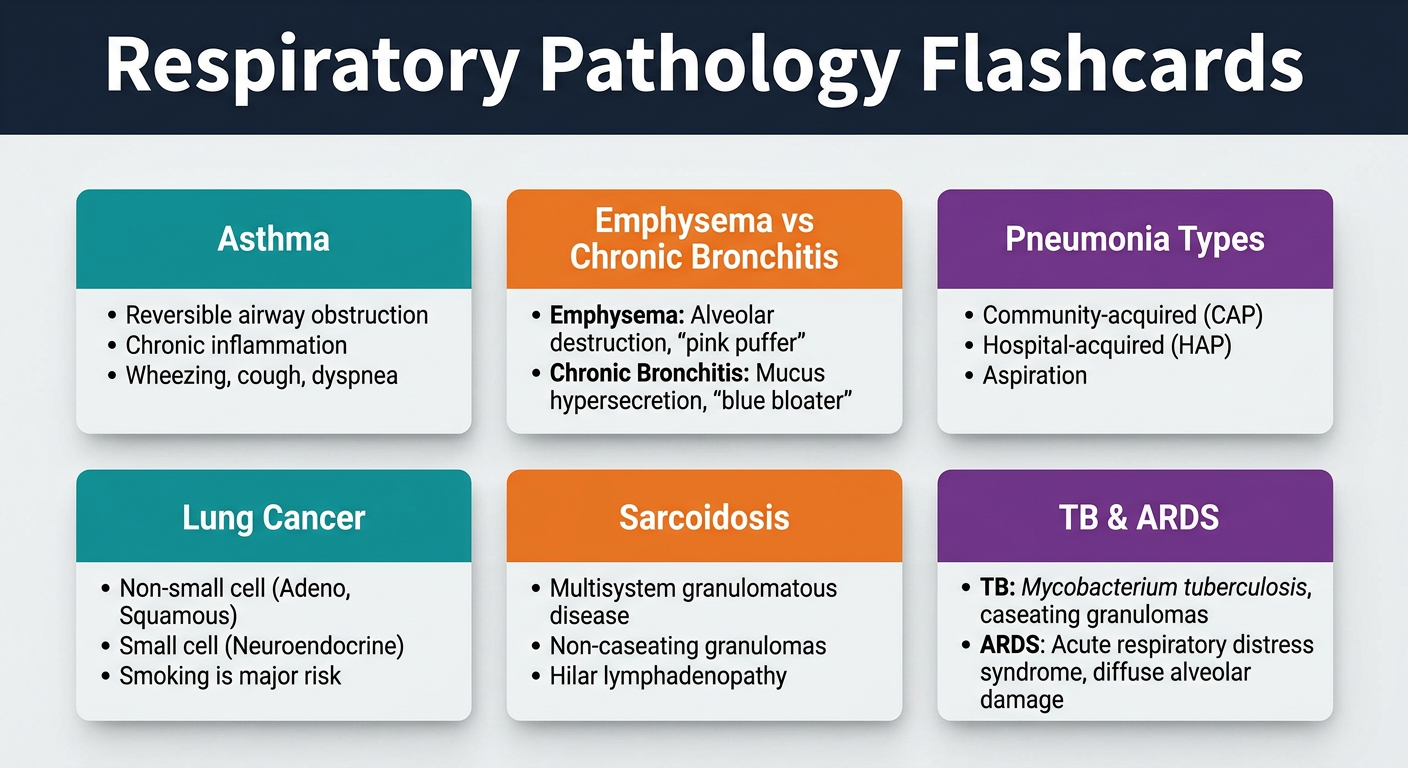
Here are your complete respiratory pathology flashcards, sourced directly from Robbins & Kumar Basic Pathology and Robbins/Cotran Pathologic Basis of Disease. Each card has a front question and back answer.
🫁 OBSTRUCTIVE LUNG DISEASES
Card 1 — Asthma: Definition & Hallmarks
Q: What are the four pathologic hallmarks of asthma?
A:
- Intermittent, reversible airway obstruction
- Chronic bronchial inflammation with eosinophils
- Bronchial smooth muscle hypertrophy and hyperreactivity
- Increased mucus secretion
Key: Episodic wheezing, dyspnea, cough - especially nocturnal/early morning Source: Robbins Basic Pathology, Ch. 11
Card 2 — Atopic vs. Nonatopic Asthma
Q: How do atopic and nonatopic asthma differ in mechanism?
A:
| Feature | Atopic | Nonatopic |
|---|---|---|
| Type | Type I IgE-mediated | Non-immunologic |
| Triggers | Allergens (pollen, dander, dust) | Infections, irritants, cold air |
| Cells | Th2 cells, IgE, mast cells | Mast cells, eosinophils |
| Key cytokines | IL-4, IL-5, IL-13 | - |
| Skin test | Wheal-and-flare positive | Negative |
- IL-4/IL-13: drive IgE production from B cells
- IL-5: recruits and activates eosinophils
- IL-13: drives mucus production
Card 3 — Obstructive Lung Disease: Quick Comparison
Q: Fill in the table: What is the anatomic site, pathology, and main symptom for each obstructive disorder?
A:
| Disease | Site | Key Pathology | Symptom |
|---|---|---|---|
| Emphysema | Acinus | Air space enlargement, wall destruction | Dyspnea |
| Chronic bronchitis | Bronchus | Mucous gland hypertrophy, hypersecretion | Cough + sputum |
| Asthma | Bronchus | Smooth muscle hypertrophy, mucus, eosinophils | Episodic wheeze |
| Bronchiectasis | Bronchus | Airway dilation and scarring | Purulent sputum, fever |
| Bronchiolitis | Bronchiole | Inflammatory scarring | Cough, dyspnea |
Card 4 — Emphysema
Q: What is the pathogenesis of emphysema and which type is most common?
A:
- Cause: Tobacco smoke - neutrophils/macrophages release elastases that destroy alveolar walls
- Centriacinar (centrilobular) = most common; affects respiratory bronchioles; upper lobes; smokers
- Panacinar = affects entire acinus; lower lobes; seen in alpha-1 antitrypsin deficiency
- Result: Loss of elastic recoil → air trapping → barrel chest, hyperresonance, pursed-lip breathing
- "Pink puffer" phenotype: dyspnea dominant, near-normal PaCO2
Card 5 — Chronic Bronchitis
Q: What is the clinical and histologic definition of chronic bronchitis?
A:
- Clinical: Productive cough for at least 3 consecutive months in at least 2 consecutive years
- Histology: Hypertrophy and hyperplasia of mucus-secreting glands in the bronchial wall
- Reid index = ratio of mucous gland thickness to total bronchial wall thickness (normal < 0.4; elevated in chronic bronchitis)
- "Blue bloater": cyanotic, hypercapnic, hypoxemic, edematous
- Cause: tobacco smoke, air pollutants
🫁 INFECTIOUS / INFLAMMATORY
Card 6 — Community-Acquired Pneumonia: Key Organisms
Q: Match the clinical scenario to the most likely pneumonia organism.
A:
| Scenario | Organism |
|---|---|
| Most common overall (lobar, rust-colored sputum) | S. pneumoniae |
| Elderly/aspiration-prone, tissue necrosis | K. pneumoniae (currant-jelly sputum) |
| Atypical, "walking pneumonia," cold agglutinins | Mycoplasma pneumoniae |
| Legionnaire's disease, water systems, hyponatremia | Legionella pneumophila |
| Flu-like, exposure to birds/droppings | C. psittaci / H. capsulatum |
| Flu followed by bacterial superinfection | S. aureus |
| Hospital-acquired / ventilator-associated | Gram-negative rods (Pseudomonas, Enterobacteriaceae), S. aureus |
Card 7 — Aspiration Pneumonia
Q: Who gets aspiration pneumonia and what are its features?
A:
- At risk: Debilitated patients, impaired gag/swallowing (post-stroke), unconscious patients
- Mechanism: Gastric acid (chemical) + polymicrobial bacteria
- Organisms: Aerobes > anaerobes (mixed flora)
- Complications: Often necrotizing; lung abscess is common in survivors
- Lower lobes most affected (gravity-dependent)
- Note: Microaspiration (in GERD) ≠ pneumonia, but can exacerbate lung disease
Card 8 — Tuberculosis: Primary vs. Secondary (Reactivation)
Q: Contrast primary and secondary TB in an immunocompetent host.
A:
| Feature | Primary TB | Secondary (Reactivation) TB |
|---|---|---|
| Prior exposure | None | Yes |
| Location | Subpleural focus + hilar nodes (Ghon complex) | Lung apices |
| Lesion type | Usually heals; calcified Ghon focus | Cavitary lesion |
| Symptoms | Usually asymptomatic | Fever, night sweats, hemoptysis |
| Immune state | Competent = contained | Competent but weakened |
- Hallmark: Caseating granulomas (Langhans giant cells + CD4+ Th1 response)
- Immunodeficiency (HIV) → miliary TB, meningitis (progressive forms)
- Stain: Ziehl-Neelsen (acid-fast, carbol fuchsin)
Card 9 — ARDS (Acute Respiratory Distress Syndrome)
Q: What are the key features, histology, and clinical outcomes of ARDS?
A:
- Triggers: Sepsis, trauma, aspiration, pancreatitis, severe pneumonia
- Onset: 85% of cases within 72 hours of insult
- Histology (DAD - diffuse alveolar damage):
- Acute phase: alveolar collapse + distension, pink hyaline membranes lining alveoli
- Healing phase: resorption of membranes, alveolar septal thickening, reactive type II pneumocytes
- Imaging: Bilateral ground-glass opacities on CT
- Prognosis: ~40% mortality; death usually from underlying cause or superinfection, not respiratory failure itself
🫁 INTERSTITIAL / DIFFUSE LUNG DISEASES
Card 10 — Sarcoidosis
Q: What are the defining features of sarcoidosis?
A:
- Hallmark: Non-necrotizing (non-caseating) epithelioid granulomas with Schaumann and asteroid bodies
- Driver: CD4+ Th1 cells; elevated IL-2, IFN-γ
- Unique: Higher prevalence in nonsmokers (rare for a lung disease)
- Distribution: Granulomas follow lymphatic routes - peribronchovascular, subpleural
- Organs: Lung (90%), hilar lymph nodes (75-90%), skin (25% - erythema nodosum), eyes (uveitis), heart
- Blood: Low circulating CD4+ T cells, elevated ACE
- 5-15% progress to honeycomb lung fibrosis
- Lymph nodes: firm, rubbery, non-matted, non-necrotic (unlike TB)
Card 11 — Idiopathic Pulmonary Fibrosis (IPF)
Q: What is the morphologic pattern and clinical course of IPF?
A:
- Pattern: Usual Interstitial Pneumonia (UIP) - temporal and spatial heterogeneity, subpleural/basal predominant
- Histology: Alternating areas of normal lung + fibrosis + honeycombing; fibroblastic foci
- Key feature: Progressive, irreversible fibrosis - median survival ~3 years from diagnosis
- Symptoms: Progressive dyspnea, dry cough, bilateral basal crackles, clubbing
- Treatment: Antifibrotics (pirfenidone, nintedanib) slow decline; lung transplant is only cure
- Differs from sarcoidosis: IPF has fibrosis without granulomas
Card 12 — Hypersensitivity Pneumonitis (Extrinsic Allergic Alveolitis)
Q: What distinguishes hypersensitivity pneumonitis from asthma?
A:
- HP: Type III (immune complex) + Type IV (cell-mediated) hypersensitivity to inhaled organic antigens
- Examples: Farmer's lung (thermophilic actinomycetes in hay), Bird-fancier's lung (avian proteins)
- Histology: Lymphocytic alveolitis + non-caseating granulomas + bronchiolitis
- Key difference from asthma: HP involves alveoli and interstitium; asthma is airway-centric
- Acute: Fever, dyspnea, crackles 4-6 hours after exposure (resolves with avoidance)
- Chronic: Irreversible fibrosis if exposure continues
🫁 LUNG TUMORS
Card 13 — Lung Cancer: Four Major Subtypes
Q: What are the four major histologic types of lung cancer and their key associations?
A:
| Type | % | Location | Key Association |
|---|---|---|---|
| Adenocarcinoma | ~40% (most common) | Peripheral | Nonsmokers, women; EGFR, ALK mutations; lepidic growth |
| Squamous Cell Carcinoma | ~25-30% | Central (hilar) | Heavy smokers; PTH-rP (hypercalcemia); cavitates |
| Small Cell Carcinoma (SCLC) | ~15% | Central | Strongest smoking link; neuroendocrine; ACTH/ADH production; TP53 + RB mutations |
| Large Cell Carcinoma | ~10% | Peripheral | Diagnosis of exclusion; poor prognosis |
Card 14 — SCLC vs. NSCLC: The High-Yield Comparison
Q: Compare SCLC and NSCLC across histology, markers, mutations, and treatment.
A:
| Feature | SCLC | NSCLC (Adeno/SCC) |
|---|---|---|
| Microscopy | Scant cytoplasm, hyperchromatic nuclei, diffuse sheets | Abundant cytoplasm, glandular/squamous architecture |
| Neuroendocrine markers (chromogranin, synaptophysin, CD56) | Present | Absent |
| Mucin | Absent | Present (adenocarcinoma) |
| TP53 mutations | ~90% | ~50% |
| RB mutations | ~90% | ~20% |
| KRAS mutations | Rare | ~30% (adenocarcinoma) |
| EGFR mutations | Absent | ~20% (adenocarcinoma, nonsmokers) |
| Chemo/RT response | Often complete, but always recurs | Incomplete response |
| Checkpoint inhibitors | Unresponsive | Responsive |
| Surgery | NOT curative (already metastatic at diagnosis) | Curative if localized |
| Median survival with Tx | ~1 year | Varies by stage |
Card 15 — Paraneoplastic Syndromes in Lung Cancer
Q: Match the paraneoplastic syndrome to the lung cancer subtype.
A:
| Syndrome | Mechanism | Cancer Type |
|---|---|---|
| Cushing syndrome (ACTH-like) | Ectopic ACTH production | SCLC |
| SIADH (hyponatremia) | Ectopic ADH production | SCLC |
| Hypercalcemia | PTH-related peptide (PTH-rP) | Squamous cell carcinoma |
| Eaton-Lambert syndrome | Anti-VGCC antibodies | SCLC |
| Clubbing / hypertrophic osteoarthropathy | Unknown mechanism | Adenocarcinoma/SCC |
| Pancoast syndrome (shoulder/arm pain, Horner's) | Apical tumor invading brachial plexus/sympathetic chain | Any (often SCC) |
🫁 VASCULAR / OTHER
Card 16 — Pulmonary Embolism (PE)
Q: What is Virchow's triad and how does it relate to PE?
A:
- Virchow's Triad: Stasis + Endothelial injury + Hypercoagulability
- Source: 95% from deep vein thrombosis (DVT) in legs
- Consequences depend on size:
- Large saddle embolus: sudden death, right heart failure
- Medium: pulmonary infarction (Hampton's hump on CXR), hemoptysis, pleuritis
- Small/recurrent: pulmonary hypertension
- Dx: CT pulmonary angiography (gold standard); D-dimer (sensitive, not specific)
- Rx: Anticoagulation (heparin → warfarin or DOAC); thrombolytics for massive PE
Card 17 — Pleural Effusion: Transudate vs. Exudate
Q: How do you distinguish a transudate from an exudate? (Light's Criteria)
A:
Exudate = ANY ONE of Light's criteria met:
- Pleural fluid protein / serum protein > 0.5
- Pleural fluid LDH / serum LDH > 0.6
- Pleural fluid LDH > 2/3 upper limit of normal serum LDH
| Transudate | Exudate |
|---|---|
| Low protein, low LDH | High protein, high LDH |
| CHF, cirrhosis, nephrotic syndrome | Pneumonia, malignancy, TB, PE |
| Hydrostatic/oncotic cause | Inflammatory/lymphatic cause |
Card 18 — Mesothelioma
Q: What is malignant mesothelioma and what causes it?
A:
- Cause: Asbestos exposure (latency 25-45 years); amphibole fibers (crocidolite) most dangerous
- Location: Pleura (most common), peritoneum, pericardium
- Presentation: Progressive dyspnea, chest pain, recurrent bloody pleural effusion
- Histology: Biphasic - epithelioid + sarcomatoid patterns; mesothelial markers (calretinin, WT-1, CK5/6); negative for CEA (distinguishes from adenocarcinoma)
- Prognosis: Very poor; median survival ~12 months
- Note: Asbestosis (interstitial fibrosis from asbestos) ≠ mesothelioma - both are asbestos-related but distinct diseases
RAPID REVIEW MNEMONICS
| Mnemonic | Meaning |
|---|---|
| "Pink Puffer vs Blue Bloater" | Emphysema (dyspnea, normal O2) vs Chronic Bronchitis (cyanosis, hypercapnia) |
| "EGFR = Ethnic/Female/No-smoking" | Adenocarcinoma EGFR mutations favor Asian women, never-smokers |
| "Ghon = Gone (healed) primary TB" | Ghon complex = subpleural focus + hilar node (primary TB, usually healed) |
| "SCLC = Surgery Contraindicated, Lethal Course" | Metastatic at diagnosis, not surgically curable |
| "ACE is high in Sarcoidosis" | Elevated serum ACE; granulomas produce it |
| "3 months × 2 years = Chronic Bronchitis" | Clinical definition |
| "Asbestosis: Ferruginous bodies, pleural plaques, mesothelioma" | All three are asbestos-related findings |
All content sourced from Robbins & Kumar Basic Pathology, 10e and Robbins, Cotran & Kumar Pathologic Basis of Disease, 10e.
This is a shared conversation. Sign in to Orris to start your own chat.