
Thyroid gland histology, physiology
thyroid gland histology follicle parafollicular cells
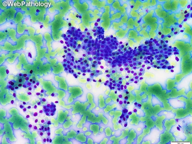
This is a light microscopy histology image of thyroid gland tissue prepared with a Romanowsky-type stain (e.g., Diff-Quik/Wright-Giemsa). The specimen shows normal-appearing follicular epithelium arranged in intact macrofollicles with abundant colloid occupying the luminal spaces. Follicular cells are evenly spaced with uniform, round to oval nuclei and scant to moderate cytoplasm, consistent with well-differentiated thyroid epithelium. The colloid background is pale blue and prominent, reflecting thyroglobulin-containing material released from follicles. The architectural pattern emphasizes large follicle units with a single layer of cuboidal to low-columnar epithelium surrounding expansive colloid pools. No prominent nuclear features of papillary carcinoma are evident, and there is no marked cellular atypia or mitotic activity in the visible fields. This image illustrates classic histology of benign thyroid tissue or colloid-rich goiter, useful for teaching follicular morphologies, thyroid folliculogenesis, and colloid dynamics. Clinically, such samples are evaluated in the context of thyroid nodules and goiter workups, with correlation to serum thyroid function tests and, when applicable, ultrasound or FNA findings. Potential educational uses include cytology-histology correlation, differential diagnosis of thyroid disease, and artifact recognition in Romanowsky-stained preparations. Images are suitable for medical education, pathology review, and database indexing. Correlative data improve student comprehension and diagnostic recall greatly.
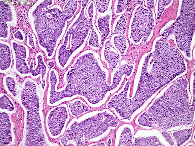
This is a light micrograph of thyroid gland tissue prepared for histopathology and stained with Hematoxylin and Eosin (H&E). The sample reveals multiple spherical to irregular follicles of varying size separated by wispy fibrous septa. The colloid-filled lumens are pink to pale pink, while the surrounding follicular epithelium is a uniform single layer of cuboidal to low-columnar cells with round to oval nuclei and inconspicuous nucleoli. No nuclear grooves, inclusions, or pseudoinclusions characteristic of papillary carcinoma are evident. The stromal background is relatively scant with no dense lymphocytic infiltrate or Hurthle cell metaplasia apparent. The architecture is reminiscent of normal thyroid parenchyma or benign nodular colloid goiter pattern, where follicle size varies and colloid predominates. There are no mitotic figures or cytologic atypia observed. This image demonstrates classic histology of endocrine gland tissue with preserved colloid and regular follicle lining, suitable as reference for normal thyroid architecture in educational, diagnostic, and research contexts. Clinically, such histology supports benign thyroid pathology when correlated with imaging and labs. In malignant differentials, absence of papillary nuclear features or tall columnar cells argues against papillary thyroid carcinoma; however, definitive diagnosis should consider capsule invasion and margin status in surgical specimens.
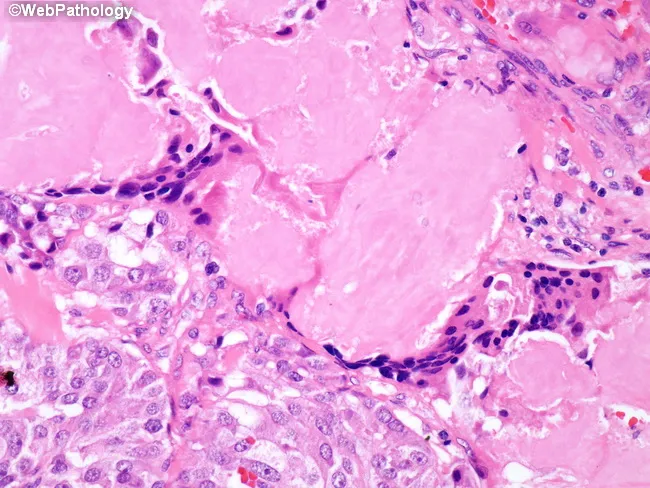
Histology image obtained from thyroid tissue showing medullary thyroid carcinoma with extracellular amyloid deposits in the tumor stroma and a foreign body-type giant cell reaction surrounding the amyloid. The slide is a paraffin-embedded tissue section stained with Hematoxylin and Eosin, visualized under light microscopy. The malignant parafollicular C-cells form nests and sheets with uniform, round to polygonal cells and granular cytoplasm, displaying salt-and-pepper nuclear characteristics. A conspicuous, pink, amorphous extracellular matrix represents calcitonin-derived amyloid within and around tumor nests; multinucleated giant cells and macrophages are present at the periphery, reflecting a foreign body-type granulomatous response to amyloid deposition. Congo red staining (not shown here) would typically reveal apple-green birefringence under polarized light, confirming amyloid. This histology is characteristic of medullary thyroid carcinoma (MTC) and helps distinguish it from papillary, follicular, or anaplastic thyroid cancers. Diagnostic significance rests on the co-occurrence of C-cell neoplasia and amyloid, a hallmark of MTC, with possible associations to calcitonin gene-related peptide and RET oncogene alterations. Clinically, MTC presents with a neck mass and elevated serum calcitonin and CEA; amyloid-rich stroma reflects tumor biology and has prognostic implications. The image supports educational and diagnostic use in pathology, endocrine oncology, and medical education.
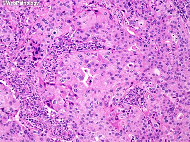
Imaging modality and technique: Light microscopy of a hematoxylin and eosin (H&E) stained thyroid tumor section, viewed at high magnification (~400x). Anatomical context: thyroid gland in the neck, parafollicular C-cell-derived medullary thyroid carcinoma. Observed architectures include diffuse growth in broad sheets, as well as solid nests and anastomosing trabeculae. Tumor cells show high-grade nuclei with conspicuous nucleoli and abundant eosinophilic cytoplasm; cytoplasmic borders are indistinct, imparting a syncytial appearance. There is little to no glandular differentiation or intraductal component. A striking, prominent lympho-plasmacytic infiltrate surrounds and permeates the tumor, a hallmark histologic feature of MTC. Background stroma may contain stromal amyloid in some cases (not always visible). Immunophenotypic expectation (not depicted): calcitonin and neuroendocrine markers positive; CEA may be expressed; TTF-1 variably positive. Diagnostic significance: the described morphology is characteristic of medullary thyroid carcinoma and helps distinguish it from papillary, follicular, anaplastic, and poorly differentiated thyroid carcinomas or metastatic neuroendocrine tumors. Clinical correlation: assess serum calcitonin and CEA; evaluate for MEN2 syndrome; RET mutation testing; imaging for primary tumor and metastasis; consider therapeutic implications and prognosis. Educational use: reference histology slide illustrating C-cell origin, syncytial tumor cells, and lymphoplasmacytic response. Correlation with amyloid deposition and calcitonin positivity may aid diagnostically.
thyroid hormone synthesis iodine organification thyroglobulin
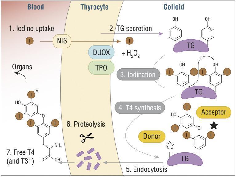
A pathophysiology diagram illustrating the seven-step process of thyroid hormone synthesis (T3 and T4) across three compartments: Blood, Thyrocyte, and Colloid. (1) Iodine uptake: Iodide (I-) enters the thyrocyte from the blood via the Sodium/Iodide Symporter (NIS). (2) TG secretion: Thyroglobulin (TG) is secreted into the colloid. (3) Iodination: Within the colloid, iodide is oxidized by Thyroid Peroxidase (TPO), dual oxidase (DUOX), and H2O2, leading to the attachment of iodine to tyrosine residues on TG. (4) T4 synthesis: A coupling reaction occurs between 'Donor' and 'Acceptor' iodinated tyrosine residues on the TG scaffold. (5) Endocytosis: The modified TG molecule is re-absorbed into the thyrocyte. (6) Proteolysis: Lysosomal enzymes break down TG, releasing individual hormones. (7) Release: Free T4 (thyroxine) and T3 (triiodothyronine) are secreted into the blood for systemic distribution to organs. The diagram includes detailed chemical structures of the phenolic rings and iodine substitutions characteristic of thyroid hormones.
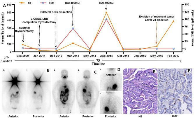
A multi-panel figure illustrating the clinical history and diagnostic findings of a patient with radioactive iodine-refractory differentiated thyroid cancer (RAIR-DTC). Panel A is a dual-axis timeline graph (Sept-2009 to Feb-2017) tracking serum Thyroglobulin (Tg) in ng/mL and Thyroid Stimulating Hormone (TSH) in uIU/mL alongside major interventions, including subtotal thyroidectomy, bilateral neck dissections, and multiple RAI treatments (100mCi each). Panels B and C show Radioactive Iodine (RAI) whole-body scans in anterior and posterior views. Scan B reveals functional iodine uptake in the neck, while Scan C demonstrates a lack of neck uptake but a new abnormal focus in the anterior-inferior mediastinum. Panel D provides localized anterior/posterior views clarifying the mediastinal focus. Panel E and F present histopathological sections: E shows Hematoxylin and Eosin (HE) staining at 200x magnification confirming papillary thyroid carcinoma morphology with papillary architecture; F shows immunohistochemical staining for Ki67, indicating a proliferative index of approximately 10%. The composite illustrates the progression of PTC from initial diagnosis through the development of RAI-refractoriness.
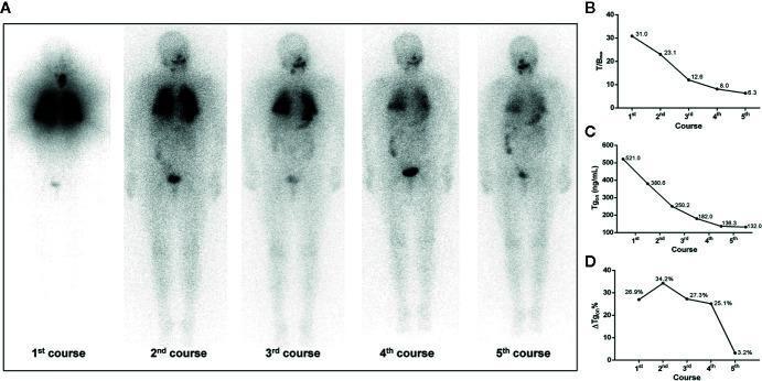
This composite educational image illustrates the longitudinal treatment response of a 68-year-old female with differentiated thyroid cancer and 131I-avid pulmonary metastases. Panel (A) displays a series of five anterior whole-body radioactive iodine (131I) scans following five successive courses of radioiodine therapy (RT). The initial scan shows intense, diffuse bilateral pulmonary uptake and thyroid bed activity. Successive scans demonstrate a progressive reduction in tracer avidity, signifying therapeutic response and decreased tumor burden. Panels (B), (C), and (D) provide quantitative correlation through line graphs. Graph B shows the maximum target-to-background ratio (T/Bmax) declining from 31.0 to 6.3. Graph C tracks the thyroid stimulating hormone-suppressed thyroglobulin (Tgon) levels, showing a significant decrease from 521.0 ng/mL to 132.0 ng/mL. Graph D illustrates the change rate of Tgon level (ΔTgon%), which peaks at 34.2% in the second course before dropping to 3.2% by the fifth course. This multimodal representation highlights the transition from biochemical remission to potential radioiodine refractoriness in late-stage treatment.
Thyroid Gland - Histology and Physiology
HISTOLOGY
Gross / Lobular Architecture
- Schwartz's Principles of Surgery, 11th Ed.
The Thyroid Follicle
| Component | Description |
|---|---|
| Follicular epithelial cells (thyrocytes) | Single layer of cuboidal to low-columnar cells lining each follicle; become columnar when active (stimulated by TSH), flat when inactive |
| Colloid | Fills the central lacuna; composed predominantly of thyroglobulin (Tg), a large (660 kDa) iodinated glycoprotein with four tyrosyl residues |
| Basement membrane | Surrounds the follicle; parafollicular C cells sit below it |
Histology image - normal thyroid follicles
Parafollicular (C) Cells
- Located individually or in small clusters in the interfollicular stroma, below the basement membrane (not in contact with the follicular lumen)
- Concentrated in the upper poles of the thyroid lobes
- Produce calcitonin (a polypeptide hormone that lowers serum calcium)
- Derived from neural crest cells (ultimobranchial body), unlike follicular cells which are endodermal in origin
- Tietz Textbook of Laboratory Medicine, 7th Ed.
Stroma
PHYSIOLOGY
1. Iodine Metabolism
- Daily iodine requirement: ~0.1 mg/day (from fish, milk, eggs, iodized salt)
- Iodine is converted to iodide in the stomach/jejunum and absorbed into the bloodstream
- Distributed uniformly in the extracellular space
- >90% of the body's total iodine is stored in the thyroid
- Remaining plasma iodide is cleared by renal excretion
- Schwartz's Principles of Surgery, 11th Ed.
2. Thyroid Hormone Synthesis - Step by Step
| Step | Process | Key Molecules |
|---|---|---|
| 1. Iodide trapping | Active ATP-dependent transport of I⁻ across the basal membrane of the thyrocyte | NIS (Na⁺/I⁻ symporter) |
| 2. TG secretion | Thyroglobulin is synthesized in the thyrocyte and secreted into the colloid | Thyroglobulin (Tg) |
| 3. Iodination (organification) | I⁻ is oxidized to I₂ and attached to tyrosine residues on Tg, forming MIT (monoiodotyrosine) and DIT (diiodotyrosine) | Thyroid peroxidase (TPO), H₂O₂ (generated by DUOX); pendrin mediates iodine efflux at the apical membrane |
| 4. Coupling | Two DIT → T4 (thyroxine); one DIT + one MIT → T3 or reverse T3 (rT3) | TPO catalyzes coupling |
| 5. Endocytosis | TSH-stimulated thyrocytes form pseudopodia to engulf Tg-containing colloid | TSH signal |
| 6. Proteolysis | Endocytosed Tg fuses with lysosomes; Tg is hydrolyzed, releasing free T3 and T4 | Lysosomal proteases |
| 7. Release + iodide recycling | T3 and T4 are secreted into blood; MIT and DIT are deiodinated, and the released iodide is recycled | Deiodinase |
3. Transport and Peripheral Conversion
- Both T3 and T4 are transported in serum bound to carrier proteins: thyroxine-binding globulin (TBG), transthyretin (T-binding prealbumin), and albumin
- Only ~0.02% is free (unbound) - the physiologically active fraction
- T3 vs. T4:
- T3 is 3-4x more potent than T4 per unit weight
- T3 half-life: ~1 day; T4 half-life: ~7 days
- T3 is less protein-bound and enters tissues more readily
- In the euthyroid state, T4 is produced entirely by the thyroid; only 20% of T3 comes directly from the thyroid - the rest is formed by peripheral deiodination of T4
- Schwartz's Principles of Surgery, 11th Ed.
4. Hypothalamic-Pituitary-Thyroid (HPT) Axis
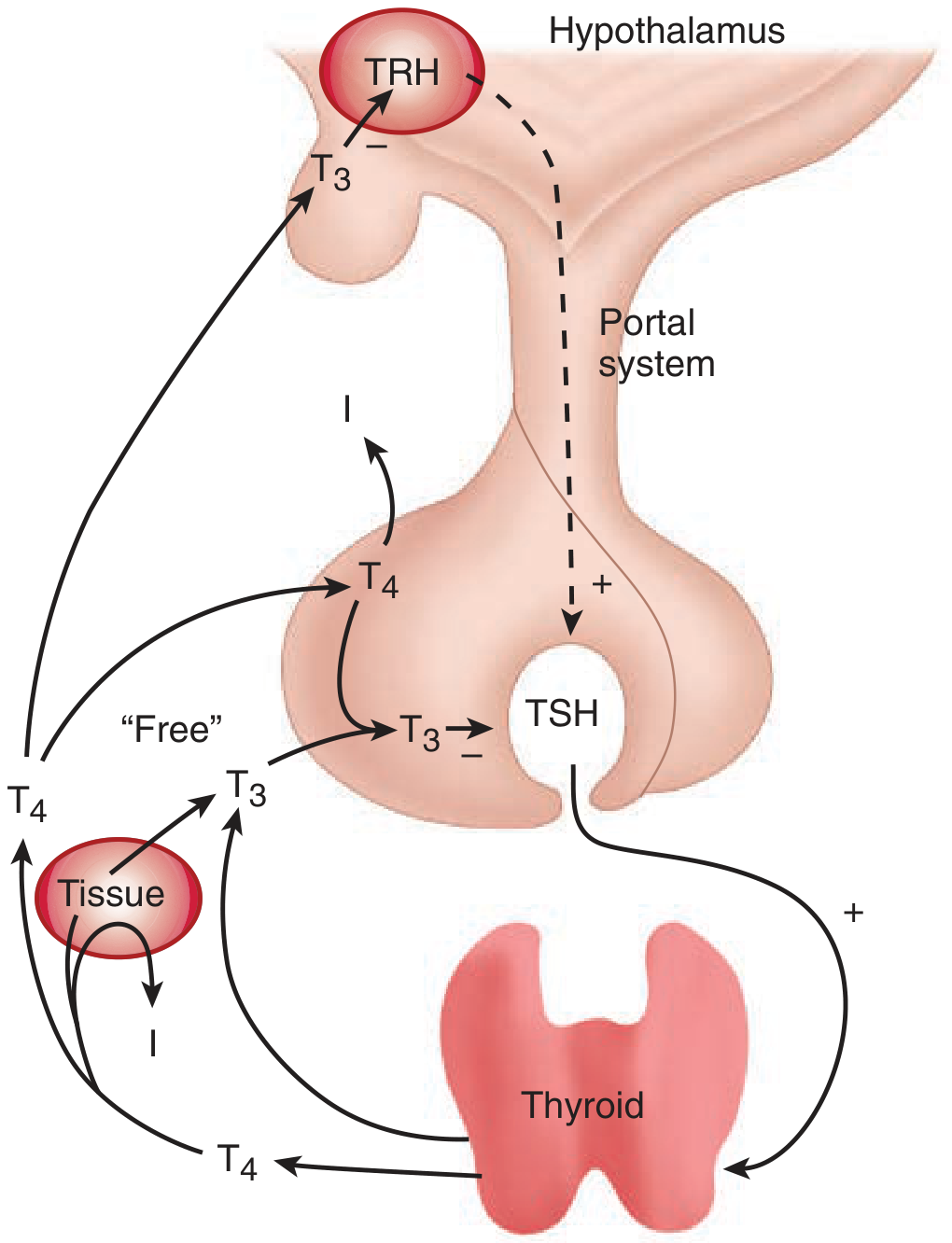
- TRH (thyrotropin-releasing hormone): tripeptide from the hypothalamus, travels via the portal circulation to stimulate TSH release from the anterior pituitary
- TSH (thyrotropin): 28 kDa glycopeptide; binds TSH-R (a G-protein-coupled receptor with 7 transmembrane domains, signals via cAMP)
- Stimulates: iodide trapping, Tg synthesis, TPO activity, hormone secretion, and thyroid cell growth/vascularity
- Negative feedback: T3 (converted from T4 in the pituitary) inhibits both TSH release from the pituitary and TRH release from the hypothalamus
- There is an inverse log-linear relationship between free T4 and TSH - small changes in free T4 cause large shifts in TSH
5. Thyroid Autoregulation
- Low iodide: preferentially synthesizes T3 over T4 (more efficient use of available iodide)
- Iodide excess: inhibits iodide transport, peroxide generation, and hormone synthesis
- Wolff-Chaikoff effect: very high doses of iodide → initial increased organification → followed by suppression of synthesis (protective mechanism; used therapeutically before thyroid surgery)
- Epinephrine and hCG stimulate hormone production (explains elevated thyroid hormones in pregnancy and hydatidiform mole)
- Glucocorticoids inhibit thyroid hormone production
6. Mechanism of Thyroid Hormone Action
- Free T4 and T3 enter cells by diffusion or specific carriers
- T4 is deiodinated to T3 intracellularly
- T3 enters the nucleus via active transport and binds to thyroid hormone receptors (TR-α and TR-β) - nuclear receptors homologous to glucocorticoid, mineralocorticoid, estrogen, vitamin D, and retinoic acid receptors
- TR-α encoded on chromosome 3; TR-β on chromosome 17
7. Physiological Effects of Thyroid Hormones
| System | Effect |
|---|---|
| Metabolism | Increase basal metabolic rate; increase O₂ consumption |
| Cardiovascular | Increase heart rate, cardiac output, speed of muscle contraction/relaxation |
| Carbohydrate | Increase glycogenolysis, hepatic gluconeogenesis, intestinal glucose absorption |
| Lipid | Increase cholesterol synthesis AND degradation (net: lower cholesterol) |
| Growth/Development | Essential for fetal brain development and maturation |
| Sympathomimetic | Upregulate beta-adrenergic receptors |
8. Calcitonin (C-Cell Product)
- Secreted by parafollicular C cells
- Lowers serum calcium by inhibiting osteoclast activity and promoting renal calcium excretion
- Clinically important as a tumor marker for medullary thyroid carcinoma (MTC)
9. Euthyroid Sick Syndrome
- Schwartz's Principles of Surgery, 11th Ed., Ch. 38 (Thyroid, Parathyroid, and Adrenal)
- Tietz Textbook of Laboratory Medicine, 7th Ed., Ch. 57
- Sabiston Textbook of Surgery - Thyroid Histology and Physiology
Create a visual comparison poster of normal vs pathological thyroid histology
Medical education poster comparing normal vs pathological thyroid histology. Professional scientific layout with dark navy blue background and white text. Title at top: "THYROID HISTOLOGY: NORMAL vs PATHOLOGICAL" in bold white letters. Two main columns side by side: LEFT COLUMN - "NORMAL THYROID" with green header: - Large detailed microscopy illustration of normal thyroid follicles - Round to oval follicles of uniform size - Single layer of cuboidal follicular epithelial cells lining each follicle - Abundant pink homogeneous colloid filling the follicular lumen - Flat nuclei, evenly spaced - Caption: "Uniform follicles • Cuboidal epithelium • Abundant colloid • No atypia" RIGHT COLUMN - "PATHOLOGICAL THYROID" with red header, divided into 3 sub-panels: Panel 1: "HASHIMOTO'S THYROIDITIS" - Illustration showing dense lymphocytic infiltrate destroying follicles - Hurthle cell (oxyphilic) metaplasia of follicular epithelium - Lymphoid germinal centers - Caption: "Lymphocytic infiltrate • Hurthle cells • Germinal centers • Follicle destruction" Panel 2: "PAPILLARY THYROID CARCINOMA" - Illustration of papillary architecture with fibrovascular cores - Overlapping nuclei with nuclear grooves and pseudo-inclusions - Ground-glass "Orphan Annie eye" nuclei - Caption: "Papillary architecture • Ground-glass nuclei • Nuclear grooves • Psammoma bodies" Panel 3: "GRAVES DISEASE / HYPERPLASIA" - Illustration of hyperplastic follicles - Tall columnar epithelium with scalloping of colloid at margins - Reduced colloid, increased epithelial height - Caption: "Tall columnar cells • Scalloped colloid • Hyperplastic follicles • Increased vascularity" Bottom section: Comparison table with key histological features: - Columns: Feature | Normal | Hashimoto's | PTC | Graves - Rows: Follicle size, Epithelium, Colloid, Nuclei, Inflammation Each cell filled with concise descriptors. Bottom footer: "C cells (parafollicular) secrete Calcitonin • Medullary carcinoma = C-cell origin with amyloid stroma" Color scheme: navy background, green for normal, red for pathological panels, yellow highlights for key diagnostic features, white body text.
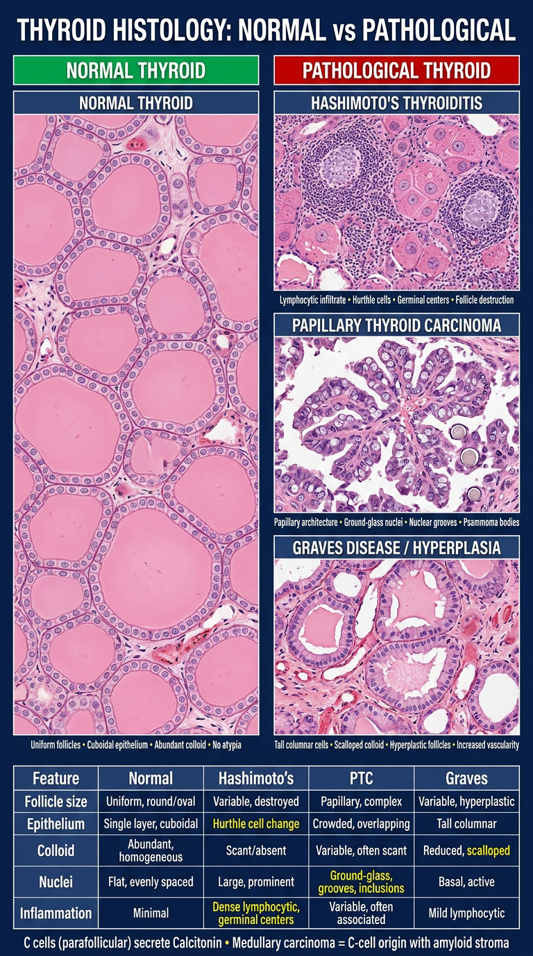
- Uniform round follicles lined by cuboidal epithelium
- Abundant homogeneous pink colloid filling the lumen
- Flat, evenly spaced nuclei, no atypia
| Condition | Key Histological Features |
|---|---|
| Hashimoto's Thyroiditis | Dense lymphocytic infiltrate, lymphoid germinal centers, Hurthle cell (oxyphilic) metaplasia, follicle destruction |
| Papillary Thyroid Carcinoma | Papillary architecture, "Orphan Annie eye" ground-glass nuclei, nuclear grooves and pseudo-inclusions, psammoma bodies |
| Graves Disease | Tall columnar hyperplastic epithelium, scalloped/pale colloid at margins, reduced colloid volume, increased vascularity |