
Hov,molluscum contagiosum ...clinical features,pathogenesis treatment
Reading File
Now let me search the medical library for molluscum contagiosum content in parallel.
Finding Sources
Finding Sources
The best source is Andrews' Diseases of the Skin. Let me read that section in parallel with other sources.
Reading File
Reading File
Reading File
Excellent. Now let me read the full Andrews' dermatology section (the most detailed) and get the pathogenesis and treatment portions:
Reading File
Searching PubMed
molluscum contagiosum treatment
Now let me read the treatment section from Andrews' and fetch a clinical image:
Reading File
Loading Image
Loading Image
I now have comprehensive information. Let me compile the full answer.
Molluscum Contagiosum
Etiology & Epidemiology
Molluscum contagiosum (MC) is caused by Molluscum contagiosum virus (MCV), a member of the Poxviridae family. There are four closely related types - MCV-1 to MCV-4:
- MCV-1 is the most common worldwide, responsible for virtually all infections in young children.
- MCV-2 predominates in HIV-infected patients (causing ~60% of HIV-associated MC).
Three main groups are affected:
- Young children (peak ages 1-4 years) - community/contact spread
- Sexually active young adults (ages 20-29) - STI route
- Immunosuppressed individuals - especially HIV/AIDS (between 10-30% of untreated AIDS patients get MC)
Prevalence affects 3-20% of the population globally.
Pathogenesis
MCV is an obligate intracellular poxvirus that infects only keratinocytes and replicates entirely in the cytoplasm. Key mechanisms:
-
Entry and replication: The virus infects epidermal keratinocytes following direct skin-to-skin contact. It does not enter the dermis under normal conditions.
-
Immune evasion - NF-κB inhibition: MC virus protein MC005 inhibits NF-κB activation by targeting NEMO-regulated IKK activation. This suppresses innate immune signaling, allowing the virus to persist for months without triggering a prompt inflammatory response.
-
Epidermal hyperplasia: Infected cells undergo ballooning degeneration and proliferate, forming a lobulated endophytic mass. As cytoplasmic viral factories grow, they compress the nucleus to the periphery, forming the pathognomonic Henderson-Patterson bodies (molluscum bodies) - large, eosinophilic, cytoplasmic inclusions visible on H&E staining.
-
Role of host immunity in resolution: When cell-mediated immunity eventually overcomes the virus, an inflammatory response is mounted - lesions become inflamed and ultimately involute spontaneously (12-18 months typically). In patients with atopic dermatitis (AD), abnormal cutaneous immunity facilitates infection, and children with AD are 4x more likely to have >50 lesions. In AIDS patients with CD4 <100, there is essentially no immune clearance, leading to hundreds of lesions and giant forms.
-
Cannot be cultured in tissue culture or animal models, complicating research.
Clinical Features
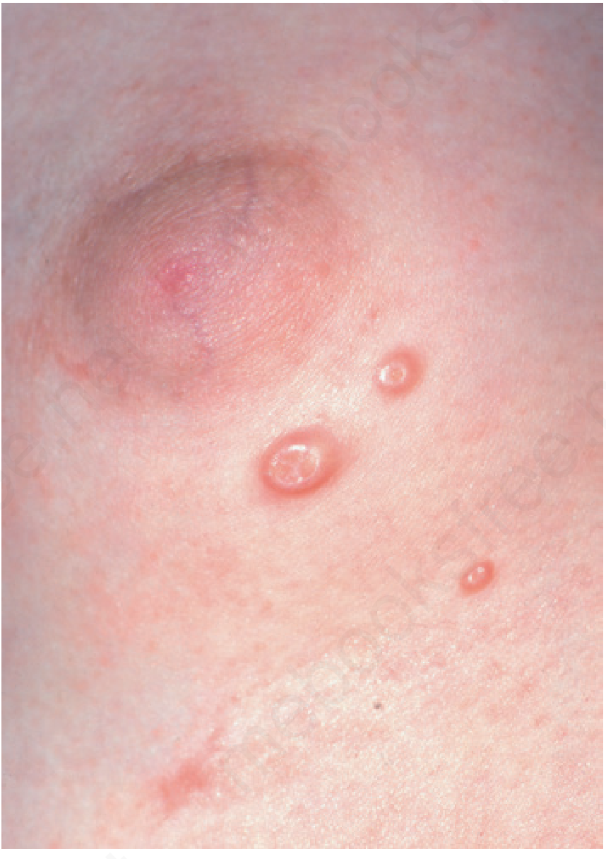
Typical Lesion
- Small (3-5 mm), smooth, firm, dome-shaped, pearly/flesh-colored papule
- Central umbilication is pathognomonic - may contain a white cheesy core (caseous plug)
- Early lesions: <1 mm; "giant" lesions: >1 cm
- Appear in clusters of 5-20 (or more)
- Painless; rarely pruritic unless inflamed
- Incubation period: 2-8 weeks
Distribution by Population
| Group | Distribution |
|---|---|
| Young children | Axillae, inguinal folds, popliteal/antecubital fossae, face; 10% have genital lesions as part of widespread infection |
| Sexually active adults | Lower abdomen, upper thighs, perineum, penile shaft |
| Atopic dermatitis | Confined to dermatitic skin; >50 lesions common; prolonged course |
| HIV/AIDS | Face (cheeks, neck, eyelids), genitalia; confluent plaques; giant lesions; may involve mucosa |
Inflammatory Variants
- Molluscum dermatitis (40% of children): Eczematous reaction surrounding individual lesions - especially in atopic individuals
- Inflamed MC (20% of children): Erythema, swelling, pustulation of individual lesions; known as the "BOTE sign" (Beginning Of The End) - signals impending spontaneous resolution
- Giannotti-Crosti-like reaction: Monomorphic papules on extensor surfaces sparing the trunk - can mimic sudden rapid spread
- Eyelid/conjunctival involvement: associated follicular conjunctivitis or keratitis
HIV-Associated MC
Between 10-30% of AIDS patients not on ART develop MC. Virtually all have CD4 <100. Features:
- Facial predominance (cheeks, neck, eyelids)
- Giant lesions, confluent plaques, possible disfigurement
- Mucosal involvement (oral/genital) indicates very advanced AIDS (CD4 <50)
Histopathology

- Lobulated epidermal hyperplasia with a central crater
- Henderson-Patterson bodies (molluscum bodies): Large eosinophilic intracytoplasmic inclusions in keratinocytes; they compress the nucleus and progressively enlarge as cells move toward the surface
- Can be seen on biopsy or by expressing the caseous core and staining with Wright, Giemsa, or Gram stain
Diagnosis
- Primarily clinical - the central umbilication of dome-shaped pearly papules is characteristic
- Light cryotherapy can highlight the round umbilical opening
- Microscopy: Expressed core squashed between two slides, stained - oval Henderson-Patterson bodies visible
- Differential includes: Cryptococcus, histoplasmosis, coccidioidomycosis (in immunocompromised), keratoacanthoma, desmoplastic trichilemmoma, condyloma acuminatum (in intergluteal region)
Treatment
Treatment decisions depend on the clinical setting:
Observation (Watchful Waiting)
- First-line for immunocompetent young children with numerous lesions
- Individual lesions last 2-4 months each; total infection duration: 12-18 months
- Aggressive treatment risks scarring and emotional trauma in young children
Physical/Destructive Methods
| Method | Notes |
|---|---|
| Cryotherapy (liquid nitrogen) | Effective; highlights umbilication; suitable for children and adults |
| Curettage | Simple disposable curette; treatment of choice especially for adults; rarely needs anesthesia |
| Cantharidin (cantharone) | Applied to body/extremities only (avoid face and perineum in children); applied by wooden end of cotton swab; left 1-6 hours then washed off; ~20 lesions per session; high patient satisfaction |
| Trichloroacetic acid (TCA) | 3.5-100% concentration |
| Comedone expression | Nick the lesion with blade/needle, squeeze with tissue forceps |
| Pulse dye laser | Reported efficacy |
Topical Agents
| Agent | Notes |
|---|---|
| Tretinoin | Applied with toothpick/Q-tip in tiny amounts to each lesion; useful on face |
| Potassium hydroxide 10% | Reported therapy |
| Hydrogen peroxide 1% cream | Reported therapy |
| Povidone-iodine | Reported therapy |
| Aloe vera | Reported therapy |
| Tape stripping | Apply tape, lift repeatedly several times/day for weeks; or surgical tape applied continuously after bathing for 16 weeks |
Immunomodulatory
| Agent | Notes |
|---|---|
| Imiquimod | Two large RCTs showed no more effective than placebo - not recommended |
| Cimetidine (oral) | Immunomodulatory effect; some benefit especially in atopic patients |
| Candidal antigen (intralesional) | Injection in up to 3 lesions; complete clearance in 5.5% of children |
Analgesia Before Procedures
- EMLA (lidocaine-prilocaine) cream applied 1 hour prior to painful procedures - but use with caution due to reports of systemic toxicity in children
HIV-Associated MC
- Initiation of antiretroviral therapy (ART) is the primary treatment - immune reconstitution leads to resolution
- Note: Immune Reconstitution Inflammatory Syndrome (IRIS) can cause paradoxical flare of MC after starting ART
- Local destructive measures (cryotherapy/curettage) are used adjunctively
Genital MC in Adults
- Cryotherapy or curettage is very effective
- Neither imiquimod nor podophyllotoxin has been shown to be effective
Recent Evidence
A 2026 systematic review with network meta-analysis (Campos et al., Paediatr Drugs 2026, PMID 42360610) evaluated MC treatments specifically in children - watch for this for updated comparative evidence. A 2025 systematic review (Naseer et al., Int Ophthalmol Clin 2025, PMID 40116404) covers ocular/periorbital MC over 20 years.
Sources:
- Andrews' Diseases of the Skin: Clinical Dermatology, Chapter 19
- Medical Microbiology 9e, Chapter 44
- Pfenninger and Fowler's Procedures for Primary Care, Chapter 12
This is a shared conversation. Sign in to Orris to start your own chat.