
Difference table between hodgkins and non hodgkins lymphoma
Hodgkin vs non-Hodgkin lymphoma comparison Reed-Sternberg cells
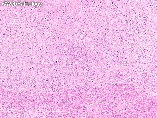
Histopathology illustration of liver tissue demonstrating lymphocyte-depleted classic Hodgkin lymphoma (cHL) with hepatic involvement. The section shows a diffuse fibrotic reticulin network (heavy background fibrosis) with hypocellular cellularity and sparse non-neoplastic inflammatory cells, in contrast to prominent Reed-Sternberg (RS) cells. RS cells are large, often binucleate with prominent nucleoli, and may exhibit sarcomatoid or bizarre mononucleate morphology; in this pattern they are the key diagnostic clue amidst scant lymphocytes. The image captures EBV-associated RS cells as typical features of lymphocyte-depleted cHL, seen in about 90% of such cases. The bottom of the field reveals normal liver parenchyma for comparison, underscoring focal hepatic involvement within otherwise normal hepatic architecture. Clinically, lymphocyte-depleted cHL frequently presents with advanced disease, fever, hepatomegaly, pancytopenia or lymphopenia, and bone marrow involvement; nodal adenopathy may be minimal or absent. Immunophenotypic confirmation (CD30+, CD15+, PAX5 weak) and EBV in situ hybridization (EBER) are commonly employed to support diagnosis. This image is valuable for education and diagnostic training, illustrating the aggressive, low-cellularity variant, the importance of recognizing RS cells in a fibrotic milieu, and the prognostic implications of liver involvement and EBV positivity in Hodgkin lymphoma. These features are essential for differential diagnosis, prognosis, and guiding systemic therapy in advanced Hodgkin lymphoma.
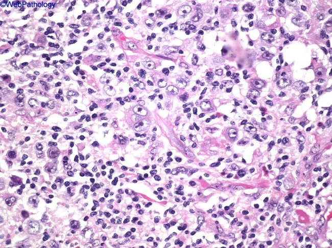
High-power light microscopy of a lymph node biopsy stained with hematoxylin and eosin (H&E) reveals classical Reed-Sternberg (RS) cells and mononuclear variants embedded in a polymorphic inflammatory background. The RS cells are large, often binucleate with prominent eosinophilic nucleoli arranged in an 'owl’s eye' configuration, accompanied by occasional multinucleated giants. In this image, RS cells demonstrate ample cytoplasm and well-defined nuclear membranes, set among a mixed admixture of lymphocytes, eosinophils, plasma cells, and histiocytes. The overall architectural pattern shows nodal effacement by neoplastic Hodgkin and reactive tissue elements rather than solid tumor mass formation. The background inflammatory milieu is characteristic of classical Hodgkin lymphoma, with background cytokine milieu promoting eosinophil recruitment and histiocytic activity. Immunophenotypically, RS cells typically express CD30 and CD15 with weak PAX5 and lack CD45, supporting a diagnosis of classical Hodgkin lymphoma (nodular sclerosis, mixed cellularity, or other subtypes). Clinically, findings correlate with painless lymphadenopathy and potential B symptoms (fever, night sweats, weight loss). This image is diagnostically significant, illustrating the defining microscopic features necessary to differentiate Hodgkin from non-Hodgkin lymphomas and infectious lymphadenitis. It informs differential diagnosis, guides immunohistochemical workup, and underpins treatment planning and prognostic assessment.
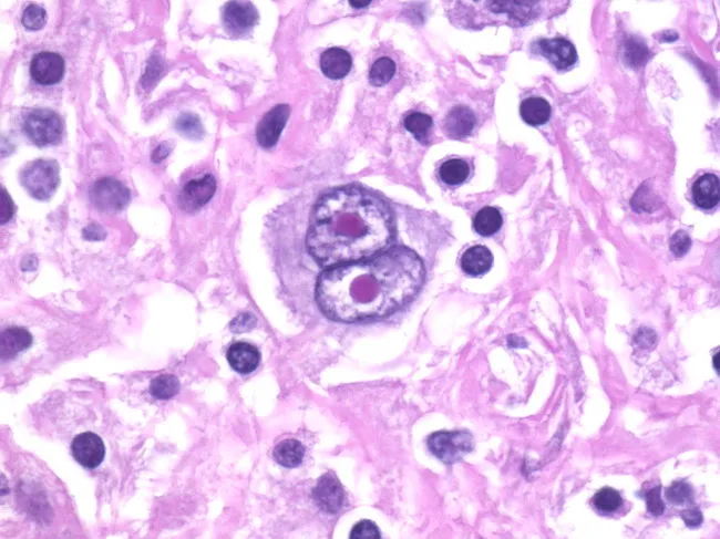
Histopathology: Light microscopy of an H&E-stained lymph node biopsy. This image highlights classic Reed-Sternberg (RS) cells within a mixed inflammatory background, a hallmark of classic Hodgkin lymphoma (cHL). The RS cells are large (up to ~100 microns) with abundant pale eosinophilic cytoplasm and prominent, bilobed to multinucleate nuclei. The nuclei show vesicular chromatin and thick nuclear membranes, with large central nucleoli creating an owl-eye appearance when two nuclei are juxtaposed. Variants, including giant RS cells and lacunar RS cells, may be seen in different HL subtypes. The surrounding milieu contains eosinophils, neutrophils, mature lymphocytes, plasma cells, and histiocytes, reflecting a characteristic reactive infiltrate. The nodal architecture is variably effaced, sometimes with formation of a rosette-like collar of T lymphocytes around RS cells. Immunophenotyping in practice typically demonstrates CD30 and CD15 positivity with weak PAX5 expression, supporting the HL diagnosis, and helps distinguish RS cells from RS-like cells in non-Hodgkin lymphomas or mimics such as anaplastic carcinoma. This histology provides essential diagnostic confirmatory evidence in suspected HL and informs prognosis and treatment planning (ABVD regimen, radiotherapy, or combined modality therapy). In some cases, classic RS cells may be absent, requiring ancillary testing and correlation with clinical findings, correlating with patient outcome.
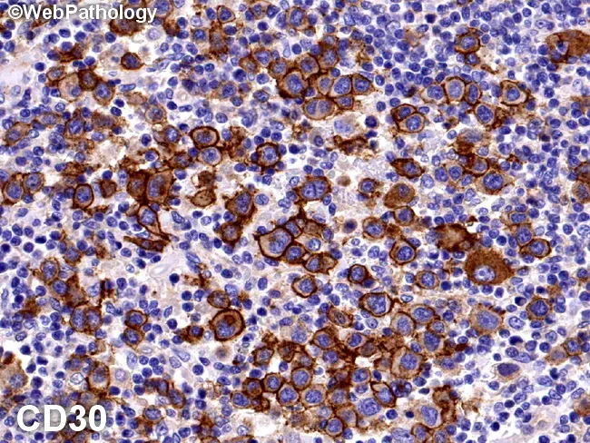
Brightfield immunohistochemistry on formalin-fixed paraffin-embedded lymph node tissue demonstrates classical Hodgkin lymphoma immunophenotype with CD30-positive Reed-Sternberg (RS) cells embedded in a dense reactive inflammatory milieu. The RS cells are rare (roughly 0.1–0.2% of cells) and display characteristic large, often multilobed nuclei with prominent nucleoli, set in a rich background of lymphocytes, eosinophils, plasma cells, and histiocytes. Immunostaining is strong and membranous with a dot-like Golgi pattern in the perinuclear zone, consistent with CD30 expression. Additional activation-associated markers may include CD25, CD40, CD71, and HLA-DR, while CD45 is typically negative, with variable expression of other lineage-associated antigens. The slide highlights the diagnostic dichotomy between RS cells and the surrounding reactive cells; in nodular lymphocyte-predominant HL, LP cells are CD30 negative but may show weak staining. The CD30 positivity supported by the current image confirms classic HL and helps distinguish from CD30-positive non-Hodgkin lymphomas such as anaplastic large cell lymphoma or certain peripheral T-cell lymphomas. This immunophenotype has therapeutic relevance, guiding consideration of CD30-targeted therapies and prognostic stratification, and is diagnostic of classic Hodgkin lymphoma in the appropriate clinical and histopathologic context. Correlation with nodal architecture, lacunar RS variants, and prominent inflammatory microenvironment; influences staging, prognosis, and eligibility for CD30-directed therapies such as brentuximab vedotin in relapsed or refractory classic Hodgkin lymphoma.
Hodgkin Lymphoma vs. Non-Hodgkin Lymphoma
| Feature | Hodgkin Lymphoma (HL) | Non-Hodgkin Lymphoma (NHL) |
|---|---|---|
| Incidence | Less common (~9,000 new cases/year in the US) | Much more common (~80,000 new cases/year in the US; ~5% of all cancers) |
| Age of onset | Bimodal: young adults (15-35 yrs) and older adults (>55 yrs) | Predominantly older adults; incidence rises sharply with age |
| Sex predominance | Slight male predominance | Male slightly > female |
| Cell of origin | Germinal center B cell (Reed-Sternberg cells) | B cells (~85-90%), T cells, or NK cells |
| Hallmark cell | Reed-Sternberg (RS) cell - large binucleate giant cell with "owl-eye" nucleoli; makes up only a small fraction of tumor mass | No RS cells; neoplastic lymphocytes predominate and constitute the bulk of the tumor |
| Immunophenotype | Classic HL: CD30+, CD15+, PAX5 (weak), CD45- | Varies widely by subtype; most B-cell NHL: CD20+, CD45+; T-cell NHL: CD3+, CD4/CD8+ |
| Spread pattern | Contiguous (stepwise) - spreads to anatomically adjacent nodes in orderly fashion | Non-contiguous - skips nodes; spreads in unpredictable, non-contiguous pattern |
| Nodal vs. extranodal | Predominantly nodal (lymph node-based) | Frequently extranodal (GI tract, CNS, skin, bone marrow, liver common) |
| Mediastinal involvement | Common (especially nodular sclerosis subtype) | Less common; seen in certain subtypes (e.g., mediastinal large B-cell lymphoma) |
| Mesenteric/abdominal nodes | Rarely involved | Commonly involved |
| B symptoms | Common (fever, night sweats, weight loss) | Present but generally less prominent |
| Bone marrow involvement | Rare at presentation | Frequent (commonly involved) |
| CNS involvement | Very rare | More common (especially in aggressive subtypes like DLBCL, Burkitt) |
| Classification subtypes | 5 subtypes: Nodular Sclerosis (most common), Mixed Cellularity, Lymphocyte Rich, Lymphocyte Depletion, Nodular Lymphocyte Predominant | >60 subtypes (WHO): Diffuse Large B-Cell (DLBCL), Follicular, Burkitt, Mantle Cell, Marginal Zone, T-cell lymphomas, etc. |
| Histology background | Reactive inflammatory background (eosinophils, plasma cells, neutrophils, lymphocytes surrounding RS cells) | Monotonous proliferation of neoplastic lymphocytes; minimal reactive background |
| EBV association | Common: EBV in RS cells in ~70% of mixed-cellularity subtype; EBV genome identical in all RS cells | Less common; EBV linked to Burkitt lymphoma, post-transplant lymphoma, CNS lymphoma |
| HIV association | Increased risk, especially mixed-cellularity | Much higher risk (>100-fold); Burkitt, DLBCL, CNS lymphoma prominent in HIV |
| Grade/behavior | Considered a single disease with predictable behavior | Ranges from indolent (follicular - years without treatment) to highly aggressive (Burkitt - doubling time hours-days) |
| Staging (Ann Arbor) | Used; spread pattern makes staging very relevant to treatment | Used, but less predictive due to non-contiguous spread |
| Treatment | Chemotherapy (ABVD regimen) ± radiation; combined modality therapy | Varies by subtype: Rituximab (anti-CD20) + CHOP for B-cell NHL; radiation, stem cell transplant for relapse |
| Prognosis | Excellent - cure rate up to 80% with treatment | Variable: indolent NHL may survive years without treatment but is generally incurable; aggressive subtypes can be cured with intensive chemo |
| Pruritus | More common | Less common (~10% of NHL patients) |
| Immune escape mechanism | RS cells express PD-1 ligands (checkpoint factors); loss of MHC class I (β2-microglobulin mutations) | Varies by subtype; often through direct immune evasion or microenvironment manipulation |
| Pathognomonic finding | Reed-Sternberg cell with owl-eye appearance on biopsy | No single pathognomonic cell; diagnosis by subtype-specific immunophenotype and morphology |
Key Memory Points
- HL spreads contiguously (like stairs, step by step) - making radiotherapy more targetable
- NHL spreads non-contiguously (skips around) - making systemic therapy the default
- RS cells define HL - large binucleate cells with owl-eye nucleoli; CD30+, CD15+
- NHL is far more common (~9x more cases) and has over 60 subtypes
- Prognosis in HL is better overall (~80% cure rate); NHL prognosis is highly subtype-dependent
- B symptoms (fever >38°C, drenching night sweats, >10% weight loss) occur in both but are classic HL indicators
Histology Images