
Patient present with sob. How to differentiate between whether it is due to copd, dcm or mi
dyspnea differential diagnosis COPD heart failure ECG chest X-ray comparison table
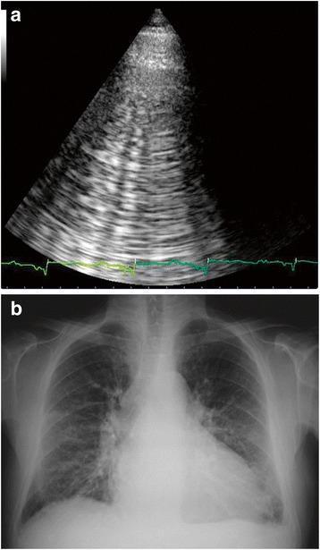
This dual-panel image displays diagnostic findings characteristic of Acute Heart Failure (AHF). Panel 'a' is a lung ultrasound using a phased-array transducer, showing multiple vertical, hyperechoic 'B-lines' (comet-tail artifacts). These laser-like lines originate from the pleural line and extend to the bottom of the screen, indicating interstitial edema. A synchronous ECG tracing is visible at the bottom. Panel 'b' is a posterior-anterior chest X-ray demonstrating cardiomegaly, evidenced by an enlarged cardiac silhouette. There is increased bronchovascular marking and bilateral interstitial opacities consistent with pulmonary venous congestion and edema. Prominent hilar regions and hazy lung fields further support the diagnosis of AHF. This comparison highlights the correlation between bedside sonographic artifacts (B-lines) and classic radiographic signs of cardiogenic pulmonary congestion, serving as a teaching tool for rapid differentiation of dyspnea in an emergency or critical care setting.
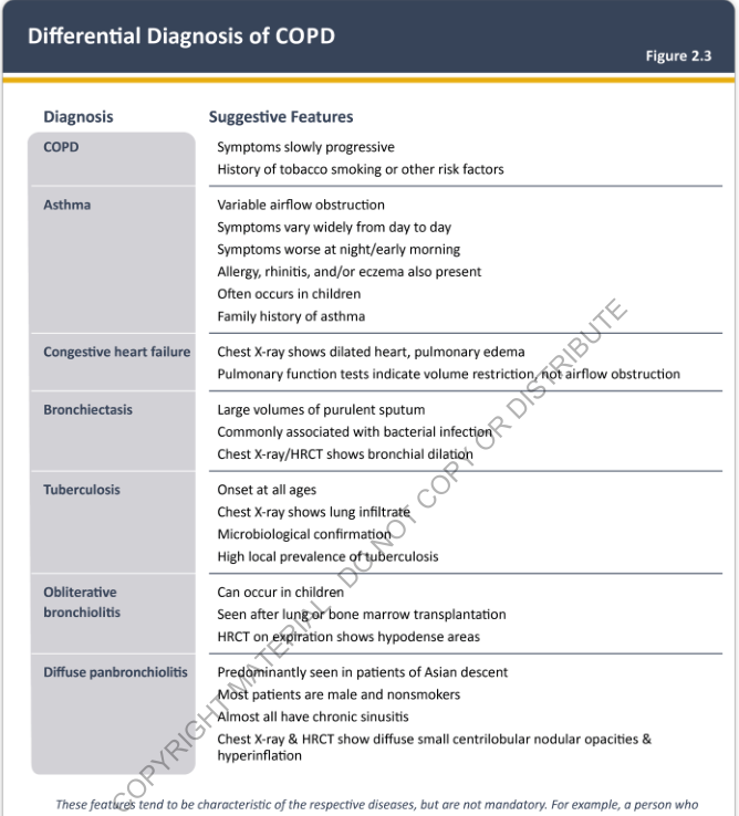
Differential Diagnosis of COPD Figure 2.3 <table><thead><tr><th>Diagnosis</th><th>Suggestive Features</th></tr></thead><tbody><tr><td>COPD</td><td>Symptoms slowly progressive<br>History of tobacco smoking or other risk factors</td></tr><tr><td>Asthma</td><td>Variable airflow obstruction<br>Symptoms vary widely from day to day<br>Symptoms worse at night/early morning<br>Allergy, rhinitis, and/or eczema also present<br>Often occurs in children<br>Family history of asthma</td></tr><tr><td>Congestive heart failure</td><td>Chest X-ray shows dilated heart, pulmonary edema<br>Pulmonary function tests indicate volume restriction, not airflow obstruction</td></tr><tr><td>Bronchiectasis</td><td>Large volumes of purulent sputum<br>Commonly associated with bacterial infection<br>Chest X-ray/HRCT shows bronchial dilation</td></tr><tr><td>Tuberculosis</td><td>Onset at all ages<br>Chest X-ray shows lung infiltrate<br>Microbiological confirmation<br>High local prevalence of tuberculosis</td></tr><tr><td>Obliterative bronchiolitis</td><td>Can occur in children<br>Seen after lung or bone marrow transplantation<br>HRCT on expiration shows hypodense areas</td></tr><tr><td>Diffuse panbronchiolitis</td><td>Predominantly seen in patients of Asian descent<br>Most patients are male and nonsmokers<br>Almost all have chronic sinusitis<br>Chest X-ray & HRCT show diffuse small centrilobular nodular opacities & hyperinflation</td></tr></tbody></table> These features tend to be characteristic of the respective diseases, but are not mandatory. For example, a person who
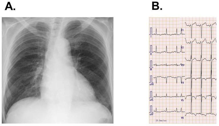
This composite medical image presents diagnostic findings for a patient with heart failure. Panel A is a posterior-anterior (PA) chest X-ray demonstrating mild cardiomegaly with a prominent cardiac silhouette and signs of bilateral pulmonary congestion, evidenced by increased bronchovascular markings in the hilar and basal regions. Panel B is a 12-lead electrocardiogram (ECG) showing normal sinus rhythm. The ECG is characterized by high-voltage QRS complexes in the precordial leads and deep T-wave inversions with a 'strain pattern' in leads V4 through V6. Together, these findings illustrate clinical signs of left ventricular hypertrophy and cardiac remodeling, consistent with a diagnosis of dilated cardiomyopathy. This material is designed for medical education regarding the correlation between radiological imaging and electrophysiological data in chronic heart failure management.
Differentiating SOB due to COPD vs. DCM vs. MI
1. History - The First Discriminator
| Feature | COPD | DCM (Dilated Cardiomyopathy) | MI |
|---|---|---|---|
| Onset | Gradual, over months-years | Gradual (weeks-months) | Acute - minutes to hours |
| Character of SOB | Exertional; "unable to get air in/out" | Exertional initially, then rest; orthopnea, PND | Sudden, often at rest; associated with chest pain |
| Chest pain | Absent (unless PE/pneumothorax complicating) | Usually absent | Present - typical crushing/pressure, radiation to jaw/left arm |
| Cough/sputum | Chronic productive cough; purulent in exacerbations | Pink frothy sputum (in acute pulmonary edema) | Usually absent |
| Orthopnea / PND | Absent | Classic - patient sleeps with multiple pillows | May occur if large MI causes acute LV failure |
| Palpitations / syncope | Rare | Common (arrhythmias from dilated ventricle) | May occur (arrhythmias post-infarct) |
| Risk factors | Smoking (>40 pack-years), occupational dust/fumes, age >40 | Alcohol excess, prior viral illness, family history, chemotherapy | Hypertension, DM, dyslipidemia, smoking, prior CAD, family history |
| Precipitating factors | Upper respiratory tract infection, pollution | Fluid overload, non-compliance with drugs | Exertion, stress, or occurs at rest |
2. Physical Examination
| Sign | COPD | DCM/Heart Failure | MI |
|---|---|---|---|
| General | Barrel chest, pursed-lip breathing, cachexia | Fluid-overloaded; may appear distressed | Diaphoretic, pale, anxious |
| Pulse | May be normal; pulsus paradoxus in severe disease | Tachycardic; may be irregular (AF) | Tachycardic; may be irregular |
| JVP | May be raised if cor pulmonale | Raised | Raised if RV infarct or LV failure |
| Trachea | Central; may shift in pneumothorax | Central | Central |
| Chest expansion | Reduced bilaterally; hyperinflation | Normal or reduced | Normal |
| Percussion | Hyperresonant | Dull at lung bases (pleural effusion) | Usually normal |
| Auscultation - lungs | Wheeze, prolonged expiration, decreased air entry | Bibasal crepitations (pulmonary edema) | May have crepitations if LV failure develops |
| Heart sounds | Normal (right heart strain: loud P2 in cor pulmonale) | S3 gallop (volume overload); S4 may be present; murmur if MR | Muffled heart sounds; new murmur (MR, VSD) |
| Peripheral edema | Ankle edema in cor pulmonale | Pitting pedal edema, ascites | Usually absent unless cardiogenic shock |
| Clubbing | Present in some COPD/lung CA patients | Absent | Absent |
3. Key Investigations
ECG
| Finding | COPD | DCM | MI |
|---|---|---|---|
| Rhythm | Sinus; multifocal atrial tachycardia | Sinus tachycardia; AF common | Sinus; ventricular arrhythmias |
| Axis | Right axis deviation (cor pulmonale) | Left axis deviation; LBBB | ST changes (elevation in STEMI, depression in NSTEMI) |
| Specific pattern | P pulmonale, right heart strain pattern | LBBB, non-specific ST-T changes; low voltage | ST elevation/depression, T-wave inversion, new Q waves |
Chest X-Ray
| Feature | COPD | DCM | MI |
|---|---|---|---|
| Heart size | Normal or cor pulmonale | Cardiomegaly (CTR >0.5) | May be enlarged acutely if chronic HF |
| Lung fields | Hyperinflation, flat diaphragm, bullae, increased AP diameter | Pulmonary venous congestion, Kerley B lines, pleural effusions | Patchy edema if LV failure develops |
| Vasculature | Peripheral pruning (in emphysema) | Upper lobe diversion, bat-wing pattern | Similar to DCM in acute failure |
Blood Tests
| Test | COPD | DCM | MI |
|---|---|---|---|
| Troponin | Normal (unless demand ischemia) | Mildly elevated (myocyte stretch) | Markedly elevated (the hallmark) |
| BNP / NT-proBNP | Mildly elevated if cor pulmonale | Markedly elevated | Elevated if LV dysfunction develops |
| ABG | Hypoxemia + hypercapnia (type 2 RF in severe) | Hypoxemia (type 1 RF) | Usually normal unless in shock |
| FBC | Polycythemia (chronic hypoxia) | May be normal or dilutional anemia | Usually normal |
| CXR/Echo | As above | As above | As above |
Pulmonary Function Tests (PFTs) / Spirometry
- COPD: Obstructive pattern - FEV1/FVC <0.70, post-bronchodilator; reduced FEV1. This is the gold standard for COPD diagnosis (as per GOLD guidelines).
- DCM: Restrictive pattern or normal PFTs. Volume restriction, NOT airflow obstruction.
- MI: Normal (unless acute pulmonary edema).
Echocardiogram
| Feature | COPD | DCM | MI |
|---|---|---|---|
| LV size | Normal or small | Dilated LV | Normal or regional WMA |
| EF | Normal | Reduced EF (<40%) | Reduced if large MI; regional WMA |
| Valves | Normal | MR (dilational); TR | New MR (papillary muscle rupture) |
| Right heart | RV hypertrophy/dilation (cor pulmonale) | Both ventricles dilated | Normal unless RV infarct |
4. Bedside Ultrasound (POCUS) - Rapid Tool in ED
- COPD: A-lines (horizontal lines) on lung US; hyperinflated lung; no B-lines
- DCM/HF: Multiple B-lines (comet-tail artifacts) bilaterally - indicate interstitial edema
- MI: B-lines if LV failure; regional WMA on cardiac POCUS
5. Quick Memory Aid - The "5C" Clue Summary
| Clue | Points to |
|---|---|
| Chronic smoker + wheeze + hyperinflation | COPD |
| Cardiomegaly + S3 gallop + orthopnea + raised JVP | DCM |
| Crushing chest pain + ST elevation + troponin spike | MI |
| Clear sputum + prolonged expiration | COPD |
| Comet-tail B-lines on POCUS | Cardiac (DCM or acute MI with LV failure) |
Important Clinical Caveat
- Harrison's Principles of Internal Medicine 22E, Table 34-3 - differential diagnosis by system
- ROSEN's Emergency Medicine - BOX 60.2, Table 2.13
- Symptom to Diagnosis: An Evidence-Based Guide, 4th ed. - Dyspnea chapter
- Braunwald's Heart Disease - Dilated Cardiomyopathy / BNP in dyspnea
- Textbook of Clinical Echocardiography - DCM echo features