
Occipital headache
"occipital neuralgia" OR "occipital headache"
PMID: 40017062
occipital neuralgia greater occipital nerve anatomy diagram
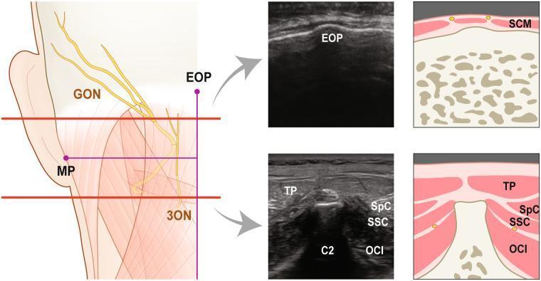
This composite educational image illustrates the topographical and sonographic anatomy of the occipital nerves for clinical blocks. The left panel features a cadaveric-style anatomical diagram of the posterior-lateral neck and scalp, highlighting the Greater Occipital Nerve (GON) and Third Occipital Nerve (3ON) in yellow. Key landmarks include the external occipital protuberance (EOP) and mastoid process (MP). Two horizontal red reference lines indicate levels for cross-sectional imaging: the upper line at the GON emergence from the trapezius (TP) and the lower line at the spinous process of the axis (C2). The right panel provides corresponding transverse ultrasound images and schematic diagrams at these two levels. The superior section identifies the EOP and sternocleidomastoid (SCM). The inferior section at C2 demonstrates the layered musculature: the superficial trapezius (TP), followed by the splenius capitis (SpC), semispinalis capitis (SSC), and the deepest layer, the obliquus capitis inferior (OCI). Small yellow dots in the diagrams pinpoint nerve locations relative to these muscle planes, aiding in ultrasound-guided anesthetic procedures.
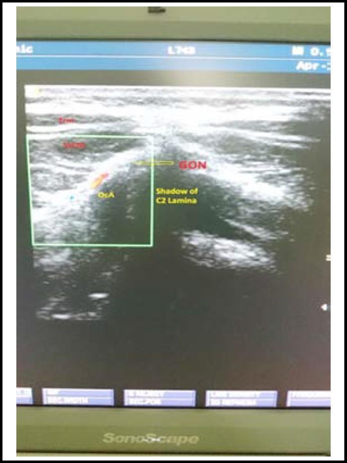
A diagnostic ultrasound image of the posterior neck region, utilized for guiding a greater occipital nerve (GON) block. The image displays the musculoskeletal and vascular anatomy at the level of the upper cervical spine. Visible layers include the trapezius muscle (TrM) and the semispinalis capitis muscle (SsCM), characterized by their striated, heterogeneous echotexture. The greater occipital nerve (GON) is identified as a small, oval structure located medial to the occipital artery (OcA), which appears as a circular hypoechoic or anechoic vascular structure. Deep to these soft tissue layers, a prominent hyperechoic bony surface with an associated posterior acoustic shadow represents the C2 lamina. This imaging modality and view are clinically significant for the precise localization of the nerve in the management of post-dural puncture headache (PDPH) or occipital neuralgia, ensuring accurate anesthetic deposition while avoiding vascular puncture.
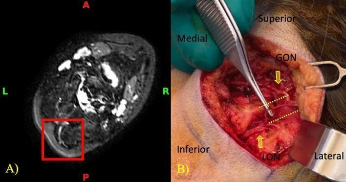
This figure provides a side-by-side comparison of diagnostic imaging and intraoperative findings related to peripheral nerve anatomy in the occipital region. Image A is a transverse NerveView MRI (Magnetic Resonance Neurography) of the neck at the level of the posterior paraspinal muscles. A red box highlights the right greater occipital nerve (GON) and lesser occipital nerve (LON), appearing as hyperintense, tubular structures with a visible anatomical connection. Image B is a corresponding intraoperative clinical photograph during a surgical decompression procedure. Yellow arrows point to the exposed GON and LON within the surgical field, while yellow dotted lines indicate a direct neural communicating branch between the two nerves. The anatomical relationship between these structures is relevant for understanding potential compression points in patients with occipital neuralgia or cervicogenic migraines. The combined images demonstrate the efficacy of high-resolution MRI in identifying fine-scale neural variations that are subsequently confirmed through direct surgical visualization.
Occipital Headache
Anatomy
| Nerve | Origin | Territory |
|---|---|---|
| Greater occipital nerve (GON) | C2 dorsal ramus | Posterior scalp from occiput to vertex |
| Lesser occipital nerve (LON) | C2–C3 ventral rami (cervical plexus) | Lateral scalp, posterior ear |
| Third occipital nerve | C3 dorsal ramus | Suboccipital, upper cervical area |
1. Occipital Neuralgia
Definition & Epidemiology
Clinical Features
- Location: Unilateral in 81% of cases; GON territory most commonly affected (98%)
- Quality: Stabbing (59%), severe intensity (54%)
- Hypoesthesia: Present in ~73% of patients in the GON distribution - an important clue to entrapment neuropathy
- Tenderness: Palpation over the GON at the superior nuchal line often reproduces or worsens the headache
- Associated features: Nausea, vomiting possible; may be confused with migraine
Causes
- Whiplash / head trauma (falls on the back of the head, closed-head injuries)
- Entrapment of GON as it pierces the trapezius
- Degenerative cervical disease
- Idiopathic
Diagnosis
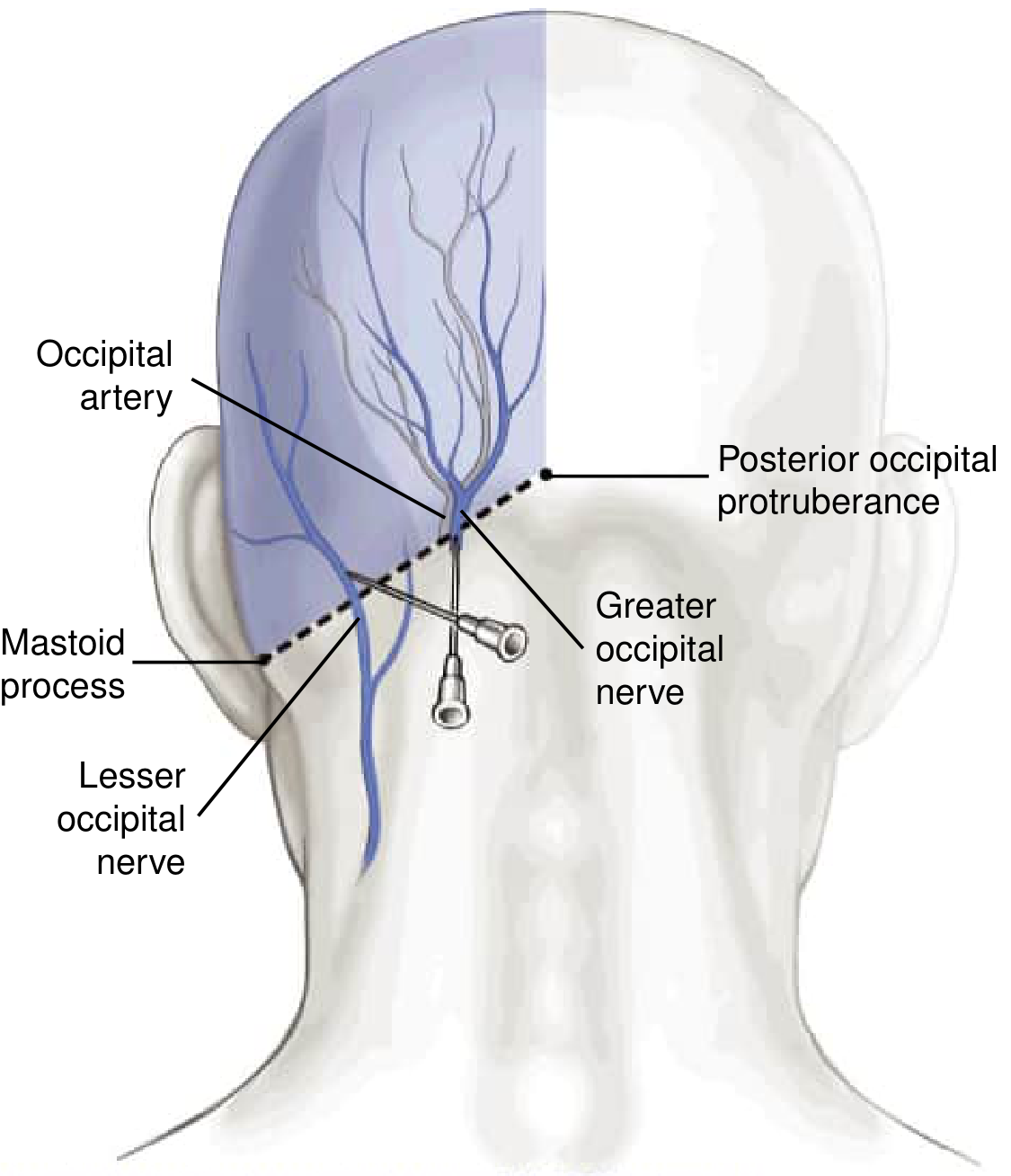
A diagnostic greater occipital nerve block (2-3 mL of 0.5% bupivacaine ± 20 mg triamcinolone, injected 1.5-2 cm lateral to the posterior occipital protuberance at the occipital artery groove) can both confirm the diagnosis and provide therapeutic relief. In whiplash-related cases, a single block provides immediate relief in ~90% of patients lasting an average of 28 days. - Bradley and Daroff's Neurology, p. 1093
Treatment
| Step | Options |
|---|---|
| Conservative | NSAIDs, gabapentin, carbamazepine, analgesics |
| Interventional | Repeated GON blocks (local anesthetic ± steroid); lesser occipital nerve block |
| Neuromodulatory | Botulinum toxin injections |
| Surgical | Occipital nerve section or C2/C3 dorsal root section (variable results; risk of anesthesia dolorosa - use with caution) |
2. "Third Occipital Nerve" Headache
- Diagnosis: Percutaneous blocking of the third occipital nerve near the facet joint under fluoroscopic guidance (diagnostic + temporarily therapeutic)
- Treatment: Radiofrequency coagulation of the nerve, steroid injections around the C2-C3 facet joint, NSAIDs
- Adams and Victor's Principles of Neurology, 12th Ed.
3. Cervicogenic Headache
Key Features
- Occipital headache, especially unilateral, with constant neck pain - strongly suspect cervicogenic origin
- Triggered or exacerbated by neck movement or sustained neck postures
- Constant pain with episodic exacerbations
- May radiate to frontal, temporal, or orbital regions (via trigeminal-cervical convergence)
- Associated nausea, vomiting, photophobia, phonophobia (overlaps significantly with migraine)
- History of head/neck trauma common
Distinguishing from Migraine
| Feature | Cervicogenic | Migraine |
|---|---|---|
| Provocation | Neck movement | Not neck movement |
| Pattern | Persistent | Episodic |
| Photophobia/nausea | Mild or absent | Prominent |
| Response to GON block | Often positive | Can also be positive (not specific) |
Treatment
- Intra-articular steroids / long-acting anesthetics (relief lasting months)
- Radiofrequency neurotomy of C2-C3 zygapophysial joint can achieve complete relief
- Physical therapy
4. Differential Diagnosis - Red Flags
| Cause | Key Features |
|---|---|
| Subarachnoid hemorrhage | "Worst headache of life," sudden onset, nuchal rigidity |
| Posterior fossa tumor | Progressive, worse in morning, associated neurological signs |
| Epidural hematoma | Trauma history, occipital injury, rapid progression with drowsiness |
| Meningitis | Fever, photophobia, nuchal rigidity |
| Vertebral artery dissection | Sudden neck pain, posterior headache, Horner syndrome, stroke symptoms |
| Hypertension | Bilateral, associated with elevated BP |
| Basilar migraine | Occipital headache with brainstem aura (diplopia, vertigo, ataxia) |
In children, occipital headache was historically associated with intracranial disease, but more recent studies have not shown an increased association with serious secondary causes. Still, those unable to localize pain or presenting with occipital location warrant careful evaluation. - Tintinalli's Emergency Medicine
Summary Approach
Occipital Headache
├── Red flags present? → Urgent imaging (CT/MRI), LP if needed
├── Sudden ("thunderclap")? → Rule out SAH first
├── Stabbing, paroxysmal, tenderness over GON? → Occipital neuralgia
│     └── Diagnostic GON block → Treatment
├── After neck injury, C2-C3 facet origin? → Third occipital nerve headache
│     └── Fluoroscopic nerve block → RF coagulation
├── Unilateral + constant + neck movement trigger? → Cervicogenic headache
│     └── Diagnostic block → RF neurotomy / PT
└── Episodic + photophobia/nausea + no neck provocation? → Migraine (occipital variant)
- Adams and Victor's Principles of Neurology, 12th Ed. (Third Occipital Nerve Headache, Occipital Neuralgia)
- Bradley and Daroff's Neurology in Clinical Practice (GON Block, Cervicogenic Headache, Headaches and the Cervical Spine)
- Tintinalli's Emergency Medicine (Location of Headache, Pediatric Headache)
- Melchior AG et al. (2025). Epidemiology and clinical features of occipital neuralgia: A systematic review and meta-analysis. Cephalalgia - PMID 40017062