
Explain me navus of ota
Reading File
Nevus of Ota is a dermal melanocytic hamartoma characterized by bluish-gray pigmentation in the distribution of the trigeminal nerve (ophthalmic and maxillary branches), predominantly affecting the periocular area, temple, and cheek. It is most common in Asian and darker-skinned individuals, with a female predominance.
Searching sources...
Finding Sources
Searching PubMed
nevus of Ota oculodermal melanocytosis
Good - I found dedicated sections in multiple dermatology textbooks. Let me read the most comprehensive ones in parallel.
Reading File
Reading File
Reading File
Now let me get the more detailed block from Fitzpatrick's which should have the fullest coverage, and also check for any clinical images.
Reading File
Reading File
Excellent data. Let me read more from the Dermatology 2-Volume Set which has the most complete coverage, and also grab the clinical images.
Reading File
Loading Image
I now have comprehensive, authoritative information from multiple dermatology textbooks. Here is the complete explanation:
Nevus of Ota (Oculodermal Melanocytosis)
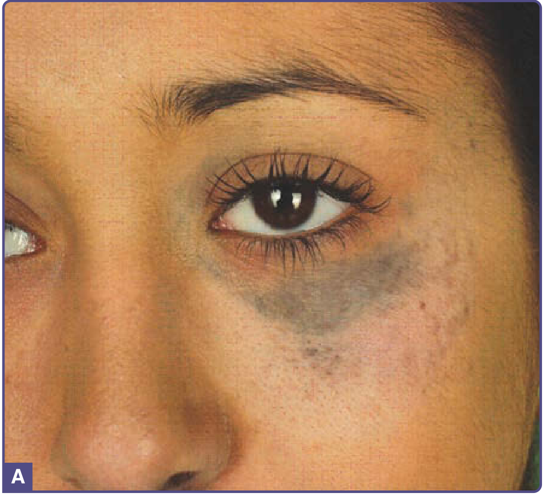
Definition and Names
Nevus of Ota, first described by Ota in 1939, is also known by several synonyms:
- Nevus fuscoceruleus ophthalmomaxillaris (most formal name)
- Oculodermal melanocytosis
- Congenital melanosis bulbi
- Oculomucodermal melanocytosis
It is a dermal melanocytic hamartoma characterized by blue-black or gray-brown pigmentation on the face.
Epidemiology
- Predominantly affects Asians (0.4%-0.8% of all dermatologic patients in Japan; 0.6% of the Asian population generally)
- About 80% of cases occur in females, though this may reflect greater cosmetic concern in women
- Has two peaks of onset:
- Infancy/birth (50-60% of cases) - most are present at birth
- Puberty (40-50% of cases)
- Rare familial forms exist; generally not hereditary
- 5-15% of cases are bilateral
Pathogenesis
The blue-to-gray color results from melanin-producing melanocytes located in the dermis (the Tyndall effect - deeper pigment appears bluish through overlying skin). The melanocyte density in nevus of Ota is greater than in simple dermal melanocytosis (Mongolian spot), supporting the concept of a hamartoma rather than a developmental aberration.
Molecular basis: Somatic activating mutations in GNA11 or GNAQ are found in up to 15% of nevus of Ota cases (compared to 65-75% in blue nevi and 80-85% in primary uveal melanomas). Hormonal influences may also play a role, explaining the female predominance and color fluctuations during menstruation, puberty, and menopause.
Triggers described include infection, trauma, UV light exposure, and hormonal changes.
Clinical Features
Distribution
- Unilateral, following the first (ophthalmic) and second (maxillary) branches of the trigeminal nerve
- Most common sites: periorbital area, temple, forehead, malar area, earlobe, pre- and retroauricular regions, nose, and conjunctivae
Appearance
- Confluence of individual macules (pinhead to several mm)
- Shape: round, oval, or serrated individual macules forming an overall irregularly demarcated, often mottled patch
- Color: shades of brown, gray, blue, black, or purple
Mucosal and Ocular Involvement
- ~60-65% have ipsilateral scleral pigmentation
- Tympanum (55%), nasal mucosa (30%), pharynx (25%), palate (20%)
- Rarely: cornea, iris, fundus oculi, retrobulbar fat, retina, optic nerve
- Iris mammillations and glaucoma (~10%) reported
Classification (Severity)
| Type | Description |
|---|---|
| Type 1 (Mild) | Limited involvement |
| Type 2 (Moderate) | Moderate involvement |
| Type 3 (Intensive) | Extensive involvement |
| Type 4 (Bilateral) | Both sides affected |
Natural History
- Persists throughout life (unlike Mongolian spots)
- Color intensity can fluctuate with hormonal changes
- Slowly enlarges over time
Histopathology
- Elongated, dendritic melanocytes scattered between collagen bundles in the upper third of the reticular dermis
- In comparison to dermal melanocytosis, cells are more numerous
- Hyperpigmentation of the basal epidermis may be present
- DOPA reactivity varies: lightly pigmented cells are often strongly reactive; heavily pigmented ones often not reactive
- Melanocytes may cluster around blood vessels and sweat/sebaceous glands
- Raised areas show larger aggregates resembling blue nevi
Complications
Malignant Transformation (rare)
- Uveal melanoma risk: estimated at 1 in 400 in White individuals (much lower in Asians)
- Cutaneous melanomas are very rare; may present as new subcutaneous nodules
- Other malignant sites: orbit, optic chiasma, meninges, choroid, ciliary body, iris, brain
- BAP1 mutations have been detected in cases that progressed to melanoma
- Malignant degeneration is more common in White patients
- The most common site of malignancy is the choroid
Other Complications
- Ipsilateral glaucoma (~10%)
- Ipsilateral sensorineural hearing loss (hypoacusia) - occasional
- Intracranial melanocytosis (association described)
Differential Diagnosis
| Condition | Distinguishing Features |
|---|---|
| Mongolian spot (dermal melanocytosis) | Lumbosacral location, fades in childhood |
| Melasma | Epidermal pigment, bilateral, hormonal trigger, no scleral involvement |
| Blue nevus | Well-circumscribed papule/nodule, not patch-like |
| Hori nevus (acquired nevus of Ota-like macules) | Bilateral, acquired, responds less well to laser |
| Venous malformation | Vascular, blanches with pressure |
| Ecchymosis | Traumatic history, resolves |
Treatment
Treatment is primarily cosmetic. Q-switched (QS) and picosecond lasers are the standard of care, with multiple sessions typically required.
| Laser | Wavelength | Notes |
|---|---|---|
| QS Nd:YAG | 1064 nm | Deepest penetration; preferred for darker skin types (Fitzpatrick IV-VI); less hypopigmentation risk |
| QS Alexandrite | 755 nm | Good efficacy; less hypopigmentation than ruby; deeper than ruby |
| QS Ruby | 694 nm | Effective but higher risk of hypopigmentation due to epidermal melanin absorption |
| Picosecond lasers | 755 nm / 1064 nm | Emerging evidence; effective in children too |
Key treatment principles:
- High-energy fluences required; space treatments at least 6 weeks apart
- Pretreatment with 0.1% tretinoin + 5% hydroquinone + 7% lactic acid can reduce epidermal melanin load before laser
- Fractional 1440-nm Nd:YAG has also shown success
- Nevus of Ota responds better to laser than Hori nevus (acquired bilateral form)
Nevus of Ota vs. Nevus of Ito
| Feature | Nevus of Ota | Nevus of Ito |
|---|---|---|
| Synonym | Nevus fuscoceruleus ophthalmomaxillaris | Nevus fuscoceruleus acromiodeltoideus |
| Location | Face (V1/V2 of trigeminal nerve) | Shoulder, neck, supraclavicular area |
| Histology | Same (dendritic dermal melanocytes) | Same |
| Treatment | Same (QS/pico lasers) | Same |
Sources: Andrews' Diseases of the Skin, p.730-733 | Dermatology 2-Volume Set (5th ed.) | Fitzpatrick's Dermatology, p.1385
This is a shared conversation. Sign in to Orris to start your own chat.