
Dangerous area of scalp 1st year mbbs
The dangerous area of the scalp is the loose areolar tissue layer (Layer 4) - the subaponeurotic layer between the epicranial aponeurosis and the pericranium. Infections here can spread freely across the entire scalp and, critically, can track intracranially via emissary veins, potentially causing meningitis, cavernous sinus thrombosis, or cerebral abscess.
Searching sources...
Reading File
Finding Sources
Finding Sources
Finding Sources
Finding Sources
Reading File
Reading File
Loading Image
Finding Sources
Reading File
Here is a complete, exam-oriented answer on the Dangerous Area of the Scalp:
Dangerous Area of the Scalp
The 5 Layers of the Scalp - Mnemonic: S.C.A.L.P.
| Layer | Structure | Key Feature |
|---|---|---|
| S | Skin | Thick, hair-bearing; contains sebaceous glands |
| C | Connective tissue (dense subcutaneous) | Contains all blood vessels and nerves; vessels held open - hence profuse bleeding in lacerations |
| A | Aponeurotic layer (Epicranial aponeurosis / Galea aponeurotica) | Connects frontalis (front) to occipitalis (back) |
| L | Loose areolar tissue | THE DANGEROUS LAYER |
| P | Pericranium (periosteum of calvaria) | Firmly attached to skull |
- Gray's Anatomy for Students, p. 1287
Why is Layer 4 (Loose Areolar Tissue) Called the "Dangerous Area"?
The 4th layer - the subaponeurotic loose areolar tissue - is the dangerous area for these reasons:
1. Unrestricted spread of infection and blood
- The loose areolar tissue has no fibrous septa or barriers. Pus, blood, or fluid in this layer can spread freely in all directions across the entire calvaria - from the orbital margins anteriorly to the nuchal lines posteriorly.
- Infections here form a "spreading cellulitis" that is not walled off.
2. Intracranial spread via Emissary Veins
- Emissary veins pass through foramina in the skull connecting the veins of this layer directly to intracranial dural venous sinuses (parasagittal, lateral/transverse, and cavernous sinuses).
- Since these veins are valveless, infection can travel in either direction - scalp infection can spread intracranially.
- This can cause:
- Meningitis
- Cavernous sinus thrombosis
- Cerebral abscess
- Osteomyelitis of the skull
3. Diploic Vein connection
- Diploic veins lie within the spongy diploe of the skull and connect pericranial veins with intracranial meningeal veins and sinuses. Septic scalp wounds can cause thrombophlebitis of cranial sinuses through these pathways.
- Parikh's Textbook of Medical Jurisprudence, Forensic Medicine and Toxicology, p. 12707
Diagram - Coronal Section of Scalp
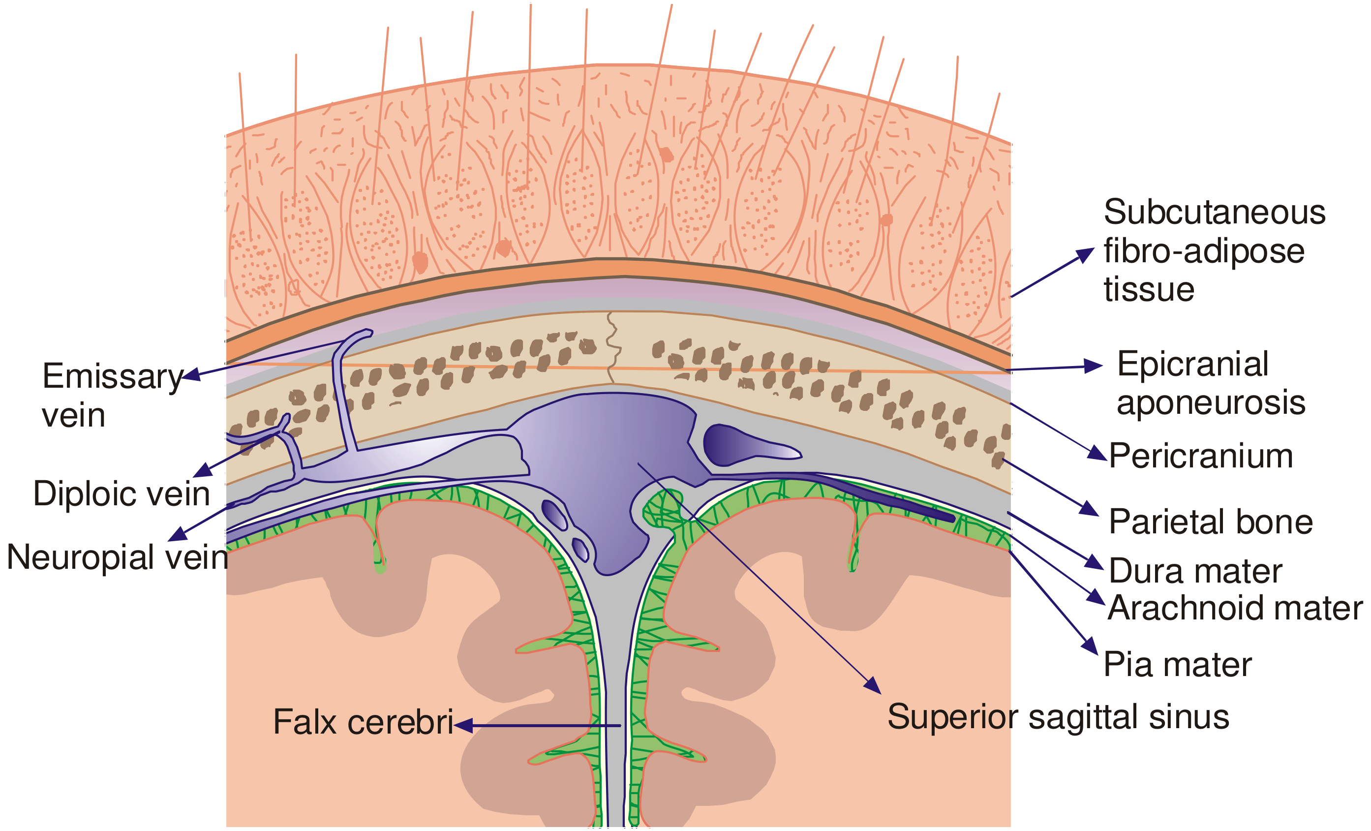
Coronal section showing the scalp layers and the critical emissary vein connections to intracranial sinuses - P.C. Dikshit Textbook of Forensic Medicine and Toxicology
Important Clinical Correlates
| Hematoma Location | Layer | Spread | Clinical Note |
|---|---|---|---|
| Subcutaneous hematoma | Layer 2 | Limited (fibrous septa) | Localised swelling |
| Subaponeurotic hematoma | Layer 4 (DANGEROUS) | Spreads widely over whole scalp | Can track to orbit / behind ear; may cause jaundice in neonates |
| Subpericranial hematoma (Cephalhematoma) | Under pericranium | Limited to one bone (sutural attachments) | Common in newborns |
Quick Exam Summary
- Dangerous area = Layer 4 = Loose areolar (subaponeurotic) layer
- Danger is from unrestricted spread of infection/blood + intracranial spread via valveless emissary veins
- Complications: Meningitis, cavernous sinus thrombosis, cerebral abscess
- Outer 3 layers (S, C, A) move together as one unit - called the "scalp proper"
- Dense connective tissue (Layer 2) is why scalp lacerations bleed profusely - vessels are held open
This is a shared conversation. Sign in to Orris to start your own chat.