
Biological width and its significance in finish line management in restorative procedures for 10 marks. Please include hand drawn images for university exam
biological width finish line management restorative dentistry crown preparation
https://pmc.ncbi.nlm.nih.gov/articles/PMC3284004
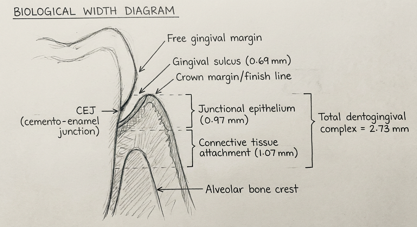
Hand-drawn style university exam diagram showing biological width of a tooth. Shows a cross-section of a tooth and surrounding gingival tissues with clear labeled measurements. Labels include: Gingival sulcus (0.69 mm), Junctional epithelium (0.97 mm), Connective tissue attachment (1.07 mm), Alveolar bone crest, Biological width = 2.04 mm (bracket spanning junctional epithelium + connective tissue), Total dentogingival complex = 2.73 mm. Also shows free gingival margin at top, CEJ (cemento-enamel junction), and the crown margin/finish line placement in relation to these structures. Pencil sketch style, clean labels with arrows, as if drawn by a dental student for exams.
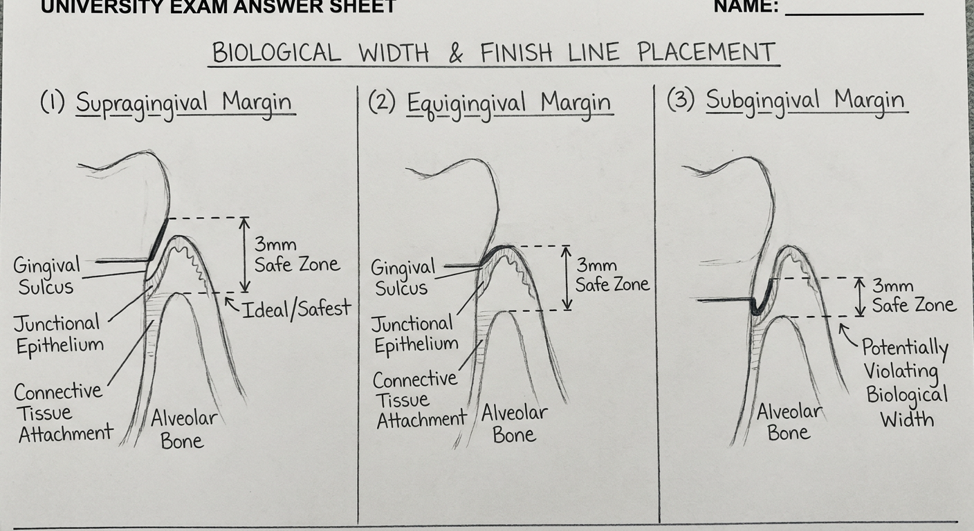
Hand-drawn university exam style diagram showing three scenarios of finish line placement in relation to biological width: (1) Supragingival margin - finish line above gingival margin, labeled as ideal/safest; (2) Equigingival margin - finish line at gingival crest; (3) Subgingival margin - finish line below gingival margin potentially violating biological width. Each tooth cross-section shows alveolar bone, connective tissue attachment, junctional epithelium, gingival sulcus. Arrows indicate the 3mm safe zone from alveolar bone to finish line. Pencil sketch diagram style with clear annotations for a dental exam answer.
Biological Width and Its Significance in Finish Line Management in Restorative Procedures
1. Introduction
2. Definition
Biological Width = Junctional Epithelium + Connective Tissue Attachment = 0.97 mm + 1.07 mm = 2.04 mm (approximately 2 mm)
3. Components of the Dentogingival Complex (Gargiulo et al.)
| Component | Average Dimension |
|---|---|
| Gingival sulcus depth | 0.69 mm |
| Junctional epithelium (JE) | 0.97 mm |
| Connective tissue attachment (CTA) | 1.07 mm |
| Total dentogingival complex | 2.73 mm |
| Biological width (JE + CTA) | 2.04 mm |
4. Anatomy of the Biological Width
- The junctional epithelium forms a collar around the tooth and attaches to the enamel or cementum via hemidesmosomes. It acts as a seal against the oral environment.
- The supracrestal connective tissue attaches directly to the root cementum above the alveolar crest via Sharpey's fibers. It provides support and maintains the gingival architecture.
- These two components together form a biological barrier protecting the alveolar bone from the oral environment.
- The dimensions are not constant - they vary from tooth to tooth, from site to site on the same tooth, and are reportedly greater on molars than on anterior teeth.
5. Significance in Finish Line Management
A. Supragingival Margin
- Placed coronal to the free gingival margin
- Easiest to prepare, impression, and maintain
- Preferred wherever esthetics are not a concern
- Does not violate biological width
- Allows self-cleansing and home care
B. Equigingival Margin
- Placed at the level of the free gingival margin
- Generally discouraged due to plaque accumulation
C. Subgingival Margin
- Placed within the gingival sulcus
- Indicated for esthetics, subgingival caries, or need for additional retention
- Most critical zone - if extended too deeply, it violates the biological width
- The finish line should be placed a maximum of 0.5-0.7 mm below the free gingival margin to remain within the sulcus and avoid encroaching on the junctional epithelium
The "3 mm Rule"
- 1 mm - Connective tissue attachment
- 1 mm - Junctional epithelium
- 1 mm - Buffer zone for finish line within the sulcus
6. Biological Width Violation
Definition
Causes
- Deep subgingival caries
- Short clinical crown
- Tooth fracture below the gingival margin
- Iatrogenic deep margin placement during crown preparation
Signs and Symptoms of Violation
- Gingival inflammation unresponsive to oral hygiene measures
- Gingival recession - the tissue migrates apically to re-establish the biological width
- Alveolar bone resorption - the body resorbs bone to create space for the biological width
- Bleeding on probing
- Periodontal pocket formation
- Repeated tissue overgrowth (hyperplasia)
7. Evaluation of Biological Width Violation
A. Bone Sounding (Transgingival Probing)
- Performed under local anaesthesia
- A periodontal probe is passed through the gingival tissue to the alveolar bone crest
- The sulcus depth is subtracted from this measurement
- If the remaining distance is less than 2 mm, biological width violation is confirmed
B. Radiographic Evaluation
- Useful for interproximal violations
- Loss of crestal bone or blurring of lamina dura may suggest violation
- Not reliable for facial/lingual sites due to superimposition
8. Management of Biological Width Violation
A. Surgical Crown Lengthening
- Most common method
- Involves apical repositioning of the gingival margin with or without osseous resection
- Creates adequate supracrestal tooth structure for margin placement
- Post-surgery waiting period: 4-6 weeks before final restorative preparation in non-esthetic areas; 4-6 months in esthetic areas for gingival stability
- Recommendation: At least 3-4 mm of tooth structure (coronal to bone) must be present after crown lengthening before margin placement
B. Orthodontic Forced Eruption (Extrusion)
- Indicated when surgical crown lengthening would compromise the adjacent periodontium or esthetics
- The tooth is extruded orthodontically so the fracture or carious margin moves supragingivally
- May need to be combined with fiberotomy to prevent bone crest from following the tooth
C. Gingivectomy
- Appropriate only when there is a pseudopocket (excess gingival tissue without bone loss)
- Simple excision of excess gingival tissue
- Not suitable when the gingival margin is at its normal position
9. Clinical Guidelines for Margin Placement
| Sulcus Depth | Margin Placement |
|---|---|
| ≤ 1.5 mm | Place margin 0.5 mm below gingival crest |
| > 2 mm | Place margin 0.7 mm below gingival crest |
| Esthetic zone with sulcus > 2 mm | Gingivectomy first, then place margin 0.5 mm subgingivally |
- For an intracrevicular restoration, there must be 5 mm of tooth structure from the incisal edge to the alveolar bone crest: 2 mm for biological width + 2 mm for intracrevicular margin placement + 1 mm for finish line within the sulcus.
10. Contraindications to Crown Lengthening
- Deep caries or fracture requiring excessive bone removal
- Inadequate crown-root ratio (ideal is 2:1)
- Risk of furcation involvement
- Non-restorable teeth
- Compromise of adjacent alveolar bone support
11. Conclusion
- Gargiulo et al. (1961) - Original histologic description of the dentogingival complex
- Biologic Width and Its Importance in Periodontal and Restorative Dentistry - J Conservative Dentistry, 2012
- AAP 2017 Classification - Supracrestal Tissue Attachment (updated terminology)