
Animia bimari kya hai kasa hoti hai titment in hindi
anemia causes symptoms treatment in Hindi
anemia red blood cells hemoglobin deficiency diagram
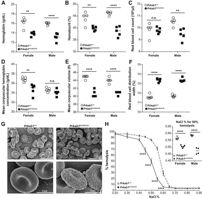
Educational figure illustrating hematological abnormalities in a murine model of Prkab1 deficiency, serving as a surrogate for studying human hemolytic anemia and microcytosis. Panels A-F present dot plots of hematological indices for wild-type (Prkab1+/+) and deficient (Prkab1tm1b/tm1b) mice, showing significant reductions in hemoglobin (A), hematocrit (B), and mean corpuscular volume (E), with a concomitant increase in red blood cell distribution width (F), indicating microcytic anemia with anisocytosis. Panel G contains high-resolution Scanning Electron Microscopy (SEM) images comparing erythrocyte morphology; wild-type cells show standard biconcave discocyte morphology, whereas Prkab1-deficient cells exhibit poikilocytosis with numerous abnormal forms, including acanthocytes, schistocytes, and echinocytes with irregular surface protrusions. Panel H displays an osmotic resistance curve, where Prkab1-deficient erythrocytes demonstrate a left-ward shift, signifying increased osmotic resistance (delayed hemolysis) compared to wild-type controls. The inset confirms a lower NaCl percentage required for 50% hemolysis in deficient cells, indicating altered membrane stability.
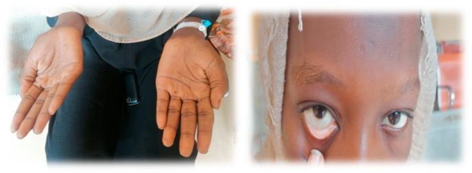
This composite clinical photograph illustrates common physical examination findings associated with severe anemia. The left panel shows the bilateral palms of a patient, demonstrating marked palmar pallor. The skin on the palms and creases appears significantly lighter and lacks the typical pinkish hue of healthy vascularized tissue, suggesting reduced hemoglobin concentration. The right panel is a close-up of the patient's face with the lower eyelid retracted to reveal the palpebral conjunctiva. Instead of the normal vibrant red or pink color, the conjunctiva exhibits conjunctival pallor, appearing whitish or very pale pink. These visual signs are critical diagnostic markers in hematology and general medicine for identifying systemic conditions such as iron-deficiency anemia or other forms of chronic blood loss. The image serves as a teaching tool for medical students and clinicians to recognize mucosal and cutaneous indicators of decreased red blood cell mass or oxygen-carrying capacity.
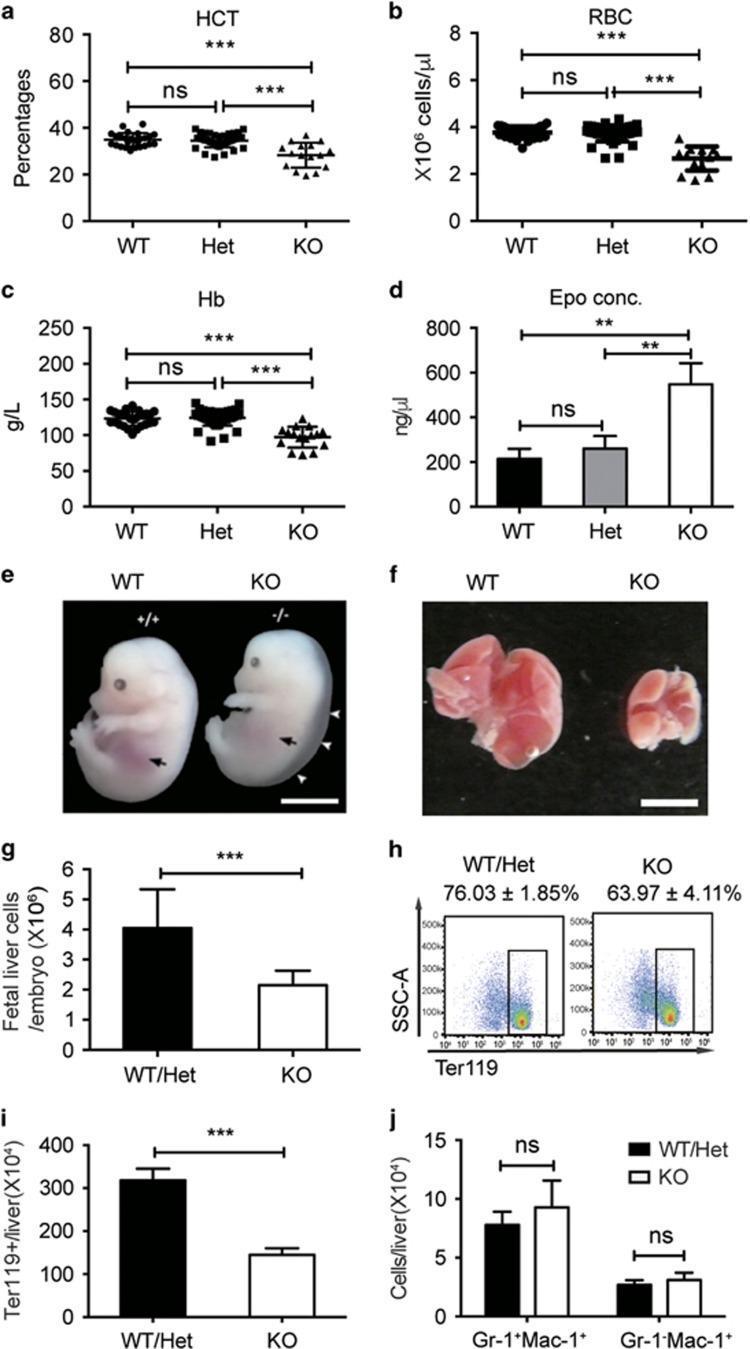
This composite educational image illustrates the hematopoietic and morphological effects of Stk40 knockout (KO) in mouse embryos. (a-d) Scatter and bar graphs display hematological indices: the KO group shows a significant reduction in hematocrit (HCT), red blood cell (RBC) count, and hemoglobin (Hb) concentration, while erythropoietin (Epo) levels are significantly elevated compared to wild-type (WT) and heterozygous (Het) controls. (e) Comparative clinical photographs of E14.5 embryos show that the Stk40 KO embryo exhibits severe subcutaneous edema (white arrowheads) and a diminished fetal liver shadow (black arrows) relative to the WT. (f) Macroscopic images of harvested fetal livers demonstrate marked hypoplasia in the KO liver compared to the WT. (g-j) Quantitative analysis and flow cytometry data show a decrease in total fetal liver cellularity and absolute numbers of Ter119+ erythroid cells in KO embryos, indicating impaired definitive erythropoiesis. Collectively, the data demonstrate that Stk40 deficiency leads to fetal anemia, edema, and hepatic hematopoietic defects.
🩸 Anemia (Khoon ki Kami) - Poori Jaankari Hindi Mein
Anemia kya hai?
- Normal Hemoglobin: Purushon mein 13.5-17.5 g/dL, Mahilaon mein 12-15.5 g/dL
- Isse kam hone par Anemia maana jaata hai
Anemia ke Prakar (Types)
| Prakar | Kya hota hai |
|---|---|
| Iron Deficiency Anemia | Sabse common - iron ki kami se |
| Vitamin B12/Folic Acid Anemia | Megaloblastic anemia - vitamin ki kami |
| Sickle Cell Anemia | Genetic bimari - RBCs ka shape bigad jaata hai |
| Thalassemia | Paida aisi genetic bimari - Hb kum banta hai |
| Aplastic Anemia | Bone marrow RBCs banana band kar deta hai |
| Hemolytic Anemia | RBCs bahut jaldi toot jaate hain |
Anemia Kyon Hoti Hai? (Karan / Causes)
1. Khoon mein Kami (Blood Loss)
- Periods mein zyada khoon aana (mahilaon mein)
- Pet ki andar andar bleeding (ulcer, piles)
- Kisi chot ya operation ke baad
2. RBCs ka kam banana
- Iron ki kami - sabse common karan
- Vitamin B12 ya Folic Acid ki kami - diet mein nahi hone par
- Bone marrow ki bimari
3. RBCs ka toot jaana (Hemolysis)
- Genetic bimariyan (Sickle cell, Thalassemia)
- Kuch dawaiyon ka side effect
- Infection
4. Diet mein kami
- Kum meat, hara saag, aur iron-yukt khana khana
Anemia ke Lakshan (Symptoms)
- 😴 Thakan aur kamzori - jaldi thak jaana
- 😰 Saans lene mein takleef - thoda kaam karne par bhi
- 💨 Chakkar aana - uthne-baithne par
- 👀 Aankhen andar se safed dikhna (conjunctival pallor)
- 🤚 Hatheli ka rang fika/safed hona (palmar pallor)
- 💗 Dil ki dhadkan tez hona (palpitations)
- 🤕 Sar dard - baar baar
- 😶 Twacha fiki ya peeli padna
- 🧊 Haath pair thande rehna
- 💅 Nakhun kamzor aur toote hue
Anemia ki Pahchaan (Diagnosis)
- CBC (Complete Blood Count) - Hemoglobin, RBCs, Hematocrit check hota hai
- Peripheral Blood Smear - RBCs ka shape dekha jaata hai
- Serum Iron, Ferritin, TIBC - Iron deficiency confirm karne ke liye
- Vitamin B12 & Folic Acid test
- Reticulocyte Count - Bone marrow kita kaam kar raha hai
Anemia ka Ilaj (Treatment)
💊 Dawai se Ilaj
| Anemia ka Prakar | Ilaj |
|---|---|
| Iron Deficiency | Iron tablets (Ferrous Sulfate 60-120 mg/din) |
| Vitamin B12 ki kami | B12 injection ya tablets |
| Folic Acid ki kami | Folic Acid 5mg tablets |
| Severe Anemia | Blood transfusion (khoon chadhaana) |
| Sickle Cell / Thalassemia | Specialist se ilaj |
Iron ki Dawai ka Note: Iron tablets khali pet ya Vitamin C ke saath leni chahiye - absorption acchi hoti hai. Chai, coffee ya dudh ke saath na lein kyunki absorption kam hoti hai.
🥗 Khane se Ilaj (Diet)
- 🥩 Mutton, liver, chicken
- 🐟 Machhi
- 🫘 Rajma, chana, masoor daal
- 🥬 Palak, methi, sarson
- 🥜 Moongfali, khajoor
- 🌾 Gehun ka aata (whole wheat)
- Chai/coffee khane ke turant baad
- Zyada doodh iron ke saath
🏥 Kab Doctor ke paas jayein?
- Bahut zyada thakan aur saans ki takleef ho
- Chehra bilkul safed ya peela lag raha ho
- Dil bahut tez dhadak raha ho
- Behoshi aa rahi ho
- Pregnancy mein Hb kam ho
Pregnancy mein Anemia
- Bachche ka wajan kam ho sakta hai (Low Birth Weight)
- Samay se pehle delivery ho sakti hai
- Bachche ko bhi nuksaan ho sakta hai
Zaroori baat: Anemia ke bahut se prakar hote hain, isliye khud dawai na lein. Doctor se CBC test karaayein aur sahi karan jaankar ilaj karwayein.