
IgE
Reading File
IgE is a broad immunology topic. Let me search the medical library comprehensively.
Searching sources...
Finding Sources
Finding Sources
Reading File
Reading File
Reading File
Reading File
Reading File
Loading Image
Reading File
Loading Image
Here is a comprehensive overview of Immunoglobulin E (IgE) drawn from multiple authoritative textbooks:
IgE (Immunoglobulin E)
Structure
IgE is the least abundant immunoglobulin in serum, comprising only ~0.0005% of total serum immunoglobulin. Its unique structural features set it apart from other isotypes:
- Heavy chain: epsilon (ε) chain - structurally distinct from other Ig isotypes
- Molecular formula: ε₂L₂ (two epsilon heavy chains + two light chains)
- Molecular weight: ~190,000 Da (ε chain alone: ~70,000 Da)
- Extra domain: Has an additional Cε4 domain, giving it higher molecular weight than IgG
- Carbohydrate content: ~18% (more heavily glycosylated than IgG)
- Valency: Functional valency of 2 (bivalent)
- No complement fixation (classic pathway)
- No placental transfer
Table 56.2, Henry's Clinical Diagnosis and Management by Laboratory Methods
Serum Half-Life and Distribution
| Property | Value |
|---|---|
| Serum half-life | 1-5 days |
| IgG serum half-life (comparison) | ~20 days |
| Catabolic rate | >70% per day |
| Tissue-bound (mast cell) half-life | Up to several months |
The very low serum concentration reflects both avid binding to effector cells and a high catabolic rate. Once bound to mast cells, IgE persists for months.
IgE Class Switching and Production
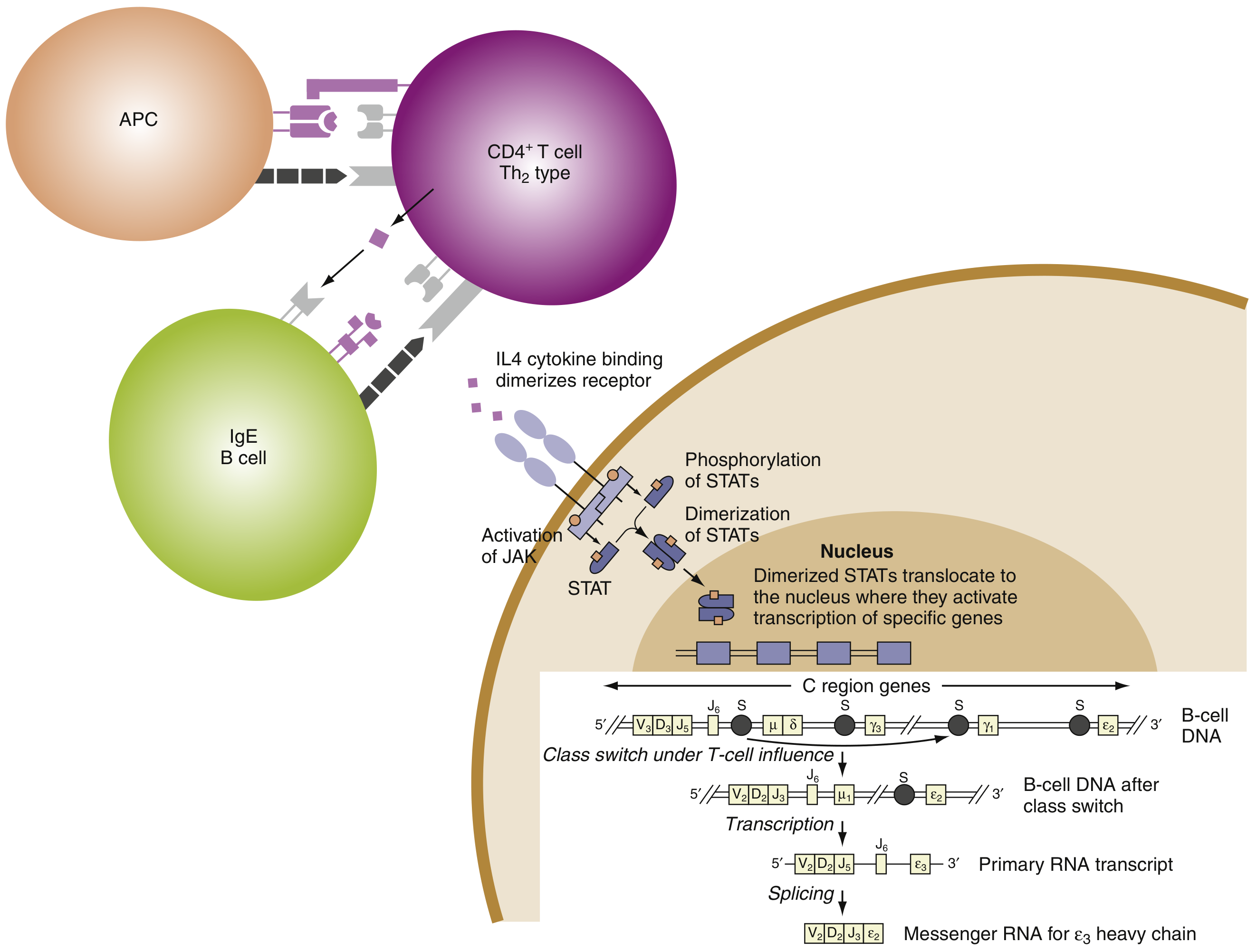
IgE production requires a two-signal B cell activation:
- IL-4 and IL-13 (from Th2/TFH cells) drive class switch recombination to the ε heavy chain via JAK-STAT6 signaling
- CD40L-CD40 interaction (T cell contact signal) provides a costimulatory signal
Key transcription factors and genes involved:
- IL-4, IL-13, IL-4Rα, STAT6 - the core molecular determinants of atopy
- GATA-3 - Th2 differentiation
- Class II HLA loci on chromosome 6p - influence sensitization to specific allergens
FcεRI - The High-Affinity IgE Receptor
IgE exerts its biological effects primarily via FcεRI (high-affinity Fc epsilon receptor I):
- Expressed on mast cells and basophils
- IgE binds to FcεRI via its Fc region (the Cε3 and Cε4 domains)
- Binding is extremely avid - this "loads" mast cells with antigen-specific IgE
- When antigen cross-links two or more FcεRI-bound IgE molecules, degranulation is triggered
There is also a low-affinity receptor FcεRII (CD23) expressed on B cells, monocytes, and eosinophils, which regulates IgE synthesis.
Mechanism of Immediate Hypersensitivity (Type I)
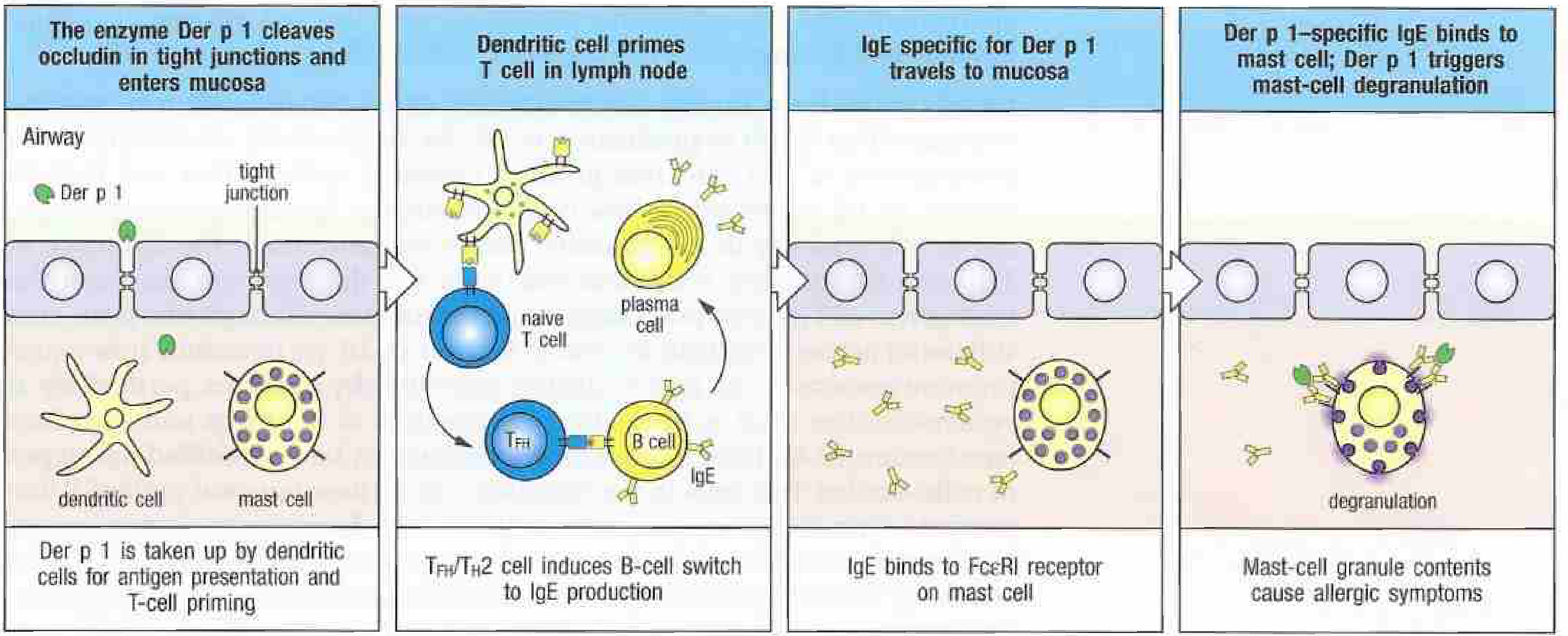
Phase 1: Sensitization
- Allergen penetrates epithelial barrier
- Dendritic cells capture allergen and traffic to lymph node
- TFH/Th2 cells are primed; IL-4 and IL-13 are produced
- B cells undergo class switch to produce allergen-specific IgE
- IgE travels to tissues and binds FcεRI on mast cells
Phase 2: Elicitation (re-exposure)
- Allergen re-enters the tissue
- Allergen cross-links mast cell-surface IgE
- Mast cell degranulation occurs within minutes
- Pre-formed mediators released: histamine, tryptase, chondroitin sulfates, TNF-α
- Newly synthesized mediators: leukotrienes (LTC4), prostaglandins (PGD2)
Janeway's Immunobiology 10e; Cellular and Molecular Immunology
Atopy and Genetic Determinants
"Atopic" individuals are genetically predisposed to produce specific IgE following allergen exposure. Key features:
- Production of high IgE depends on a propensity to generate allergen-specific TFH cells secreting IL-4 and IL-13
- Candidate genes: IL4, IL13, IL4R, STAT6 (chromosome 5q), HLA class II loci (chromosome 6p), FcεRI α chain locus (chromosome 1q23)
- Allergen characteristics that favor IgE responses: MW 5-70 kDa, stable, glycosylated, soluble, often enzymatic activity (e.g., house dust mite cysteine protease, bee venom phospholipase A2)
- Repeat exposure is required - first exposure sensitizes (IgE produced), second exposure triggers reaction
- Non-protein antigens (e.g., penicillin) can cause IgE responses by acting as haptens bound to self proteins
Mast Cell Subtypes Relevant to IgE
| Feature | MCT (mucosal) | MCTC (connective tissue) |
|---|---|---|
| Contains | Tryptase only | Tryptase + chymase |
| Location | Mucosal surfaces, near T cells | Submucosa, connective tissue |
| Allergic role | Yes - at sites of allergic inflammation | Less associated |
| Mediators | High LTC4, PGD2 | Less LTC4 |
| IL-4 content | Low | Higher |
Clinical Diseases Associated with IgE
IgE-mediated (Type I Hypersensitivity)
- Allergic rhinitis (hayfever)
- Allergic asthma
- Atopic dermatitis (eczema)
- Urticaria (hives)
- Food allergy
- Anaphylaxis - systemic, life-threatening; IgE cross-linking in vascular system
Hyper-IgE Syndrome (Job Syndrome)
Characterized by markedly elevated IgE + eczema + eosinophilia + recurrent pulmonary infections + "cold" staphylococcal skin abscesses:
- Autosomal dominant form: Heterozygous dominant-negative STAT3 mutations - impairs IL-6, IL-10, IL-17, IL-21, IL-22 signaling; reduces Th17 responses; increases IL-4/IL-13/Th9 driving IgE overproduction
- Autosomal recessive form: DOCK8 mutations - reduced T, B, NK cells; defective lymphocyte signaling similar to Wiskott-Aldrich syndrome
Cellular and Molecular Immunology
Anti-IgE Therapy
Omalizumab (anti-IgE monoclonal antibody) targets free IgE and is used in:
- Severe allergic asthma
- Chronic urticaria
- Other IgE-driven conditions
By binding free IgE, it reduces loading of mast cells and basophils, thereby blunting allergic responses.
Goodman & Gilman's Pharmacological Basis of Therapeutics
Laboratory Measurement
- Total serum IgE: Normal reference range is low (typically < 100 IU/mL in adults); elevated in atopic disease, parasitic infections, Hyper-IgE syndrome
- Specific IgE (sIgE): ImmunoCAP assay measures IgE directed against individual allergens; used in allergy diagnosis
- Nasal specific IgE: Can be detected locally even when serum IgE is normal (entopy)
Henry's Clinical Diagnosis & Management by Laboratory Methods; Cummings Otolaryngology
Sources: Janeway's Immunobiology 10e | Cellular and Molecular Immunology | Henry's Clinical Diagnosis and Management by Laboratory Methods | Goodman & Gilman's Pharmacological Basis of Therapeutics | Cummings Otolaryngology
This is a shared conversation. Sign in to Orris to start your own chat.