xray view name to see mandible fracture
Reading File
Searching the Web
X-ray views mandible fracture radiographic assessment
Finding Sources
Reading File
Reading File
Loading Image
Loading Image
Here is a complete summary of the X-ray views used to assess mandible fractures, sourced from Bailey and Love's Short Practice of Surgery and Grainger & Allison's Diagnostic Radiology:
X-ray Views for Mandible Fractures
Primary Views (always obtain both)
| View | What it Shows |
|---|---|
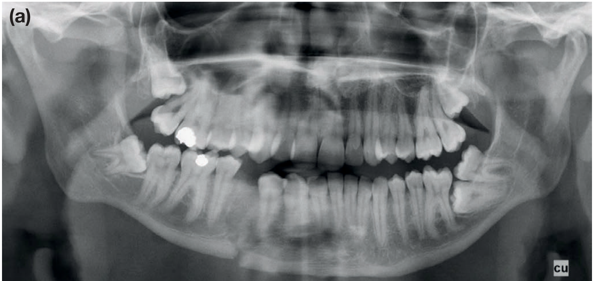
| OPG / OPT (Orthopantomogram / Dental Panoramic Radiograph) | Single panoramic image of the entire mandible - best overall view; shows body, angle, ramus, and condyles |
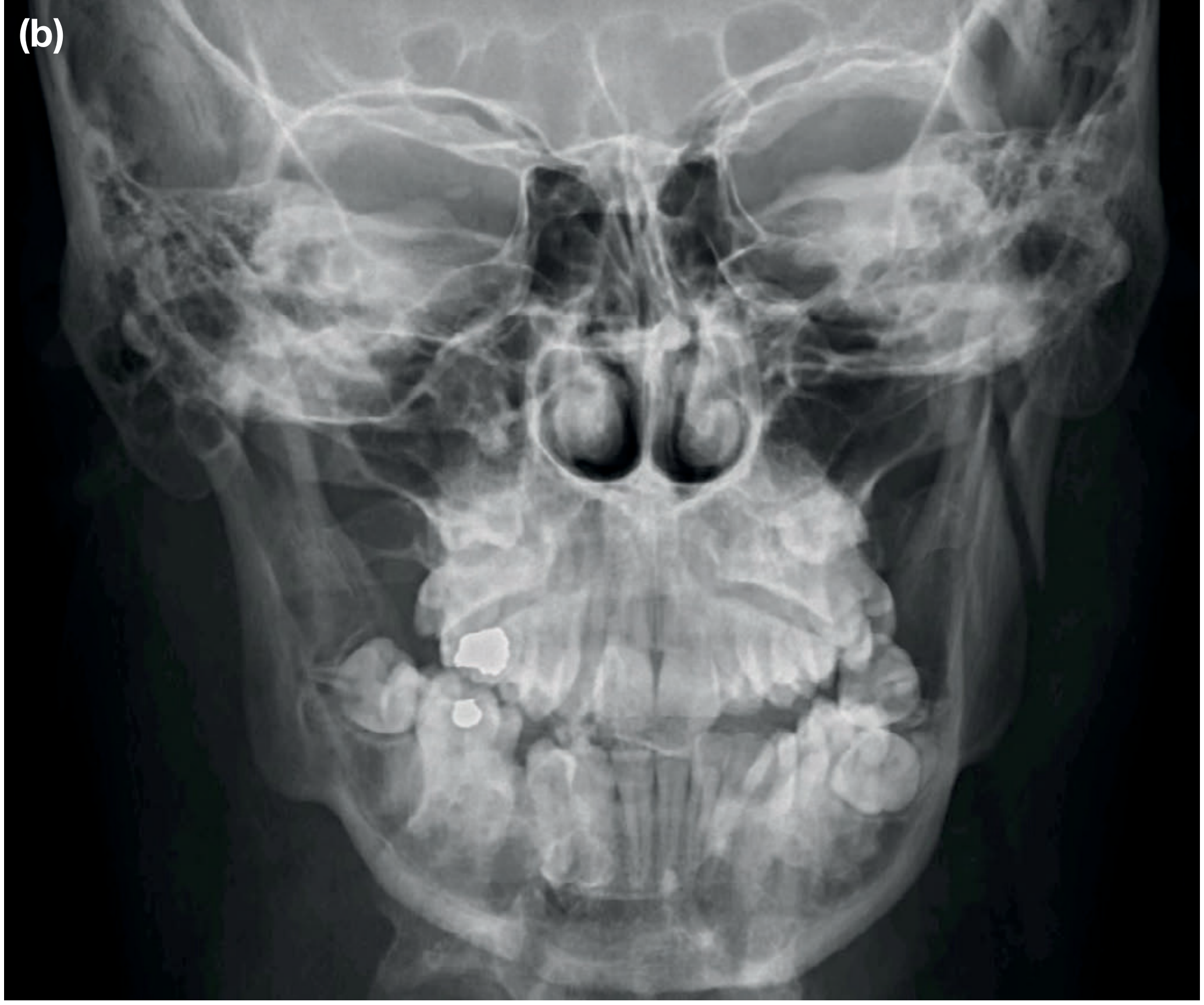
| PA Mandible (Posteroanterior) | Shows displacement of fractures; picks up condylar and symphysis/parasymphyseal fractures that may be subtle on OPG |
It is critical to have two views at right angles to each other to avoid missing fractures. - Bailey and Love's, p. 429
Supplementary Views
| View | Indication |
|---|---|
| Oblique lateral views (right + left) | Look at the angle, body, and ramus; used when OPG is unavailable |
| Towne's view (modified AP with 30° caudal tilt) | Best for condylar fractures - shows the condylar heads and necks clearly |
| Intraoral / periapical views | Parasymphyseal fractures; when anterior teeth are also suspected fractured |
| Occlusal view | Useful for symphysis; underutilized but helpful |
Real Radiograph Examples (from Bailey and Love)
(a) OPT - right mandibular body and left condylar fracture:
(b) PA mandible radiograph - reveals the left condylar fracture more clearly:
Key Points
- The mandible is a ring of bone - a fracture in one place is accompanied by a second fracture or TMJ dislocation in ~50% of cases (especially body fracture + contralateral condylar neck fracture after a blow to the chin).
- CT scan is the modality of choice in trauma settings - it has near 100% sensitivity and allows 3D reconstruction for surgical planning.
- If a patient is too unstable for CT, ultrasound can detect fractures at bedside.
- Parasymphyseal fractures can look like a missing tooth on OPG - a PA view helps distinguish.
Sources: Bailey and Love's Short Practice of Surgery (28th ed.), p. 429; Grainger & Allison's Diagnostic Radiology; Radiology Masterclass - Mandible; Medscape - Mandibular Fracture Workup
This is a shared conversation. Sign in to Orris to start your own chat.