
Controversies in management of ameloblastoma
ameloblastoma management controversy treatment
ameloblastoma surgical treatment recurrence
ameloblastoma BRAF targeted therapy
PMID: 37231111
PMID: 37115331
PMID: 37872712
PMID: 40335195
ameloblastoma jaw mandible radiograph multilocular soap bubble
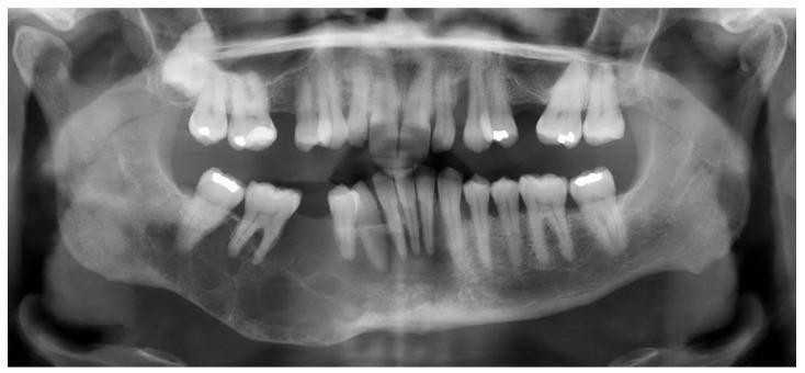
This diagnostic panoramic radiograph (orthopantomogram) demonstrates a large, approximately 10 cm, expansile multilocular radiolucency involving the right body and angle of the mandible. The lesion exhibits a characteristic 'soap bubble' appearance with internal bony septa and poorly demarcated, ill-defined borders. There is significant thinning of the inferior cortical border of the mandible and evidence of bone expansion. Clinically significant features include the disruption of normal trabecular bone patterns and the displacement or 'floating' appearance of adjacent teeth, particularly in the right mandibular molar and premolar regions. The radiographic presentation is highly suggestive of an aggressive odontogenic neoplasm, such as clear cell odontogenic carcinoma or ameloblastoma. The image is intended for dental and oral surgery education, illustrating the appearance of extensive intraosseous jaw lesions and their impact on surrounding anatomical structures.
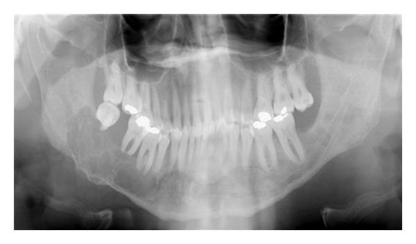
This diagnostic image is a panoramic radiograph (orthopantomogram) of the human jaws, illustrating a significant pathological lesion in the right mandible. The primary finding is a well-defined, multilocular radiolucency located in the right mandibular body and angle. The lesion exhibits a 'soap bubble' or 'honeycomb' internal appearance, characteristic of odontogenic tumors such as ameloblastoma or odontogenic keratocysts. Anatomically, the radiolucency extends horizontally from the second premolar region to approximately one centimeter posterior to the third molar, and vertically from the alveolar crest to the inferior border of the mandible. Notable clinical features include the displacement of the mandibular second and third molars, which appear 'floating' within the lesion, and the apparent obliteration or displacement of the inferior alveolar nerve canal. Several teeth demonstrate radiopaque dental restorations. This radiograph serves as a key educational tool for identifying expansive mandibular lesions, assessing their impact on surrounding dental and neural structures, and understanding the differential diagnosis for multilocular radiolucent jaw pathologies.
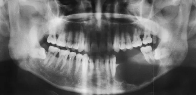
This is a two-dimensional panoramic dental radiograph (Orthopantomogram) of the mandible displaying a large, radiolucent lesion in the left hemi-mandible. The lesion is expansile and predominantly multilocular, with classic soap-bubble (when locules are large) or honeycomb (when locules are small) appearance described for ameloblastoma on plain films. The borders are well-demarcated and scalloped, and there is no radiopaque matrix or calcification within the lesion. Cortical expansion is evident with cortical breach and thinning; the lesion displaces and resorbs adjacent teeth, producing tooth mobility and in this image a floating or absent left second molar region. An associated unerupted or impacted third molar is frequently observed with ameloblastoma and may be present here, contributing to crowding and dentition loss. The radiographic differential includes odontogenic cysts and tumors; however, the combination of a large multilocular radiolucency with cortical perforation and tooth displacement strongly favors ameloblastoma. Clinically significant due to local aggressiveness and potential for recurrence, these lesions typically require surgical planning with margin resection. This image is illustrative for educational radiology, maxillofacial pathology, and dental tumor management, aiding recognition of mandibular ameloblastoma patterns, differential diagnoses, and treatment implications. Correlate with clinical findings and consider biopsy for definitive histopathology confirmation and management planning.
Controversies in Management of Ameloblastoma
Radiographic Context
1. Conservative vs. Radical Surgery for Conventional (Solid/Multicystic) Ameloblastoma
- En bloc resection with 1 cm bone margin past the radiographic limits is the most predictable treatment
- Resected specimens allow intraoperative radiographic assessment and postoperative histologic margin confirmation - neither is possible with conservative approaches
- Even after well-planned resections, recurrence rates of 10-15% are reported; conservative methods fare far worse
- Modern series report some success with enucleation + aggressive curettage (peripheral osteotomy with rotary bur)
- The apparent success may be explained by relatively efficient medullary bone removal with aggressive curettage
- For small mandibular lesions, a conservative approach (enucleation + curettage + close follow-up) may be considered
2. What Constitutes an Adequate Margin?
- The widely accepted recommendation is 1 cm of bone past the radiographic tumor margin in the medullary (proximal-distal) dimension
- Lingual and buccal cortical plates are sacrificed, but the 1-cm rule does NOT mandate soft tissue outside the periosteum when it is intact
- Critically, as Cummings notes: "there are no well-controlled studies of radiographs of the resected specimen compared to the postsurgical defect to actually confirm what margins were achieved" - the entire evidence base for the 1-cm rule is expert opinion
- Whether histologic tumor extension past the radiographic margin has ever been rigorously mapped is unknown; Marx attempted early data collection but a definitive study has not been published
3. Unicystic Ameloblastoma (UA): The Type III / Intramural Debate
| UA Subtype | Histology | Recommended Treatment |
|---|---|---|
| Type I (Luminal) | Ameloblastomatous lining only | Enucleation + curettage |
| Type II (Intraluminal) | Polypoid nodule into lumen | Enucleation + curettage |
| Type III (Intramural/Mural) | Invasion into fibrous wall | Treat as conventional ameloblastoma (resection) |
- Ackerman and Shear's "Type 3" classification with connective tissue invasion is listed as unicystic by some authors, leading to inappropriate conservative treatment and higher recurrences
- The definition itself has been diluted: some authors allowed multilocularity within the unicystic category, which Cummings calls "spurious at best" since multilocular lesions recur at higher rates with enucleation
- Eversole and Leider found lesions larger than 2 cm recur more often even with enucleation, suggesting size is an independent risk factor regardless of histologic subtype
- Some authors still allow a plexiform unicystic variant, which Cummings describes as "uncommon and perhaps somewhat controversial"
- The plexiform variant diagnosis is also complicated by inflammation that can disrupt the Vickers and Gorlin diagnostic criteria
4. The Role of Adjuvant Techniques
- Applied to bone cavity after enucleation to fix residual tumor cells
- Penetrates ~1.5 mm into bone
- Controversy: risk of inferior alveolar nerve damage, limited penetration depth, no high-quality RCT data
- The network meta-analysis ranked "curettage + cryotherapy" second only to segmental resection (SUCRA 66.9)
- Mechanism: freeze-thaw cycles disrupt tumor cells while preserving bone architecture
- Controversy: technical difficulty, risk of pathologic fracture, delayed healing, and no standardized protocol
- Preferred by many conservative proponents as more reproducible than simple curettage
- No comparative data against resection
- A 2025 systematic review and meta-analysis (Suárez-Pérez et al.) found no statistically significant difference in recurrence between piezoelectric and conventional techniques for jaw cysts and tumors [PMID: 40121687]
5. Maxillary Ameloblastoma: The High-Stakes Site
- The mandible: maxilla ratio is approximately 5:1, so maxillary cases are rare and series are small
- Maxillary ameloblastomas can extend into the sinonasal complex, pterygopalatine fossa, infratemporal fossa, and skull base - all of which make complete resection progressively more dangerous
- Scott-Brown explicitly states: "All maxillary lesions should be treated radically since the consequences of recurrence involving the skull base are severe with a mortality of up to 60%"
- Once a recurrence tracks into the skull base, the surgeon is operating near the cavernous sinus, internal carotid artery, and optic nerves - the debate shifts from "how much to remove" to "whether reoperation is feasible at all"
- Desmoplastic ameloblastoma is more common in the maxilla and anterior jaw, and has an unusual mixed radiolucent-radiopaque appearance that can delay diagnosis
6. BRAF V600E and Molecular Stratification
- Approximately 66% of ameloblastomas harbor a BRAF V600E somatic mutation (in the MAPK signaling pathway); some series put the figure for mandibular lesions as high as 80-90%
- Maxillary and desmoplastic ameloblastomas are enriched for non-BRAF mutations (FGFR2, SMO, KRAS, etc.)
| Role | Evidence | Controversy |
|---|---|---|
| Neoadjuvant (shrink then resect) | Case series only | Can "downstaging" reduce morbidity? Does it mask residual disease? |
| Recurrent/unresectable disease | Best current evidence for use | Longest follow-up is only 38 months |
| Monotherapy (avoid surgery) | No evidence | Risk of acquired resistance (MEK escape); concern about incomplete eradication |
| Adjuvant post-resection | No evidence | Role in BRAF+ cases with positive margins? |
7. Tooth Preservation vs. Extraction
8. Peripheral Ameloblastoma
- They behave much more favorably than intraosseous types (no bone invasion by definition)
- Simple local excision is curative in most cases
- The controversy is whether underlying bone should be routinely curetted (some advocate it; data are sparse)
- Rare cases show bone involvement on imaging, which reclassifies them to intraosseous behavior and requires corresponding management
9. Reconstruction Timing
- Immediate free flap reconstruction (fibula free flap being the gold standard) is now standard at most major centers
- Controversy: patients in resource-limited settings or with uncertain margins may be better served by delayed reconstruction after confirmed negative margins and disease-free interval
- Implant-supported prosthetic rehabilitation after ameloblastoma resection: a 2023 systematic review (Alarcón-Sánchez et al., BMC Oral Health) found acceptable implant survival rates in reconstructed jaws, supporting rehabilitation as a realistic goal [PMID: 38110933]
10. Surveillance and Follow-up
- No consensus exists on follow-up interval or duration
- Most centers recommend clinical + radiographic (OPG or CBCT) surveillance every 6-12 months for at least 5 years, and annually for at least 10 years
- Late recurrences (>10 years) are documented for conventional ameloblastoma, making lifelong follow-up an argument by some
Summary of Key Controversies
| Controversy | Status |
|---|---|
| Resection vs. conservative surgery for conventional SMA | Resection favored; conservative debated for small mandibular lesions |
| Adequate margin width (1 cm rule) | No high-quality data; expert consensus only |
| UA Type III treatment | Agreed: treat as conventional; controversy over how to classify borderline lesions |
| Role of Carnoy's/cryotherapy/bur | No RCT data; cryotherapy second-best in one NMA |
| Maxillary ameloblastoma extent of resection | Aggressive resection unanimous; extent limited by anatomy |
| BRAF inhibitor indications | Neoadjuvant/unresectable only; no surgery-replacing role yet |
| BRAF status as recurrence predictor | Does NOT predict overall recurrence (2023 meta-analysis) |
| Immediate vs. delayed reconstruction | Immediate standard at most centers; delayed acceptable where margins uncertain |
| Tooth preservation | Trending toward extraction; insufficient ameloblastoma-specific data |
| Follow-up duration | No consensus; minimum 10 years widely recommended |
- Cummings Otolaryngology Head and Neck Surgery (Cummings), pp. 1616-1622
- Scott-Brown's Otorhinolaryngology Head & Neck Surgery, pp. 454-459
- K.J. Lee's Essential Otolaryngology, pp. 3990-4032
- Hendra et al. 2023 - Network meta-analysis on surgical approaches [PMID: 37231111]
- Singh et al. 2023 - BRAF mutation and recurrence meta-analysis [PMID: 37872712]
- Ebeling et al. 2023 - BRAF inhibitors systematic review [PMID: 37115331]
- Mariani et al. 2025 - Tooth management and recurrence meta-analysis [PMID: 40335195]