
Moebius syndrome presentation at birth
Moebius syndrome facial palsy newborn
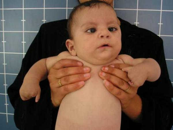
A clinical photograph of an infant displaying dysmorphic features and limb anomalies, characteristic of Moebius syndrome or related orofacial-digital-limb syndromes. The facial features demonstrate bilateral strabismus (suggestive of abducens nerve palsy) and a mask-like, expressionless face (suggestive of facial nerve palsy). Other cranial findings include a prominent forehead, a depressed nasal bridge, and low-set ears. The most striking abnormality is terminal transverse limb deficiency, specifically a subtotal absence of the phalanges. The hands show adactyly/hypoglossia-hypodactyly spectrum characteristics: the central digits are largely absent, while the thumbs and fifth fingers are preserved but hypoplastic. The infant also exhibits a short neck. This image serves as a clinical reference for identifying congenital multiple cranial nerve palsies associated with significant distal limb reduction defects, emphasizing the diagnostic triad of facial paralysis, ocular misalignment, and digital anomalies.
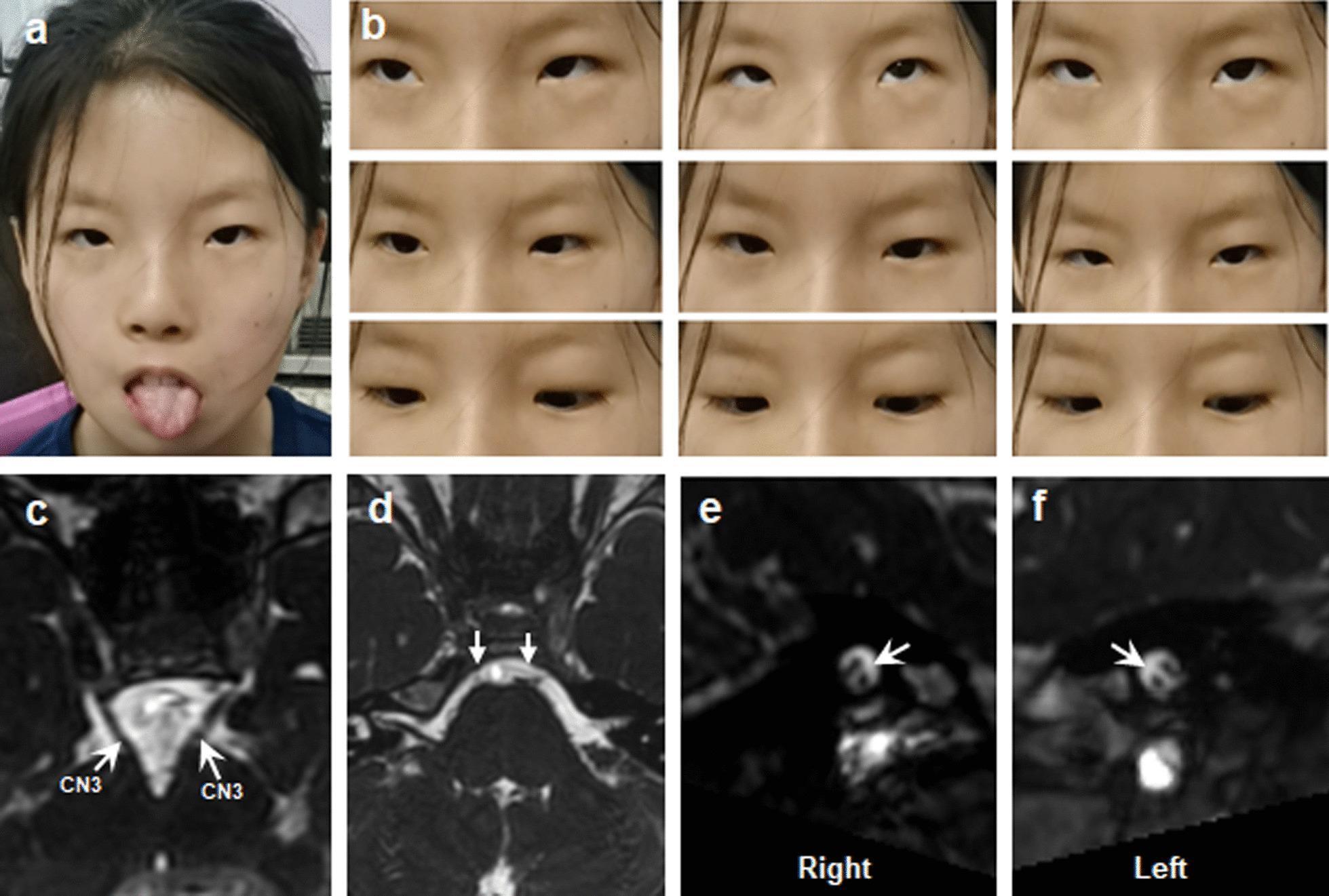
This composite educational graphic illustrates the clinical and radiological hallmarks of Moebius Syndrome (MBS). Panel (a) is a clinical photograph of a young patient demonstrating bilateral congenital facial weakness, characterized by a masked facies and hypoplasia of the tongue upon protrusion. Panel (b) consists of a nine-gaze photograph series showing significant limitations in horizontal eye movements, consistent with abducens nerve palsy. Panels (c–f) present T2-weighted cranial MRI sections focused on the brainstem. White arrows in panel (c) identify normal appearances of the oculomotor nerves (CN3). In contrast, panels (d–f) demonstrate hypoplasia of the bilateral abducens (CN6) and facial (CN7) nerves, visible as significantly reduced or absent neural signals at their respective brainstem exit zones. This figure serves as a diagnostic reference for Congenital Cranial Dysinnervation Disorders (CCDDs), correlating external physical signs like facial paralysis and restricted motility with neuroanatomical deficits of the cranial nerves.
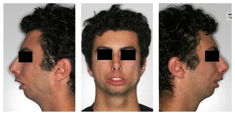
This set of clinical photographs displays the extraoral facial features of a male patient in frontal and bilateral profile views, illustrating classic manifestations of Moebius syndrome and severe skeletal class II malocclusion. The profile views demonstrate a markedly convex profile characterized by severe micrognathia and a retrognathic mandible, leading to a receding chin and an obtuse nasolabial angle. The frontal view reveals a 'mask-like' facies with reduced expression, secondary to congenital bilateral facial nerve palsy. Key visible findings include lip incompetence with a significant interlabial gap at rest, exposing the maxillary incisors, and reduced lower anterior facial height. The soft tissue presentation shows lack of muscular tension in the perioral region and slight flatness of the cheeks. These images serve as educational material for maxillofacial surgery and orthodontics, highlighting the relationship between skeletal hypoplasia and neuromuscular dysfunction in complex craniofacial conditions.
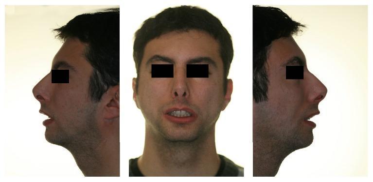
Post-operative clinical photographs of a 23-year-old male patient with Moebius syndrome, presented in three panels: a right profile view, a frontal view, and a left profile view. The images demonstrate the aesthetic and functional outcomes of orthognathic surgery, including mandibular advancement, genioplasty, and lip augmentation via fat injection. In the profile views, there is a corrected convex profile with increased chin projection and a more defined jawline, addressing pre-operative severe micrognathia. The frontal view shows improved lip symmetry and enhanced lip volume, aimed at correcting congenital lip incompetence and reducing interlabial separation at rest. The primary educational focus is the multidisciplinary management of craniofacial dysmorphism and facial nerve palsy manifestations through surgical and orthodontic intervention.
Moebius syndrome neonatal presentation clinical features
Moebius syndrome presentation at birth neonatal features clinical
Moebius Syndrome - Presentation at Birth
Definition and Pathophysiology
- Scott-Brown's Otorhinolaryngology Head & Neck Surgery Vol 2, p. 277
The Cardinal Signs Present at Birth
1. Facial Diplegia (Mask-Like Face)
- Complete expressionless face - no smiling, frowning, or grimacing even when crying
- Absence of nasolabial folds
- Forehead immobility - no wrinkling
- Incomplete eyelid closure (lagophthalmos) - the infant cannot fully close eyes during sleep, risking corneal exposure and drying
- Inability to close the mouth - undue prominence of the upper lip, drooling
2. Lateral Gaze Palsy (Abducens Palsy - CN VI)
- Inability to abduct the eyes (move eye away from midline/nose) - eyes cannot move horizontally outward
- Results in a fixed, forward-staring gaze
- Convergent strabismus (esotropia) is common at birth
- Vertical eye movements are typically preserved
Feeding Problems - A Key Neonatal Alert
- Poor or absent sucking - incomplete mouth closure prevents adequate suction
- Swallowing difficulties - involvement of CN IX, X, XII
- Need for nasogastric tube or gastrostomy in ~5.5% of severe cases
- Poor weight gain / failure to thrive in the newborn period
Additional Cranial Nerve Involvement
| Cranial Nerve | Finding at Birth |
|---|---|
| CN V (trigeminal) | Reduced jaw movement, weak bite |
| CN IX (glossopharyngeal) | Swallowing difficulty, absent gag |
| CN X (vagus) | Palatal weakness, dysphonia |
| CN XII (hypoglossal) | Tongue deviation, reduced tongue movement |
| CN III/IV (oculomotor/trochlear) | Ptosis, additional gaze restriction |
Craniofacial Structural Anomalies
- Micrognathia - small, receding chin/jaw
- Microstomia - small mouth opening
- Short or abnormally shaped tongue
- High arched or cleft palate - contributes to feeding and later speech problems
- Dental anomalies - missing or misaligned teeth (apparent later)
Limb and Musculoskeletal Anomalies
- Club foot (talipes equinovarus) - most common limb defect
- Syndactyly or brachydactyly
- Poland sequence (pectoral muscle aplasia + ipsilateral limb defects) - when present, called Poland-Moebius syndrome
- Transverse limb deficiencies (absent digits or terminal reduction defects) - seen in a subset
Neurological and Developmental Features
-
Autism spectrum disorder and intellectual disability are present in approximately one-third of patients, though cognitive development is normal in the majority
-
Hypotonia may be present
-
Emotional and social difficulty - inability to express emotion through the face has significant developmental impact
-
Scott-Brown's Otorhinolaryngology Head & Neck Surgery Vol 2, Table 112.8
Differential Diagnosis of Neonatal Facial Palsy
| Condition | Key Distinguishing Feature |
|---|---|
| Birth trauma (forceps injury) | Unilateral; EMG responses present; >90% resolve spontaneously |
| Dystrophia myotonica | Bilateral palsy WITHOUT abducens palsy; progressive wasting |
| CHARGE association | Colobomata, choanal atresia, heart defects, ear anomalies |
| Asymmetric crying facies | Only mouth corner affected; normal forehead and eyes |
- Scott-Brown's Otorhinolaryngology Head & Neck Surgery Vol 2, Table 112.8
When Is the Diagnosis Made?
- Congenital facial diplegia (or uniplegia), lower motor neuron type
- Paralysis of lateral eye movement with strabismus
Summary - Neonatal Red Flags for Moebius Syndrome
- Expressionless face, no grimacing even with crying
- Incomplete eye closure during sleep
- Poor sucking and feeding from birth
- Fixed forward gaze, inability to move eyes laterally
- Convergent squint (esotropia)
- Micrognathia, small mouth
- Club foot or digital anomalies