
Which nerve is involved in claw hand
- The ulnar nerve innervates the intrinsic hand muscles (interossei and medial two lumbricals)
- Loss of these muscles causes unopposed action of the long flexors and extensors, producing the classic "claw" posture
- The ring and little fingers are most affected (ulnar two digits)
claw hand ulnar nerve deformity
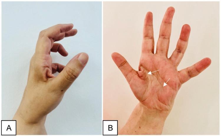
Two-panel clinical photograph demonstrating the late sequelae of a peripheral nerve injury in the hand, specifically illustrating features of ulnar nerve palsy. Image A (lateral view) shows a classic 'claw hand' deformity, characterized by hyperextension at the metacarpophalangeal (MCP) joints and flexion at the interphalangeal (IP) joints, most prominent in the ring and little fingers. The thumb appears adducted and flexed. Image B (anterior/palmar view) displays generalized muscle atrophy of the hand, with significant hollowing of the hypothenar eminence and interosseous spaces (indicated by white arrows). Notably, the thenar eminence remains relatively spared, suggesting the median nerve's innervation to the thenar muscles is intact. This combination of clawing and selective intrinsic muscle wasting is an important educational marker for localizing nerve lesions to the distal ulnar nerve (e.g., Guyon's canal or post-surgical complication). The images illustrate the clinical presentation of chronic motor deficit and the characteristic postural changes resulting from unbalanced muscle pull in the hand.
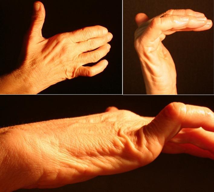
This composite of clinical photographs demonstrates a 'claw hand' (main en griffe) deformity, a characteristic sign of ulnar nerve neuropathy. The images show a right hand from dorsal, palmar, and lateral perspectives. Key visual features include hyperextension at the metacarpophalangeal (MCP) joints and flexion at the proximal and distal interphalangeal (IP) joints, most pronounced in the fourth and fifth digits. There is noticeable atrophy of the hypothenar eminence and hollowing of the interosseous spaces on the dorsal surface, indicating wasting of the intrinsic hand muscles (interossei and ulnar-innervated lumbricals). The thenar eminence appears relatively preserved, though subtle flattening of the adductor pollicis region may be present. This presentation is consistent with distal ulnar nerve compression, such as within Guyon’s canal, leading to motor deficit and muscle denervation. These clinical signs are essential for diagnosing peripheral nerve entrapment syndromes and distinguishing them from proximal lesions or central nervous system pathologies.
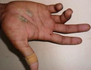
Clinical photograph of a right hand demonstrating ulnar claw hand (main en griffe) deformity, secondary to traumatic ulnar nerve injury. The image shows characteristic hyperextension at the fourth and fifth metacarpophalangeal (MCP) joints, accompanied by fixed flexion at the proximal and distal interphalangeal (PIP and DIP) joints of the ring and little fingers. In addition to the neurological deficit, the palmar surface exhibits multiple signs of acute trauma, including two irregular areas of pale/abrasive discoloration on the palm and a small punctate wound near the base of the index finger. An adhesive bandage is visible on the thumb. This visual presentation is classic for a distal or high ulnar nerve palsy where the loss of intrinsic muscle function (lumbricals and interossei) leads to an imbalance in the hand's extrinsic musculature. The image serves as an educational tool for identifying physical signs of peripheral nerve compression or transection following orthopedic trauma, such as a humeral shaft fracture.
Claw Hand - Nerve Involved
Primary Nerve: Ulnar Nerve
Mechanism
- The interossei (which flex the MCP joints and extend the IP joints) are lost
- The long flexors (FDP, FDS) and extensor digitorum remain intact and act unopposed
- Result: hyperextension at the metacarpophalangeal (MCP) joints + flexion at the proximal and distal interphalangeal (PIP/DIP) joints = classic claw
The "Ulnar Paradox"
| Lesion Level | Claw Severity | Reason |
|---|---|---|
| High (proximal) ulnar lesion - at elbow | Less severe claw | FDP to ring/little fingers is also paralyzed, so less pull into flexion |
| Low (distal) ulnar lesion - at wrist | More severe claw | FDP is intact, producing stronger finger flexion |
Levels of Ulnar Nerve Injury
| Level | Common Cause | Features |
|---|---|---|
| Proximal (at elbow) | Fracture, cubital tunnel syndrome, sulcus ulnaris syndrome | Claw hand + sensory disturbances (ulnar 1½ fingers) |
| Midlevel (at wrist) | Lacerations, ulnar tunnel syndrome | Claw hand + sensory disturbances sparing the hypothenar (palmar branch intact) |
| Distal (deep branch in palm) | Chronic pressure (air hammer, tools) | Claw hand with no sensory disturbances (superficial branch intact) |
Associated Signs of Ulnar Nerve Palsy
- Froment sign - positive: patient flexes the thumb IPJ to grip paper because adductor pollicis is paralyzed; they use FPL (median nerve) instead
- Hypothenar atrophy - wasting of the medial palm
- Interosseous wasting - hollowing of the dorsal interosseous spaces, most conspicuous in the first web space
- Sensory loss - ulnar half of the ring finger and entire little finger
Clinical Images
Important Note: "All Claw" (Combined Nerve Injury)

Explain this
Physiotherapy Management of PIN Palsy - Explained
What is PIN Palsy?
- Extensor Digitorum Communis (EDC) - cannot extend fingers at MCPs
- Extensor Pollicis Longus (EPL) - cannot extend the thumb IPJ
- Extensor Pollicis Brevis (EPB) - cannot extend thumb MCJ
- Extensor Carpi Ulnaris (ECU), Abductor Pollicis Longus (APL), Extensor Indicis
Point-by-Point Explanation
1. Splinting - Finger extension splint (MCP extension assist); wrist splint not required if ECRL intact
- PIN palsy causes finger drop (inability to extend at MCP joints) but the wrist can still extend because ECRL is spared
- A dynamic MCP extension assist splint holds the MCP joints in extension/neutral, allowing the patient to use the hand functionally while the nerve recovers
- Because wrist extension is preserved via ECRL, a wrist cock-up splint is NOT needed - this distinguishes PIN palsy from high radial nerve palsy (where the wrist also drops)
- The splint also prevents flexion contractures from developing at the finger joints during the recovery period
2. PROM - MCP and IP joint ROM; prevent flexion contractures of fingers
- Passive Range of Motion exercises are done by the therapist/patient to keep all finger joints supple
- Since the extensor muscles are paralyzed, the flexors will gradually pull the fingers into a fixed flexed posture
- Daily PROM through full range at the MCP, PIP, and DIP joints prevents this irreversible flexion contracture from setting in
- Think of this as "maintaining the joint while the nerve grows back"
3. NMES - EDC, EPL, EPB - retard atrophy
- Neuromuscular Electrical Stimulation (NMES) is applied over the paralyzed extensor muscles
- Targets: EDC (finger extensors), EPL (extensor pollicis longus), EPB (extensor pollicis brevis)
- A denervated muscle begins to atrophy and undergo fibrotic change within weeks; NMES sends electrical impulses to cause muscle contractions even without nerve input
- This retards (slows down) disuse atrophy, preserves muscle bulk and fiber integrity, and keeps the muscle "ready" for when reinnervation eventually occurs
- It does NOT regenerate the nerve - it simply maintains the target muscle
4. Activity Modification - Avoid repetitive forearm rotation; ergonomic assessment
- PIN palsy is commonly caused by compression neuropathy at the radial tunnel, often from repetitive forearm pronation-supination (e.g., screwdriver work, throwing, assembly line tasks)
- Avoiding repetitive forearm rotation reduces ongoing compression on the nerve at the arcade of Frohse (entry to supinator)
- Ergonomic assessment involves reviewing the patient's workstation/daily activities and modifying positions or tools to reduce nerve stress
- This removes the ongoing mechanical cause and allows the nerve to recover
5. Progressive Strengthening - Finger extension exercises as reinnervation occurs
- Once nerve regeneration starts (evidenced by returning EMG signals or muscle flicker), active assisted and active resisted exercises are started
- Exercises focus on finger and thumb extension against graduated resistance
- The principle: nerve regenerates at ~1 mm/day from the injury site; as motor endplates are reinnervated, progressive loading encourages muscle fiber recruitment and strength rebuilding
- Starting this too early (before reinnervation) is useless; the timing follows clinical/EMG evidence of recovery
6. Prognosis - Compression neuropathy (neurapraxia) - good recovery with conservative management in 3-6 months
- Neurapraxia is the mildest class of nerve injury (Seddon's classification) - the myelin sheath is disrupted but the axon itself is intact
- There is a conduction block but no Wallerian degeneration
- This recovers completely and spontaneously once the compressive cause is removed
- For PIN compression neuropathy (commonest cause: radial tunnel syndrome, Frohse arcade compression), conservative management gives good recovery within 3-6 months
- If there is no improvement by 3-4 months, surgical decompression of the radial tunnel is considered
- Neuroanatomy through Clinical Cases: "By 4 months after the injury, strength had returned to 4+/5 in the affected muscles and was continuing to improve gradually."
Summary Table
| Intervention | Goal | Rationale |
|---|---|---|
| Finger extension splint | Functional use + prevent contracture | ECRL intact so wrist OK; finger MCPs need support |
| PROM | Maintain joint mobility | Prevents fixed flexion contracture |
| NMES (EDC, EPL, EPB) | Retard muscle atrophy | Keeps muscle viable until reinnervation |
| Activity modification | Remove causative compression | Repetitive rotation compresses PIN at supinator |
| Progressive strengthening | Rebuild muscle strength | Timed to reinnervation, not before |
| Prognosis | Full recovery expected | Neurapraxia resolves spontaneously in 3-6 months |
Create a clinical reference guide on PIN palsy vs high radial nerve palsy
radial nerve anatomy branches spiral groove posterior interosseous nerve
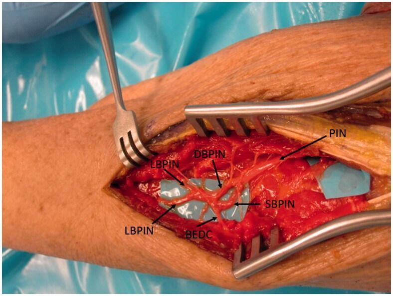
This clinical photograph displays a surgical dissection of the human forearm, focusing on the peripheral nervous system anatomy of the radial nerve. The primary structure shown is the Posterior Interosseous Nerve (PIN) as it courses distally through the extensor compartment. The image highlights a complex branching pattern: the main PIN trunk bifurcates into the Deep Branch (DBPIN) and the Superficial Branch (SBPIN). Further distal arborization is visible, including the lateral branches (LBPIN) and the specific branch to the extensor digitorum communis (BEDC). The nerves are meticulously isolated and placed over blue background material for visual contrast against the red muscular and soft tissue. Surgical retractors are in situ, maintaining exposure of the deep forearm musculature. This anatomical specimen demonstrates the terminal motor innervation of the wrist and finger extensors, critical for understanding PIN palsy, nerve entrapment syndromes, and reconstructive nerve transfer procedures such as AIN-to-PIN transfers.
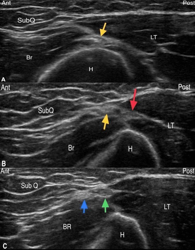
A series of three transverse axis sonographic images (A, B, and C) illustrating the diagnostic tracking of the radial nerve and the posterior cutaneous nerve of the forearm (PCNF) in the lateral brachium. Image A shows the radial nerve (yellow arrow) at the level of the spiral groove, positioned posterolateral to the humerus (H) in the fascial plane between the brachialis (Br) and the long head of the triceps (LT). Image B demonstrates the radial nerve (yellow arrow) distally as it branches into the PCNF (red arrow) after exiting the spiral groove. Image C captures the distal third of the humerus, where the PCNF has subdivided into anterior (blue arrow) and posterior (green arrow) divisions within the lateral triceps-brachioradialis (LT-BR) interval. The ultrasound highlights the hyperechoic surface of the humerus with posterior acoustic shadowing, the intermediate echogenicity of the musculature (Br, LT, BR), and the relatively hypoechoic nerve structures as they move more superficially into the subcutaneous tissue (SubQ) plane.
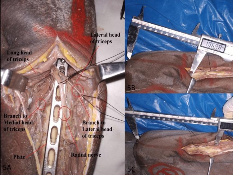
A composite of three clinical photographs demonstrating an anatomical dissection and surgical fixation of the humerus. Panel A provides a high-detail posterior view of the proximal arm. A distal humeral locking compression plate is fixed to the posterior humerus. The dissection clearly identifies the radial nerve as it traverses the spiral groove, showing its anatomical relationship to the surgical hardware. Labels highlight the long and lateral heads of the triceps brachii muscle, as well as specific muscular branches of the radial nerve to the medial and lateral heads. Panels B and C illustrate the quantitative morphometric analysis of the radial nerve's course. A digital Vernier caliper is shown measuring distances from a proximal bony landmark (identified as the posterolateral tip of the acromion) to the points where the radial nerve crosses the medial and lateral borders of the humerus. These images illustrate surgical anatomy relevant to posterior humeral plating and the prevention of iatrogenic radial nerve injury during orthopedic procedures.
ls -la /tmp/workspace/ 2>/dev/null || echo "workspace empty or missing"