
Morning human body cell
Reading File
Good morning! The human body cell is the fundamental unit of life. Let me pull together a rich overview from the medical library.
Searching sources...
Finding Sources
Finding Sources
Finding Sources
Reading File
Loading Image
Finding Sources
Finding Sources
Reading File
Reading File
Good morning! Here is a complete overview of the Human Body Cell - the fundamental unit of all life - sourced from Medical Physiology and Junqueira's Basic Histology:
🔬 The Human Body Cell
The human body contains nearly 40 trillion cells. Every cell is the smallest living part of the body - each enclosed by a membrane, eukaryotic (with a true nucleus), and capable of performing the core functions of life.
Ultrastructure of a Typical Animal Cell
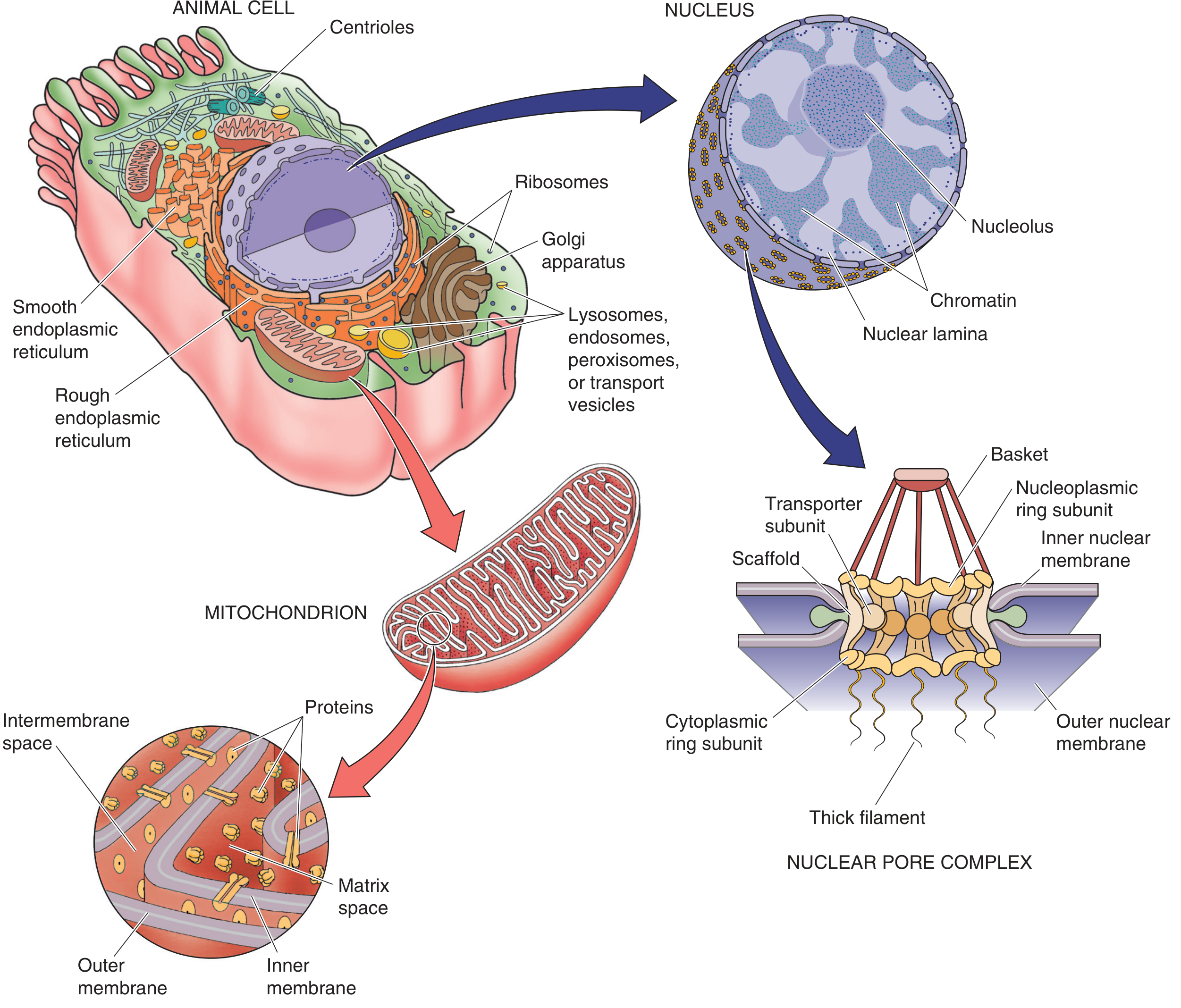
Figure: Ultrastructure of a typical animal cell - Medical Physiology, p. 34
1. Plasma Membrane (Cell Membrane)
The outermost boundary of the cell. It is a lipid bilayer made of amphipathic phospholipids stabilized by cholesterol, with many embedded proteins.
| Feature | Details |
|---|---|
| Lipid bilayer | Phospholipids + cholesterol; creates a selectively permeable barrier |
| Integral proteins | Receptors, ion channels, and pumps for active transport |
| Peripheral proteins | Attached to cytoplasmic surface; involved in signaling |
| Lipid rafts | Cholesterol-rich microdomains with reduced protein mobility |
| Endocytosis | Phagocytosis (particles), pinocytosis (fluids), receptor-mediated endocytosis |
| Exocytosis | Vesicles fuse with membrane to release contents outside the cell |
- Junqueira's Basic Histology, p. 139
2. The Nucleus
The largest organelle in most cells (2-20 µm diameter). It is the control center housing the cell's DNA.
- Enclosed by a double membrane (nuclear envelope) with nuclear pore complexes that regulate traffic of molecules in/out
- Contains chromatin (DNA + histone proteins)
- The nucleolus inside the nucleus is where ribosomal RNA (rRNA) is synthesized
- Most cells have one nucleus; exceptions include skeletal muscle (multinucleated) and red blood cells (no nucleus)
3. Endoplasmic Reticulum (ER)
A convoluted network of membrane-enclosed cisternae extending from the nucleus to the plasma membrane.
| Type | Features | Function |
|---|---|---|
| Rough ER (RER) | Studded with ribosomes; basophilic | Protein synthesis and processing; secretory proteins |
| Smooth ER (SER) | No ribosomes | Lipid synthesis, glycogen metabolism, detoxification, Ca²⁺ storage |
The ER also acts as a major calcium ion reservoir - Ca²⁺ is pumped in via an ATP-dependent pump and released in response to signaling molecules. - Medical Physiology, p. 34
4. Ribosomes
- Made of two subunits (each = rRNA + proteins)
- Attach to mRNA and translate it into protein
- Can be free in the cytosol or bound to RER
- Multiple ribosomes on one mRNA = polyribosome (polysome)
- Cells actively secreting proteins have abundant RER
5. Golgi Complex (Golgi Apparatus)
- Looks like a stack of pancakes (flattened saccules/cisternae)
- A processing and sorting station - receives proteins from RER, modifies them (glycosylation, phosphorylation), and directs them to their final destinations (lysosomes, secretory vesicles, plasma membrane)
6. Mitochondria
The powerhouse of the cell.
- Structure: "balloon within a balloon" - outer membrane + inner membrane
- The inner membrane folds inward to form cristae (dramatically increases surface area)
- Two compartments: intermembrane space and matrix space
- Function: ATP production via oxidative phosphorylation; also involved in many catalytic reactions
- ~0.2 µm in diameter (limit of light microscope resolution)
7. Lysosomes
- The cell's digestive organelle
- Contain hydrolytic enzymes that break down worn-out organelles, pathogens, and cellular debris (autophagy)
- Often contain smaller round vesicles called exosomes within their interior
8. Other Organelles
| Organelle | Function |
|---|---|
| Peroxisomes | Breakdown of fatty acids and H₂O₂; detoxification |
| Endosomes | Sorting compartments for endocytosed material |
| Centrioles | Organize the mitotic spindle during cell division |
| Proteasomes | Degrade misfolded or ubiquitinated proteins |
| Secretory granules | Store proteins for regulated exocytosis |
9. Cytoskeleton
The internal scaffold that gives the cell its shape and drives movement.
| Component | Composition | Function |
|---|---|---|
| Microtubules | Tubulin dimers | Cell shape, intracellular transport, mitotic spindle |
| Microfilaments (Actin filaments) | Actin | Cell motility, muscle contraction, cytokinesis |
| Intermediate filaments | Various (keratin, vimentin, etc.) | Mechanical strength, nuclear support |
10. Cell Differentiation
- The adult body has ~40 trillion cells of hundreds of distinct types, all derived from a single zygote
- Differentiation = specialization process in which cells express specific gene sets to perform particular functions
- For example, muscle cells elongate and develop large arrays of actin/myosin; breast fibroblasts become sensitive to sex hormones due to unique receptor profiles
- Junqueira's Basic Histology, p. 70
Quick Summary Table
| Organelle | Key Function |
|---|---|
| Plasma membrane | Boundary, transport, signaling |
| Nucleus | DNA storage, gene expression |
| Rough ER | Protein synthesis/processing |
| Smooth ER | Lipid synthesis, Ca²⁺ storage |
| Ribosomes | Translation (protein assembly) |
| Golgi complex | Protein modification and sorting |
| Mitochondria | ATP production (energy) |
| Lysosomes | Intracellular digestion |
| Cytoskeleton | Shape, movement, division |
Good morning! The human cell is remarkably organized - every organelle has a distinct job, and together they keep you alive and functioning. Let me know if you want to go deeper into any specific organelle or cell type!
This is a shared conversation. Sign in to Orris to start your own chat.