
Lung carcinoma
lung carcinoma histology types squamous adenocarcinoma small cell
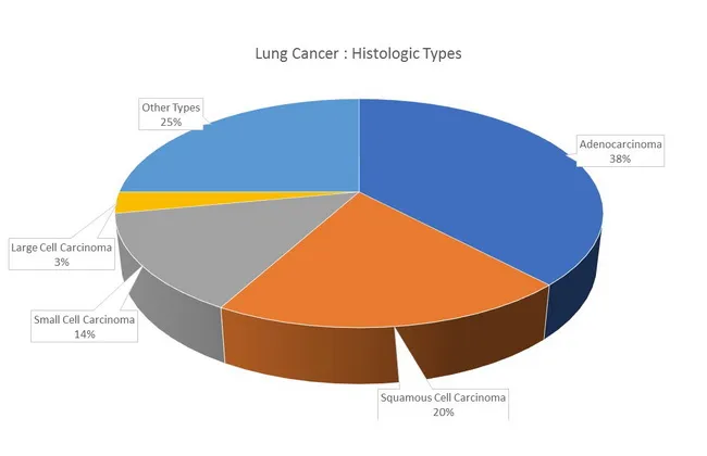
Lung cancer histologic types distribution infographic presenting major histologies and their relative frequencies. The chart is a non-imaging visualization with labeled segments. It shows adenocarcinoma as the most common histology (~38%), followed by squamous cell carcinoma (~20%), small cell carcinoma (~14%), large cell carcinoma (~3%), and other types (~25%). This representation highlights epidemiology: adenocarcinoma is more frequent in women and never-smokers and tends to arise in peripheral airways, whereas squamous and small cell carcinomas originate in proximal airways and show strong smoking associations. Mixed histologies occur in about 10% of cases (e.g., adenocarcinoma with squamous cell). In never-smokers, adenocarcinoma frequently harbors EGFR mutations, while KRAS mutations are more linked to smoking history; TP53 mutations are more common in smoking-related cancers. Clinical relevance includes informing diagnostic workups, guiding biopsy targeting, and shaping research into histology-specific biology and molecular profiles. This image serves educational, epidemiologic, and research contexts in pathology, oncology, pulmonology, and medical education, and supports discussions about tumor heterogeneity, histology-driven therapy implications, and population-based cancer statistics.
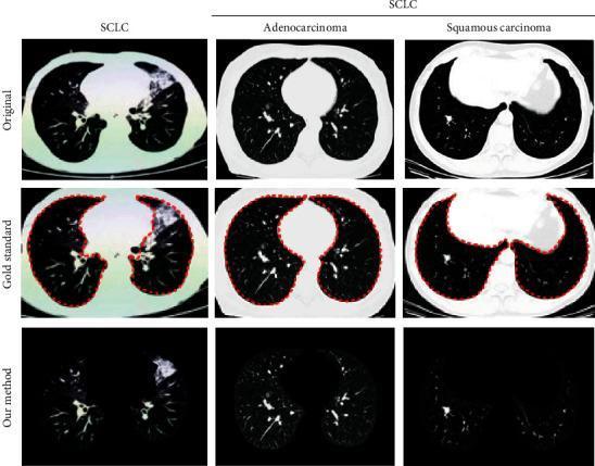
This diagnostic imaging set presents axial thoracic CT scans illustrating three types of lung cancer: Small Cell Lung Cancer (SCLC), Adenocarcinoma, and Squamous Cell Carcinoma. The content is organized into three comparative rows: 'Original' scans, 'Gold Standard' manual segmentations, and automated segmentation results ('Our method'). The SCLC image displays an irregular, large area of increased density in the upper right lung field, consistent with a consolidated mass. The adenocarcinoma scan reveals a more localized, peripheral rounded nodule in the left lung parenchyma. The squamous cell carcinoma scan shows a central lesion with significant soft tissue density near the mediastinum. The 'Gold Standard' row utilizes a red dotted contour to delineate the lung parenchyma boundaries, including the identified pathological regions. This comparison highlights the capability of deep learning algorithms in Lung Parenchyma Segmentation (LPS), specifically the extraction of texture features and nodule morphology for differential diagnosis between SCLC and Non-Small Cell Lung Cancer (NSCLC) subtypes in clinical oncology.
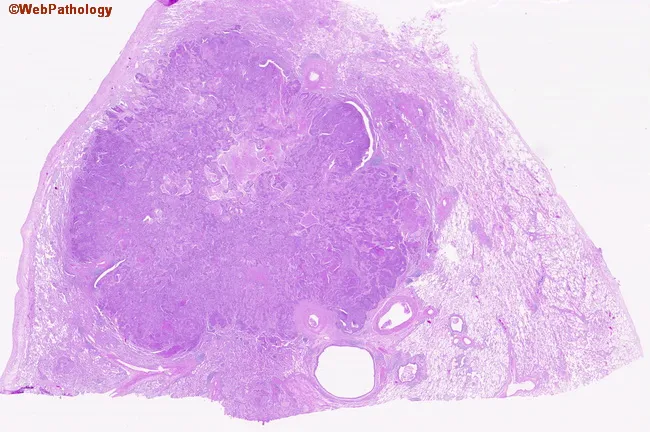
Digital whole-slide histopathology image of a peripherally located lung squamous cell carcinoma captured on a hematoxylin and eosin stained section. The specimen represents peripheral parenchyma with a bronchogenic carcinoma infiltrating the lung periphery. On low- and high-power inspection, nests and irregular clusters of polygonal tumor cells extend from the capsule into adjacent stroma. Tumor cells exhibit eosinophilic cytoplasm, distinct cell borders, and prominent nuclei with coarse chromatin; intercellular bridges may be visible, and occasional keratinization features can be present in well-differentiated foci. The surrounding lung parenchyma shows desmoplastic stromal response and inflammatory infiltrate. The lesion demonstrates peripheral localization, an infiltrative growth pattern, and disruption of normal alveolar architecture. The image supports a histopathologic diagnosis of squamous cell carcinoma due to squamous differentiation, keratinization in places, and intercellular keratin bridges; such morphology is typical in smokers with bronchogenic carcinoma. Clinically, this histology correlates with a smoking history and indicates a solid, non-small cell lung cancer variant with squamous lineage. This image would be valuable for educational purposes, pathology review, differential diagnosis with adenocarcinoma or large cell carcinoma, and in teaching about tumor-stroma interactions in peripheral lung cancers. Correlation with clinical history enhances interpretive accuracy and guides multidisciplinary care in treatment planning.
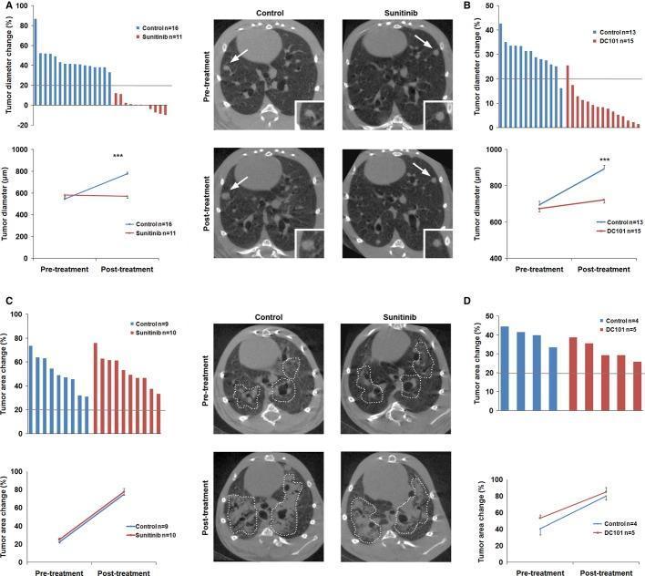
This composite educational graphic illustrates the differential response of lung adenocarcinoma (ADC) and squamous cell carcinoma (SCC) to anti-VEGFR therapies (Sunitinib and DC101) using preclinical mouse models. The data is presented in four panels (A-D). Panels A and B focus on ADC, featuring waterfall plots and line graphs of tumor diameter change. Representative transverse respiratory-gated micro-CT images of the thoracic cavity show ADC lesions (white arrows and high-magnification insets) at pre-treatment and post-treatment stages. In the control group, micro-CT shows visible lesion enlargement, while the Sunitinib-treated group exhibits stabilization of lesion size, corroborated by the flat line plot and waterfall bars below the 20% progression threshold. Panels C and D focus on SCC. Waterfall plots and line graphs depict tumor area change. Micro-CT images demonstrate diffuse tumor masses (outlined by dotted lines). In both control and Sunitinib-treated SCC groups, post-treatment images show significant expansion of the tumor-occupied area, indicating primary resistance to anti-VEGFR2 treatment. This highlights the clinical concept of histology-dependent therapy response in non-small cell lung cancer subtypes.
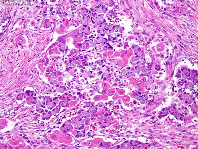
This histopathology slide depicts lung tumor tissue examined by light microscopy following hematoxylin and eosin staining. The specimen is derived from pulmonary parenchyma in a region containing tumor cells. The architecture shows a hybrid neoplasm composed of mucin-producing cells arranged in gland-like clusters and adenomatoid glands intermingled with nests of squamous cells, characterized by dense eosinophilic cytoplasm and rounded to polygonal nuclei. Mucinous cells exhibit bluish cytoplasm and intracytoplasmic mucin; squamous cells display keratinization features and abundant eosinophilic cytoplasm; together they confirm a combined histology with two distinct subtypes. The background stroma contains desmoplastic reaction and inflammatory infiltrate in places. This pattern is typical of adenosquamous carcinoma of the lung, observed in roughly 10% of lung cancers, where prognosis and therapeutic decisions hinge on the more aggressive component. Clinically, such tumors portend a poorer prognosis than pure adenocarcinoma or pure squamous cell carcinoma and may influence choices between surgical resection and systemic therapy. Immunophenotypic profiling (TTF-1 and Napsin A positivity for glandular components; p40/p63 positivity for squamous components) can assist subtyping. Recognize that combined histology carries implications for staging, prognosis, and potential targeted regimens; differential considerations include pure adenocarcinoma with mucin production, pure squamous cell carcinoma, or small cell variants.
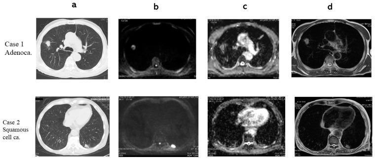
Multi-modal chest imaging comparison of two primary lung cancer types: Adenocarcinoma (Case 1) and Squamous Cell Carcinoma (Case 2). The 4x2 grid displays axial sections across four modalities: (a) Computed Tomography (CT), (b) Diffusion-Weighted Imaging (DWI), (c) Apparent Diffusion Coefficient (ADC) maps, and (d) T2-Weighted Imaging (T2WI). In Case 1, the adenocarcinoma presents as a focal nodule in the right lung field with hyperintensity on DWI and corresponding hypointensity on the ADC map (1.39 x 10^-3 mm^2/s), indicating restricted diffusion. In Case 2, the squamous cell carcinoma is located in the left posterior lung periphery, demonstrating stronger signal suppression on the ADC map (1.04 x 10^-3 mm^2/s) compared to Case 1. T2WI reveals heterogeneous internal architecture for the adenocarcinoma and a more homogeneous appearance for the squamous cell carcinoma. This comparative imaging set illustrates the diagnostic utility of combining structural (CT) and functional (DWI/ADC) MRI sequences to characterize pulmonary nodules and differentiate histological subtypes based on diffusion restriction and T2 contrast ratios.
"lung cancer" AND "non-small cell" AND treatment
Lung Carcinoma - Comprehensive Overview
Epidemiology & Risk Factors
- Radon gas exposure
- Asbestos (synergistic with smoking)
- Occupational carcinogens (arsenic, chromium, nickel, polycyclic hydrocarbons)
- Inherited predisposition: first-degree relatives of probands have a 2-3 fold excess risk
- Germline mutations in RB1, TP53 (Li-Fraumeni), and EGFR T790M (never-smokers)
- Loci at 5p15, 6p21, and 15q25 linked to susceptibility
WHO Classification
| Type | Frequency | Key Features |
|---|---|---|
| Adenocarcinoma | ~38% | Most common; peripheral; women/non-smokers; EGFR/KRAS/ALK mutations |
| Squamous cell carcinoma | ~20% | Central/hilar; keratinization; intercellular bridges; smokers |
| Small cell carcinoma (SCLC) | ~14% | Neuroendocrine; paraneoplastic syndromes; metastatic at presentation |
| Large cell carcinoma | ~3% | Diagnosis of exclusion; <10% of cases |
| Others | ~25% | Adenosquamous, sarcomatoid, carcinoid, NUT, salivary gland-type |
Histopathology
Precursor Lesions
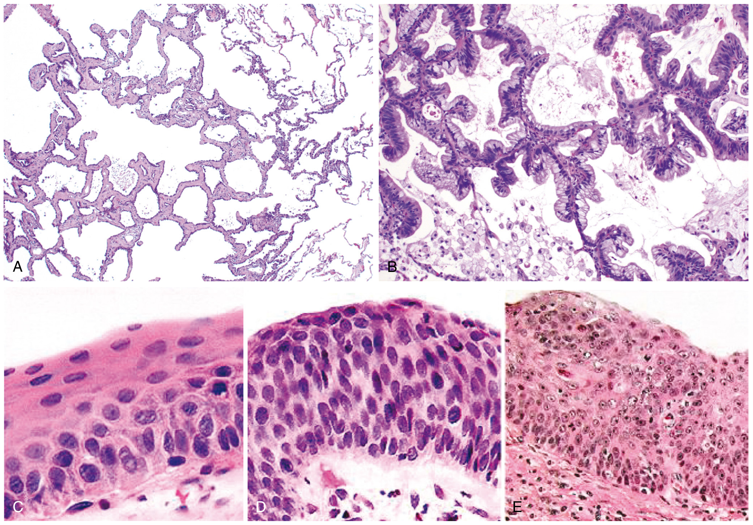
Adenocarcinoma
- Arises from atypical adenomatous hyperplasia (AAH) and adenocarcinoma in situ (AIS)
- Growth patterns: lepidic (noninvasive, crawling along alveolar septa), acinar, papillary, micropapillary, solid
- Usually peripheral; expresses TTF-1 and Napsin-A
- Microinvasive adenocarcinoma (≤3 cm, ≤5 mm invasion): far better prognosis
- Mucinous adenocarcinoma spreads aerogenously, forming satellite tumors
- Associated with EGFR, KRAS, ALK, ROS1, BRAF, MET driver mutations
Squamous Cell Carcinoma
- Arises from squamous metaplasia/dysplasia in bronchial epithelium
- Usually central/hilar; keratinization, intercellular bridges, keratin pearls
- Strongly associated with smoking
- Peripheral localization is also possible
- Molecular targets: FGFR1 amplification, DDR2 mutations, PIK3CA mutations/PTEN loss
Small Cell Carcinoma (SCLC)
- Neuroendocrine origin; small cells with scant cytoplasm, granular chromatin, absent nucleoli, high mitotic count
- Markers: CD56, synaptophysin, chromogranin, NCAM, INSM1
- >90% harbor mutations in TP53 and RB1
- Nearly always metastatic at presentation
- Treated primarily with chemotherapy (not surgery)
- Prone to paraneoplastic syndromes (SIADH, Cushing, Lambert-Eaton, cerebellar degeneration)
Molecular Driver Mutations (NSCLC)
| Target | Mutation/Alteration | Therapy |
|---|---|---|
| EGFR (exon 19 del, L858R) | Activating mutation | Osimertinib, erlotinib, gefitinib |
| ALK | Chromosomal rearrangement | Alectinib, crizotinib, brigatinib |
| KRAS G12C | Point mutation | Sotorasib, adagrasib |
| ROS1 | Rearrangement | Entrectinib, crizotinib |
| BRAF V600E | Point mutation | Dabrafenib + trametinib |
| MET exon 14 skip | Splice mutation | Capmatinib, tepotinib |
| RET | Rearrangement | Selpercatinib, pralsetinib |
| NTRK | Fusion | Larotrectinib, entrectinib |
| HER2 | Mutation/amplification | T-DXd (trastuzumab deruxtecan) |
Clinical Features
Local/Regional Symptoms
- Cough (new or changed) - most common
- Hemoptysis
- Dyspnea, wheezing, stridor
- Post-obstructive pneumonia/recurrent pneumonia
- Hoarseness (left recurrent laryngeal nerve palsy)
- Superior vena cava (SVC) syndrome: facial swelling, arm edema, venous distension - especially SCLC
- Pancoast syndrome (superior sulcus tumor): shoulder/arm pain, Horner's syndrome (ptosis, miosis, anhidrosis)
- Phrenic nerve palsy: diaphragm elevation
Distant Metastases
- Brain: headache, focal neurological deficits, seizures
- Bone: pain, pathological fractures (hypercalcemia in SqCC from PTHrP)
- Liver: jaundice, RUQ pain
- Adrenal: incidental finding on CT
Paraneoplastic Syndromes
| Syndrome | Associated Histology |
|---|---|
| SIADH (hyponatremia) | SCLC |
| Cushing syndrome (ectopic ACTH) | SCLC |
| Lambert-Eaton myasthenic syndrome | SCLC |
| Hypercalcemia (PTHrP) | Squamous cell |
| Hypertrophic osteoarthropathy | Adenocarcinoma |
| Cerebellar degeneration | SCLC |
| Dermatomyositis/polymyositis | Any |
Staging
NSCLC - TNM (8th Edition AJCC/UICC)
| Stage | Description | 5-yr Survival (approx.) |
|---|---|---|
| IA1-IA3 | T1, N0, M0 - Tumor ≤3 cm | 68-92% |
| IB | T2a, N0, M0 | ~60% |
| IIA-IIB | T1-2, N1 or T3, N0 | 35-55% |
| IIIA | T1-3, N2 or T3-4, N1 | 10-35% |
| IIIB-IIIC | T any, N3 or T4, N2-3 | 5-15% |
| IV | M1 (any distant metastasis) | <5% |
Staging Investigations
- CT chest/abdomen/pelvis - initial imaging
- PET-CT - preferred for mediastinal and extrathoracic staging; SUV >2.5 suspicious for malignancy
- MRI brain - brain metastases (especially SCLC and adenocarcinoma)
- EBUS (endobronchial ultrasound) - mediastinal lymph node staging
- Mediastinoscopy/thoracoscopy - invasive staging when required
- Bone marrow biopsy - rarely needed in SCLC now
SCLC Staging (simplified)
- Limited disease (LD): confined to one hemithorax + ipsilateral nodes, encompassable in radiation field
- Extensive disease (ED): beyond LD, including contralateral nodes, distant mets, malignant effusion
Treatment
NSCLC
- Surgical resection (lobectomy preferred; pneumonectomy if needed)
- Sublobar resection (segmentectomy) for Stage IA with tumors ≤2 cm
- Adjuvant osimertinib for Stage IB-IIIA with EGFR mutations (FDA approved)
- Adjuvant chemotherapy (cisplatin-based) for Stage II-IIIA
- Stereotactic body radiotherapy (SBRT) for medically inoperable patients
- Concurrent chemoradiation (cisplatin + etoposide or carboplatin + paclitaxel)
- Consolidation durvalumab (PD-L1 inhibitor) after CRT (PACIFIC regimen)
- Neoadjuvant nivolumab + chemotherapy for resectable Stage III (CheckMate 816)
- Perioperative pembrolizumab + chemotherapy (KEYNOTE-671)
- Targeted therapy first-line when driver mutation present (osimertinib for EGFR, alectinib for ALK, etc.)
- Immunotherapy: pembrolizumab monotherapy if PD-L1 ≥50%; combination chemo-immunotherapy if PD-L1 <50%
- Carboplatin/paclitaxel or cisplatin/pemetrexed doublet for non-squamous without driver mutation
- Bevacizumab (anti-VEGF) added in non-squamous, non-bleeding patients
SCLC
- Cisplatin/etoposide + concurrent thoracic radiation
- Prophylactic cranial irradiation (PCI) in complete/partial responders
- Addition of atezolizumab is being evaluated
- Carboplatin/etoposide + atezolizumab (IMpower133) - first-line standard
- Carboplatin/etoposide + durvalumab ± tremelimumab (CASPIAN)
- Response rates are initially high (~70%) but relapse is nearly universal
- Second-line: topotecan, lurbinectedin
Screening
- Age 50-80 years
- ≥20 pack-year smoking history
- Current smoker or quit within the past 15 years
Recent Evidence (2024-2026)
- Dual CTLA-4/PD-L1 blockade in advanced NSCLC shows improved long-term overall survival in biomarker-defined subgroups (PMID 41038207, Lancet Oncol 2025)
- ICIs after EGFR TKI failure in EGFR-mutated NSCLC: network meta-analysis shows modest efficacy with immune checkpoint inhibitors in this setting (PMID 39159630, Lancet Oncol 2024)
- Cardiovascular toxicity of EGFR TKIs is a significant concern: systematic review identifies class-specific CV adverse events that require monitoring (PMID 40897431, BMJ 2025)
Key Immunohistochemistry Markers
| Marker | Adenocarcinoma | SqCC | SCLC |
|---|---|---|---|
| TTF-1 | + | - | + (weak) |
| Napsin-A | + | - | - |
| p40/p63 | - | + | - |
| CK5/6 | - | + | - |
| CD56/Synaptophysin/Chromogranin | - | - | + |
| Ki-67 | Variable | Variable | Very high (>80%) |