
Explain subdural and epidural hemorrhage
subdural hematoma CT scan brain
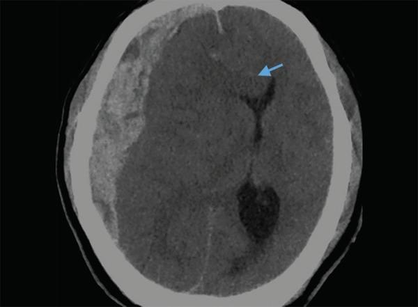
This diagnostic axial non-contrast CT scan of the brain demonstrates an acute subdural hematoma (SDH) in the right frontoparietal region. The visual features include a hyperdense, crescentic (concavo-convex) extra-axial collection extending along the inner table of the calvarium. The hematoma exerts significant mass effect on the underlying brain parenchyma, resulting in a marked leftward midline shift. A blue arrow identifies subfalcine herniation, where the cingulate gyrus is displaced across the midline beneath the falx cerebri. There is also evident effacement of the ipsilateral (right) lateral ventricle and cortical sulci, accompanied by surrounding hypodense areas consistent with cerebral edema. This image serves as an educational example of acute traumatic intracranial hemorrhage and secondary brain herniation syndromes, highlighting the critical radiological signs used in neurosurgical triage and emergency medicine.
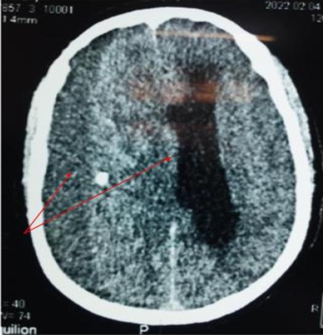
This diagnostic image is an axial non-contrast computerized tomography (CT) scan of the brain. It demonstrates a voluminous chronic subdural hematoma in the right hemisphere, characterized by a large, crescent-shaped collection that conforms to the inner table of the skull. The hematoma exhibits heterogeneous density, indicating areas of liquified blood and potentially acute-on-chronic components. Significant mass effect is evident, manifested by a marked midline shift to the left, subfalcine herniation, and severe compression of the right lateral ventricle. The image highlights the clinical significance of intracranial pressure and brain displacement, which in this case leads to subfalcine herniation and suggests a potential Kernohan-Woltman notch phenomenon due to compression of the contralateral cerebral peduncle against the tentorium. This educational material is suitable for neurosurgical and radiological training regarding acute-on-chronic subdural hemorrhage and its secondary mechanical effects on brain structures.
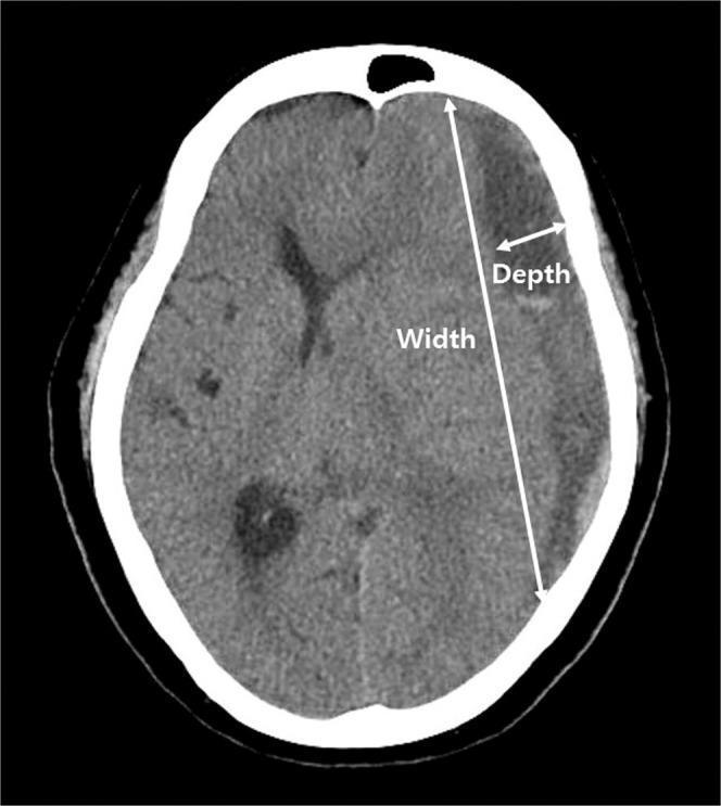
An axial non-contrast computed tomography (CT) scan of the head demonstrating a chronic subdural hematoma (SDH) located along the right convexity. The hematoma presents as a crescent-shaped, hypodense collection relative to the brain parenchyma, situated between the inner table of the skull and the brain surface. The image highlights the methodology for calculating depressed brain volume through two annotated measurements: 'Depth' and 'Width'. Depth is indicated by a short double-headed arrow representing the maximal transverse diameter between the inner skull table and the displaced brain surface. Width is indicated by a long double-headed arrow representing the linear distance between the superior and inferior margins where the hematoma meets the skull. The SDH causes visible mass effect, resulting in the effacement of cortical sulci and inward displacement of the underlying brain tissue. This visual serves as an educational reference for neuroradiological assessment of intracranial fluid collections and the quantitative evaluation of brain compression.
epidural hematoma CT scan brain
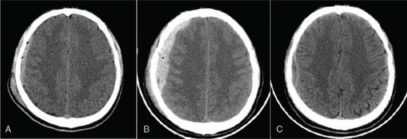
This comparison chart consists of three axial non-contrast computed tomography (CT) scans of the brain, labeled A, B, and C, illustrating the progression and resolution of an acute epidural hematoma (EDH) in the right frontal region. Scan A shows an initial, relatively thin hyperdense (bright) biconvex collection along the inner table of the right frontal skull, consistent with an acute EDH. Scan B demonstrates a significant interval increase in the volume and thickness of the hyperdense collection, which now exerts more pronounced mass effect on the underlying right frontal lobe parenchyma. Scan C is a follow-up image obtained after arterial embolization, showing a dramatic reduction in the size and density of the hematoma, which has evolved into a thin, nearly isodense crescent, indicating successful absorption and relief of previous mass effect. The series serves as an educational example of hematoma expansion post-trauma and subsequent resolution following interventional radiological management.
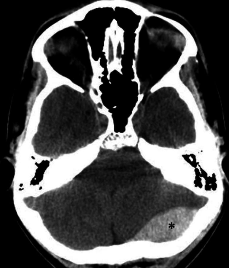
An axial non-contrast computerized tomography (CT) scan of the brain demonstrating an acute epidural (extradural) hematoma. The primary finding is a well-defined, biconvex (lens-shaped), hyperdense fluid collection located in the left occipital region, marked with an asterisk. This hyperdensity is characteristic of an acute hemorrhage within the epidural space, which typically does not cross cranial sutures. Secondary mass effect is visible, characterized by a mild left-to-right midline shift and compression of adjacent brain parenchyma. Anatomical landmarks include the skull base, posterior fossa structures, and the cerebellum. The image illustrates a classic neurosurgical emergency often associated with traumatic skull fractures and arterial bleeding, typically from the middle meningeal artery. This diagnostic image is intended for educational use in emergency medicine, radiology, and neurology to distinguish epidural hematomas from crescent-shaped subdural hematomas.
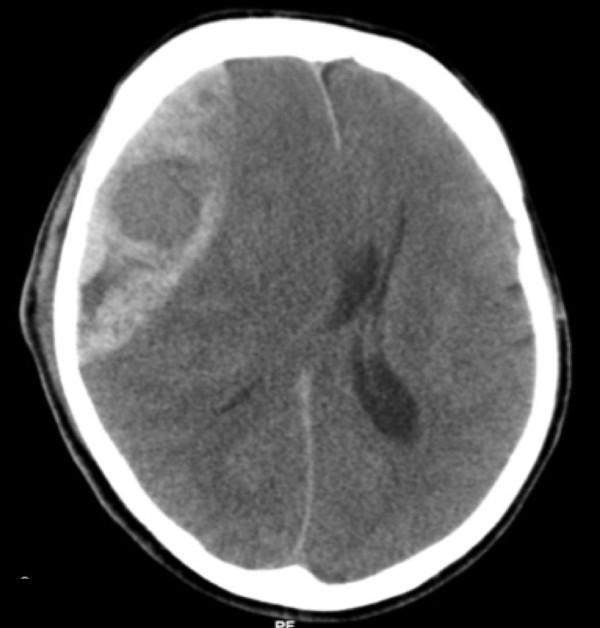
This diagnostic image is an axial non-contrast Computed Tomography (CT) scan of the brain, demonstrating a large, acute epidural (extradural) hematoma. The lesion is located on the right side of the skull and exhibits a classic biconvex or lens-shaped (lentiform) morphology, characterized by a hyperdense appearance relative to the brain parenchyma. The hematoma is clearly demarcated and constrained by the cranial sutures, positioned between the inner table of the skull and the dura mater. The image illustrates a significant mass effect, resulting in the effacement of adjacent sulci, partial compression of the right lateral ventricle, and a notable leftward midline shift of the falx cerebri. These findings are highly characteristic of an acute arterial bleed, typically involving the middle meningeal artery. This scan serves as a critical educational tool for identifying neurosurgical emergencies, understanding intracranial pressure dynamics, and recognizing the radiological hallmarks that differentiate epidural hematomas from crescent-shaped subdural hematomas.
Subdural and Epidural Hemorrhage
Anatomical Context
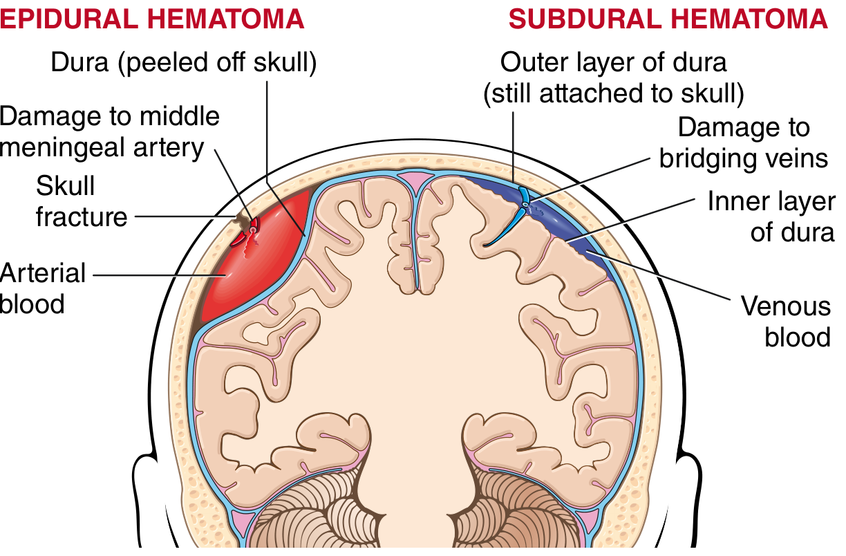
EPIDURAL HEMORRHAGE (EDH)
Location and Space
Mechanism and Source
- The dura is fused with the periosteum of the skull and is supplied by the middle meningeal artery, which runs in a groove on the inner surface of the temporal bone.
- Trauma - typically a blow to the temporal or temporoparietal region - causes a skull fracture that crosses the middle meningeal artery groove, lacerating the vessel.
- This produces high-pressure arterial bleeding that strips the dura away from the periosteum, expanding the hematoma rapidly.
- In children, temporary deformation of the skull can lacerate a vessel even without a frank fracture.
- Occasionally, venous sinus tears (parieto-occipital or posterior fossa trauma) can cause an EDH - these bleed more slowly and behave more like subdurals.
Classic Clinical Presentation
- Initial loss of consciousness (brief, from the concussive blow)
- Lucid interval - patient recovers and may seem normal (minutes to hours)
- Rapid neurologic deterioration - worsening headache, vomiting, declining consciousness
- Uncal herniation signs - ipsilateral pupil dilation, then complete ophthalmoparesis, then contralateral motor signs
Associated Signs
- Battle's sign - ecchymosis behind the ear (mastoid area), indicating basal skull fracture
- Raccoon eyes - periorbital ecchymosis from anterior skull base fracture
- These signs indicate the skull fracture that caused middle meningeal artery injury
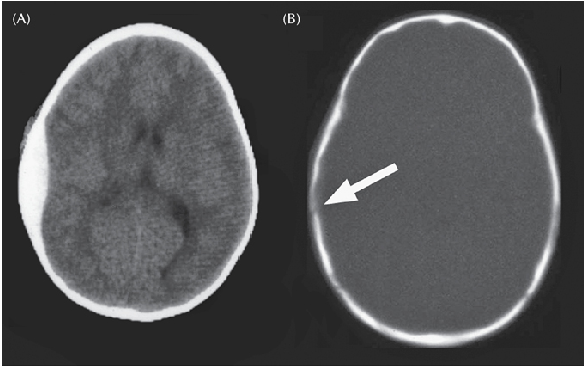
CT Appearance

Prognosis and Treatment
SUBDURAL HEMORRHAGE (SDH)
Location and Space
Mechanism and Source
- Bridging veins travel from the cortical surface of the cerebral hemispheres, across the subarachnoid space and through the dura, to empty into the dural venous sinuses (particularly the superior sagittal sinus).
- The brain is suspended in CSF and moves freely with trauma; the dural sinuses are fixed. Sudden acceleration/deceleration shears the bridging veins at the point they penetrate the dura.
- The extravasated blood dissects through the dural layers, producing a subdural hematoma.
- Bleeding is venous - lower pressure, slower accumulation, often self-limiting.
Who is at Risk?
- Elderly individuals with brain atrophy: as the brain shrinks, bridging veins stretch over a greater distance, making them more vulnerable - even minor trauma can tear them.
- Infants (non-accidental injury / "shaken baby"): thin-walled bridging veins are very susceptible.
- Anticoagulated patients: bleeding is worse and may occur with trivial trauma.
- Patients with coagulopathies, alcohol use disorder (falls + brain atrophy).
Classification by Time
| Type | Time frame | CT appearance |
|---|---|---|
| Acute | <3 days | Hyperdense (bright) crescent |
| Subacute | 3 days - 3 weeks | Isodense (may be hard to see) |
| Chronic | >3 weeks | Hypodense (dark) crescent |
Morphological Progression
- ~1 week: Lysis of the clot
- ~2 weeks: Fibroblasts grow in from the dural surface
- 1-3 months: Hyalinized connective tissue forms; organized hematoma attaches to inner dura, free from the arachnoid
- Lesion may retract to a thin "subdural membrane"
- Alternatively, thin-walled granulation tissue vessels may re-bleed repeatedly → chronic subdural hematoma
Clinical Presentation
- Symptoms typically manifest within 48 hours of injury.
- Most common over the lateral aspects of cerebral hemispheres; bilateral in ~10% of cases.
- Clinical features are often nonlocalizing: headache, confusion, gradually progressive neurologic deterioration.
- Focal signs can occur from pressure on adjacent brain.
- Acute decompensation may supervene on a subacute/chronic course.
- Chronic SDH in the elderly may present with weeks of progressive cognitive decline, sometimes mistaken for dementia.
CT Appearance
Treatment
Head-to-Head Comparison
| Feature | Epidural Hematoma | Subdural Hematoma |
|---|---|---|
| Space | Between skull and dura | Between dura and arachnoid |
| Bleeding source | Middle meningeal artery (arterial) | Bridging veins (venous) |
| Cause | Skull fracture (temporal blow) | Acceleration-deceleration; brain atrophy |
| Speed | Rapid (hours) | Slower (hours to weeks) |
| CT shape | Biconvex / lens-shaped | Crescent-shaped |
| Crosses sutures? | No | Yes |
| Lucid interval | Classic (but not always present) | Less typical |
| At-risk population | Young adults, MVAs, sports | Elderly, infants, anticoagulated |
| Underlying brain injury | Often absent | Brain often compressed/injured |
| Urgency | Extreme - herniation in hours | Variable - may progress over days-weeks |
| Prognosis if treated promptly | Excellent | Good, but depends on underlying brain injury |