
Write a short note on forearm movements for Ms orthopedics theory examination gold medal level
Reading File
Now let me search the medical library for forearm movements content in parallel:
Finding Sources
Finding Sources
Finding Sources
Good hits. Let me now read the key sections from Gray's Anatomy and the imaging anatomy book in parallel:
Reading File
Reading File
Finding Sources
Excellent. Now let me read more from Gray's Anatomy for the muscles section, Miller's Review for the orthopaedic-specific details, and fetch the key diagram:
Reading File
Reading File
Loading Image
I now have rich content from Gray's Anatomy, Miller's Review of Orthopaedics, and the Imaging Anatomy atlas. Let me also quickly get the Rockwood & Green section on deforming forces related to forearm rotation (relevant for orthopaedic examinations - forearm fractures) and search for nerve supply details:
Reading File
Finding Sources
I now have comprehensive, gold-medal level content from multiple authoritative orthopaedic and anatomy sources. Let me compose the full answer.
Forearm Movements
A Gold-Medal Level Short Note for MS Orthopaedics Theory Examination
1. Introduction
The forearm possesses a unique biomechanical design that allows it to function as a rotational unit. Its two principal movements are pronation and supination, collectively termed pronosupination. These occur entirely within the forearm and are distinct from shoulder rotation, though shoulder rotation supplements them.
- Supination: Palm faces anteriorly (anatomical position) - "palm up"
- Pronation: Palm faces posteriorly - "palm down"
Normal range: 80-85 degrees in each direction from neutral (total arc ~180 degrees). Combined with shoulder rotation, a total of ~270 degrees of upper limb rotation is achievable. The functional range clinically required for most ADLs is 50 degrees of pronosupination (50° pronation + 50° supination). (Miller's Review of Orthopaedics, 9th Ed.)
2. Articulations Involved
Pronation and supination occur simultaneously at two trochoid (pivot) joints that function as a single "forearm joint unit":
a. Proximal Radio-Ulnar Joint (PRUJ)
- Located within the elbow joint capsule
- The radial head spins on the capitulum while its articular surface slides against the radial notch of the ulna
- Stability provided by the annular ligament and a strong osteoligamentous cavity formed by the radial notch of the ulna
b. Distal Radio-Ulnar Joint (DRUJ)
- The ulnar notch of the radius (sigmoid notch) slides anteriorly over the convex head of the ulna during pronation
- Stability provided by the triangular fibrocartilage complex (TFCC): consisting of the articular disc, dorsal and palmar radioulnar ligaments, and the meniscus homologue
c. Connecting Structure - Interosseous Membrane (IOM)
- Fibrous sheet connecting medial border of radius to lateral border of ulna
- Fibers run obliquely downward from radius to ulna
- Does not restrict pronation/supination
- Transfers axial load from radius to ulna
- A thicker central band and proximal/distal oblique cords maintain forearm stability
- The IOM prevents proximal migration of the radius (Imaging Anatomy, Vol. 3)
d. The Axis of Rotation
A longitudinal axis runs from the center of the radial head proximally to the fovea of the ulnar head distally. The ulna remains relatively stationary - it is the primary load-bearing bone. The radius rotates around it. During pronation, the radius crosses over the ulna; during supination, the two bones are parallel.
3. Muscles of Pronation and Supination
Supinators (2 muscles)
| Muscle | Origin | Insertion | Nerve Supply | Key Feature |
|---|---|---|---|---|
| Biceps brachii | Supraglenoid tubercle (long head); coracoid process (short head) | Radial tuberosity | Musculocutaneous nerve (C5, C6) | Most powerful supinator; most effective with elbow flexed at 90° |
| Supinator | Lateral epicondyle + supinator crest of ulna + related ligaments | Lateral surface of upper 1/3 of radius (above oblique line) | Deep branch of radial nerve (posterior interosseous nerve) C6 | Works at all elbow angles; sole supinator when elbow extended |
Key point: The biceps brachii and supinator tendons both wrap around the proximal radius during pronation. Contraction unwraps them, generating supination torque.
Pronators (2 muscles)
| Muscle | Origin | Insertion | Nerve Supply | Key Feature |
|---|---|---|---|---|
| Pronator Teres (PT) | Humeral head: medial epicondyle; Ulnar head: coronoid process | Lateral surface of radius (~midshaft) | Median nerve (C6, C7) | Primary pronator; also assists elbow flexion |
| Pronator Quadratus (PQ) | Anterior surface of distal ulna (lower 1/4) | Anterior surface of distal radius (lower 1/4) | Anterior interosseous nerve (branch of median; C8, T1) | Works at all speeds; the "workhorse" of pronation; deep to all tendons |
Key point: Both pronators pull the distal radius over the ulna, producing pronation. The PQ acts throughout the entire arc; the PT adds power particularly against resistance. (Gray's Anatomy for Students)
Accessory Muscle
- Anconeus: During pronation, the distal end of the ulna abducts slightly to keep the palm axis central. The anconeus (lateral epicondyle to proximal ulna; radial nerve) controls this abduction of the ulna.
4. Biomechanics of the Axis
- In neutral position: styloid process of ulna aligns with the sigmoid notch
- In pronation: ulnar head rotates, rolling the styloid process to the palmar side; the radius migrates proximally - this increases ulnar variance (clinically important in DRUJ pathology and wrist fractures)
- In supination: the styloid process moves to the dorsal side; negative ulnar variance
Clinical pearl: For forearm rotation to occur, the axes of the PRUJ and DRUJ must be coaxially aligned. Any deformity (malunion, Monteggia, DRUJ injury) that disrupts this coaxial alignment blocks rotation. (Imaging Anatomy, Vol. 3)
5. Deforming Forces - The Orthopaedic Significance
This is a high-yield topic for MS examinations in the context of radius and both-bone forearm fractures:
- In the intact forearm, rotational forces are balanced in pronation
- Main supinating forces: Biceps brachii + Supinator
- Main pronating forces: Pronator teres (PT) + Pronator quadratus (PQ)
Fracture Level Determines Rotation Deformity:
| Fracture Level | Proximal Fragment | Distal Fragment | Reasoning |
|---|---|---|---|
| Above PT insertion (above midshaft) | Supinated (biceps + supinator unopposed) | Pronated (PT + PQ acting) | Severe deformity - maximal rotational mismatch |
| Below PT insertion (below midshaft) | Less supination (PT counteracts supinators on proximal fragment) | Pronated (PQ acting) | Lesser deformity |
| Distal radius (Colles/Smith) | N/A | Deformity determined by PQ | PQ pulls distal fragment into pronation and palmarly |
Surgical principle: When reducing and plating radial shaft fractures, the forearm must be held in the appropriate rotational position matching the estimated deformity to restore the normal radial bow and prevent malunion. (Rockwood & Green's Fractures in Adults, 10th Ed., 2025)
6. Nerve Supply Summary
| Nerve | Muscles Supplied |
|---|---|
| Musculocutaneous (C5, C6) | Biceps brachii (supinator) |
| Deep radial / PIN (C6) | Supinator |
| Median nerve (C6, C7) | Pronator teres |
| Anterior interosseous nerve (C8, T1) | Pronator quadratus |
Mnemonic: "Biceps Supinates, Pronators Pronate" - Biceps (musculocutaneous), Supinator (radial), PT (median), PQ (AIN)
7. Clinical Correlations (Gold-Medal Points)
-
Essex-Lopresti injury: Radial head fracture + IOM disruption + DRUJ instability. Loss of IOM integrity allows proximal radial migration (radius shortens relative to ulna), causing positive ulnar variance and DRUJ pain. Demonstrates the IOM's role in maintaining radial length.
-
Monteggia fracture-dislocation: Ulnar shaft fracture + radial head dislocation disrupts PRUJ. Bado classification based on direction of radial head dislocation. Rotational movement is immediately lost.
-
Galeazzi fracture: Radial shaft fracture + DRUJ disruption (the "reverse Monteggia"). Treatment: ORIF of radius + DRUJ stabilization.
-
Paralysis patterns:
- Loss of AIN: PQ paralysis - weak pronation against resistance; confirmed by loss of tip pinch (FPL + FDP index paralyzed too)
- Loss of musculocutaneous nerve: Supination weak (only supinator remains) but not absent; elbow flexion lost
- Loss of radial/PIN nerve: Supinator paralyzed; biceps now the sole supinator (most effective only with elbow flexed)
-
Position of immobilization: Forearm fractures in children are immobilized in supination for proximal-third fractures (to bring the distal fragment to match the proximally supinated fragment) and in neutral-to-pronation for distal-third fractures.
Key Diagram
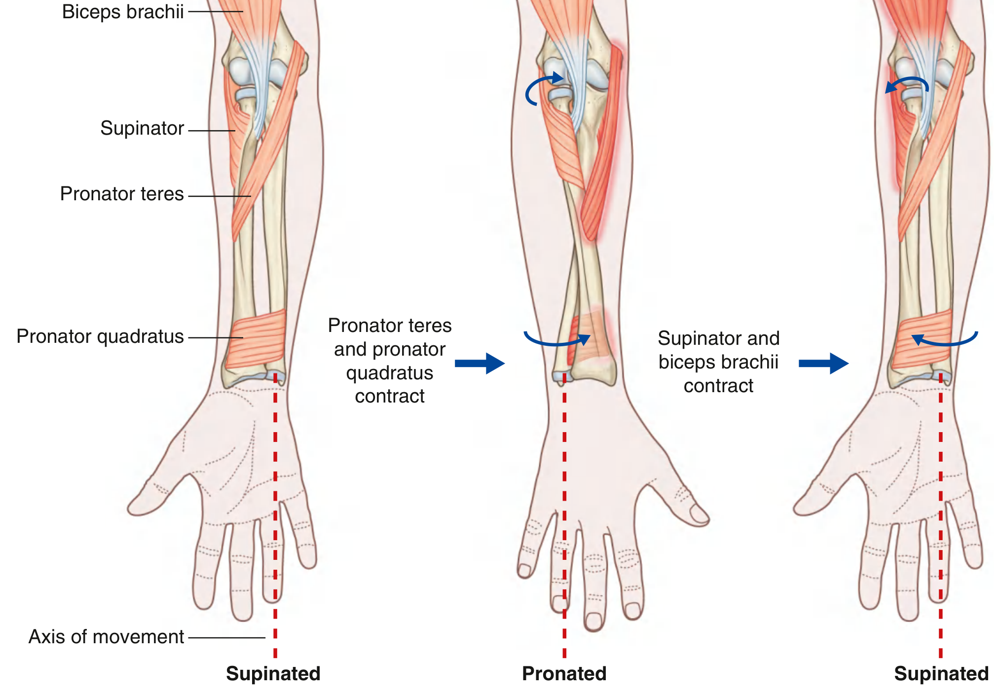
Fig. Pronation and Supination showing the axis of movement, with pronator teres + pronator quadratus contracting to produce pronation, and supinator + biceps brachii contracting to produce supination. (Gray's Anatomy for Students)
Summary Box
| Feature | Pronation | Supination |
|---|---|---|
| Definition | Palm faces posteriorly | Palm faces anteriorly |
| Muscles | PT, PQ | Biceps brachii, Supinator |
| Nerve | Median (PT), AIN (PQ) | Musculocutaneous (biceps), Radial/PIN (supinator) |
| Range | 80-85° | 80-85° |
| PRUJ movement | Radius rotates medially | Radius rotates laterally |
| DRUJ movement | Sigmoid notch slides anteromedially | Sigmoid notch returns posteriorly |
| Ulnar variance | Increases (positive) | Decreases (negative) |
| Daily function | Typing, door handle turning (down) | Carrying soup bowl, screwdriver (out) |
Sources: Gray's Anatomy for Students; Miller's Review of Orthopaedics, 9th Ed.; Rockwood & Green's Fractures in Adults, 10th Ed. (2025); Imaging Anatomy Text and Atlas, Vol. 3
This is a shared conversation. Sign in to Orris to start your own chat.