
ABG analysis
Reading File
ABG (arterial blood gas) analysis is a systematic process of interpreting pH, PaCO2, PaO2, HCO3, and base excess to identify acid-base disorders and oxygenation status. The key steps are: determine if acidemic or alkalemic (pH), identify primary disorder (respiratory vs metabolic), check for compensation, and identify mixed disorders.
Searching sources...
Finding Sources
Finding Sources
Reading File
Reading File
Reading File
Reading File
Reading File
Now I have comprehensive content. Let me fetch the anion gap diagram image and compile the full answer.
Loading Image
Here is a complete, structured guide to ABG analysis.
Arterial Blood Gas (ABG) Analysis
Normal Values
| Parameter | Normal Range |
|---|---|
| pH | 7.35 - 7.45 |
| PaCO2 | 35 - 45 mmHg |
| PaO2 | 70 - 95 mmHg (age-dependent) |
| HCO3- | 22 - 26 mEq/L |
| Base Excess (BE) | -2 to +2 mEq/L |
| SaO2 | 95 - 100% |
Step-by-Step Approach
Step 1 - Check the pH
| pH | Interpretation |
|---|---|
| < 7.35 | Acidemia |
| 7.35 - 7.45 | Normal |
| > 7.45 | Alkalemia |
A normal pH does not rule out a mixed disorder.
Step 2 - Identify the Primary Disorder
Look at PaCO2 and HCO3- together with the pH:
| Disorder | pH | PaCO2 | HCO3- |
|---|---|---|---|
| Metabolic Acidosis | ↓ | ↓ (compensation) | ↓ (primary) |
| Metabolic Alkalosis | ↑ | ↑ (compensation) | ↑ (primary) |
| Respiratory Acidosis | ↓ | ↑ (primary) | ↑ (compensation) |
| Respiratory Alkalosis | ↑ | ↓ (primary) | ↓ (compensation) |
Key rule: If pH and PaCO2 move in opposite directions → primary respiratory disorder. If they move in the same direction → primary metabolic disorder.
Step 3 - Check for Appropriate Compensation
Compensation always moves in the same direction as the primary disturbance, but never fully corrects the pH.
Metabolic Acidosis - Respiratory Compensation (Winter's Formula)
Expected PaCO2 = (1.5 × HCO3-) + 8 ± 2
- PaCO2 above expected = concurrent respiratory acidosis
- PaCO2 below expected = concurrent respiratory alkalosis
Metabolic Alkalosis - Respiratory Compensation
Expected PaCO2 = (0.7 × HCO3-) + 21 ± 2
Respiratory Acidosis - Renal Compensation
- Acute: HCO3- rises by 1 mEq/L per 10 mmHg rise in PaCO2
- Chronic: HCO3- rises by 3.5 mEq/L per 10 mmHg rise in PaCO2
Respiratory Alkalosis - Renal Compensation
- Acute: HCO3- falls by 2 mEq/L per 10 mmHg fall in PaCO2
- Chronic: HCO3- falls by 5 mEq/L per 10 mmHg fall in PaCO2
If compensation falls outside the expected range, a mixed disorder is present.
Step 4 - Calculate the Anion Gap (if metabolic acidosis present)
Anion Gap = Na+ - (HCO3- + Cl-) Normal = 8-12 mEq/L (some sources cite 8-16)
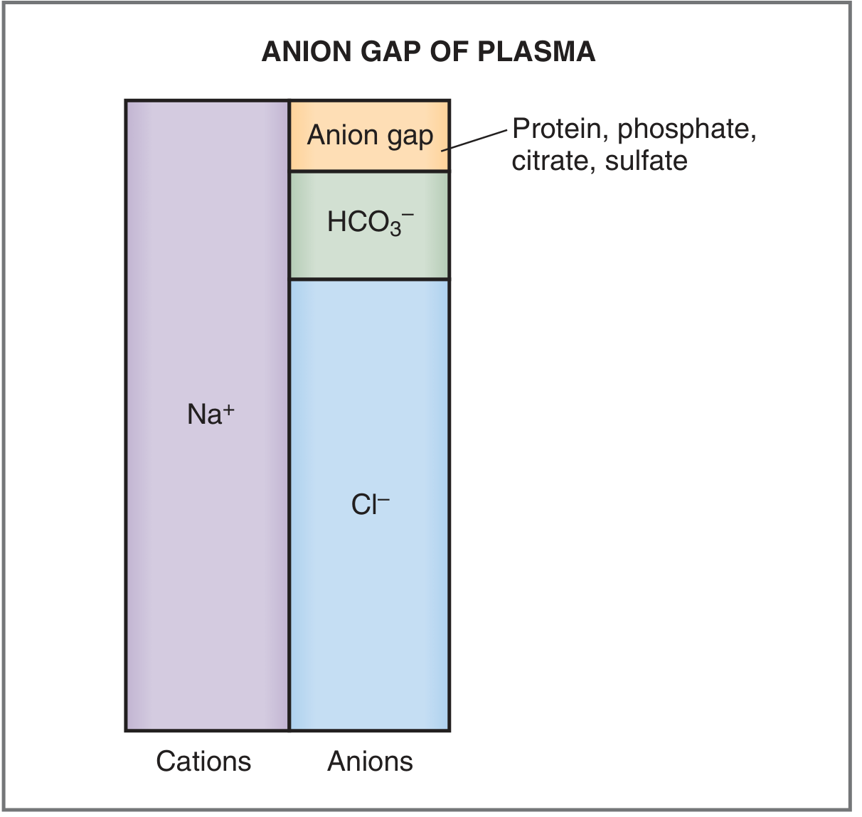
The anion gap = unmeasured anions (plasma proteins, phosphate, citrate, sulfate).
High Anion Gap Metabolic Acidosis (HAGMA)
Mnemonic: MUDPILES (or GOLDMARK)
- M - Methanol
- U - Uremia (chronic renal failure)
- D - Diabetic ketoacidosis (DKA)
- P - Propylene glycol / Paracetamol
- I - Iron, Isoniazid
- L - Lactic acidosis
- E - Ethylene glycol
- S - Salicylates
Normal Anion Gap Metabolic Acidosis (NAGMA / Hyperchloremic)
Mnemonic: HARDUPS
- H - Hyperalimentation
- A - Addison's disease
- R - Renal tubular acidosis (RTA)
- D - Diarrhea
- U - Ureteroenteric fistula
- P - Pancreatic fistula
- S - Saline excess (dilutional)
Step 5 - Delta-Delta Ratio (for HAGMA)
If there is a high anion gap, use the delta-delta to check for a hidden concurrent metabolic disorder:
Delta-Delta = (AG - 12) / (24 - HCO3-)
| Ratio | Interpretation |
|---|---|
| < 0.4 | Concurrent normal AG metabolic acidosis |
| 0.4 - 1.0 | Mixed HAGMA + NAGMA |
| 1.0 - 2.0 | Pure HAGMA (expected) |
| > 2.0 | Concurrent metabolic alkalosis |
Step 6 - Assess Oxygenation
PaO2 normal: 70-95 mmHg (decreases with age)
PaO2 sitting = 104.2 - (0.27 × age in years)
Alveolar-Arterial (A-a) Gradient:
P(A-a)O2 = [FiO2 × (PB - 47)] - (PaCO2 / 0.8) - PaO2 Normal A-a gradient = (Age/4) + 4
| A-a Gradient | Mechanism of Hypoxemia |
|---|---|
| Normal | Hypoventilation, high altitude |
| Increased | V/Q mismatch, diffusion impairment, shunt |
Summary Table of Four Simple Disorders
| Disorder | pH | PaCO2 | HCO3- | Respiratory Compensation | Renal Compensation |
|---|---|---|---|---|---|
| Metabolic Acidosis | ↓ | ↓ | ↓ | Hyperventilation | ↑ HCO3- reabsorption |
| Metabolic Alkalosis | ↑ | ↑ | ↑ | Hypoventilation | ↑ HCO3- excretion |
| Respiratory Acidosis | ↓ | ↑ | ↑ | None | ↑ HCO3- reabsorption |
| Respiratory Alkalosis | ↑ | ↓ | ↓ | None | ↓ HCO3- reabsorption |
(Bold = primary disturbance)
A Quick Note on Venous Blood Gases (VBG)
VBGs are increasingly used as a safer alternative. Venous pH averages ~0.03 lower than arterial. PvCO2 runs 3-8 mmHg higher than PaCO2 and correlates less reliably. VBGs are unreliable in hypotensive patients or severe hypercapnia, but a normal PvCO2 effectively excludes hypercapnia.
Sources: Costanzo Physiology 7th Ed., Goldman-Cecil Medicine
This is a shared conversation. Sign in to Orris to start your own chat.