
Systemic lupus erythematosus in brief for famous questions in an exam.
systemic lupus erythematosus butterfly rash malar rash
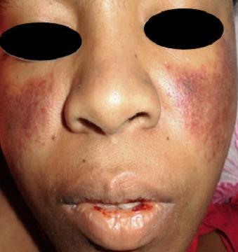
A clinical photograph of a patient's face demonstrating dermatological and mucosal manifestations of Systemic Lupus Erythematosus (SLE). A classic malar rash (butterfly rash) is present, characterized by symmetric, erythematous-to-violaceous patchy lesions over the malar eminences and the bridge of the nose, notably sparing the nasolabial folds. Additionally, the perioral region exhibits significant erythema and mucosal involvement. The lips show evidence of hemorrhagic lesions, with visible blood crusting and a small fissure on the lower lip, suggestive of vasculitis or active systemic inflammation. These visual findings are key diagnostic indicators for SLE, particularly when associated with hematological abnormalities such as thrombocytopenia. The photograph provides a clear example of cutaneous lupus manifestations for clinical diagnosis and medical education.
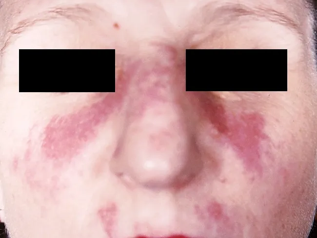
This clinical dermatology photograph displays the classic malar or butterfly rash of systemic lupus erythematosus (SLE) on the central face. Modality is clinical photography using standard white-light illumination; frontal/anterior view; color-balanced, high-resolution capture to depict superficial erythema and patchy hyperemia across the malar eminences and nasal bridge. The rash forms a bilateral, 'butterfly' distribution that typically reaches the cheeks and bridge of the nose while sparing the nasolabial folds. The observed features include confluent to patchy, erythematous macules and plaques with uniform erythema, mild perivascular edema, and subtle textural change without overt crusting or scaling in this image. The clinical morphology is characteristic for acute cutaneous lupus erythematosus; photosensitivity may exacerbate lesions. This cutaneous finding is one of the diagnostic criteria for SLE when aligned with serologic abnormalities (ANA, anti-dsDNA) and systemic features; its presence increases diagnostic probability in a compatible patient. Differential considerations include rosacea, seborrheic dermatitis, contact dermatitis, and dermatomyositis rash; however, the malar distribution and nasal bridge involvement help distinguish lupus. Clinically, this image supports SLE workup and educational reference for recognizing lupus-associated facial rash in medical students, residents, and researchers; useful for pattern-recognition training and multimodal data repository indexing. This image emphasizes clinical-context interpretation and education.
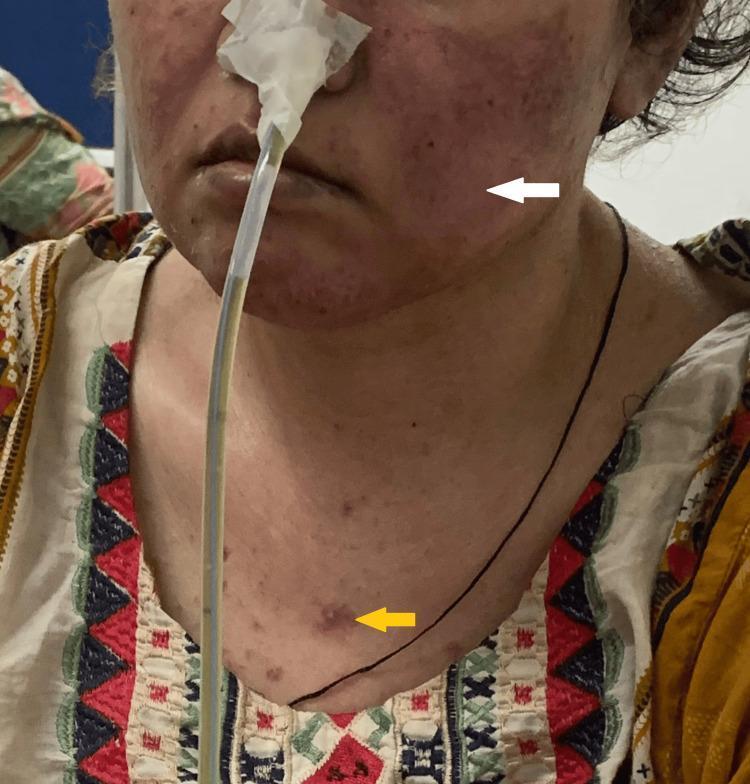
Clinical photograph of a female patient presenting with characteristic dermatological manifestations of Systemic Lupus Erythematosus (SLE). A white arrow points to a malar rash, also known as a butterfly rash, which is characterized by fixed erythema and slight edema across the bridge of the nose and the malar eminences of the cheeks, typically sparing the nasolabial folds. A yellow arrow indicates a discoid rash on the upper chest, appearing as a well-demarcated, erythematous, raised papule/plaque with a slightly hyperpigmented or scarred center. The patient also has a nasogastric tube in place, secured with tape, and appears to have thin hair, potentially representing non-scarring alopecia. The clinical significance of these visual findings is their role as diagnostic criteria in rheumatology for SLE. The presentation is suitable for intermediate to advanced medical education regarding autoimmune connective tissue diseases and cutaneous lupus manifestations.
lupus nephritis histology wire loop lesions glomerulonephritis
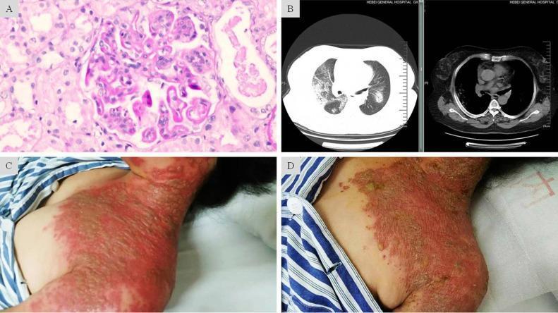
A composite medical image featuring four panels (A-D) illustrating clinical, radiological, and pathological findings of a complex case involving Lupus Nephritis and severe Herpes Zoster. Panel A is a light microscopy image (H&E stain) of a renal biopsy showing glomerular endothelial and mesangial hypercellularity with characteristic 'wire-loop' lesions, consistent with Class IV-G Lupus Nephritis. Panel B presents axial chest CT scans (lung and mediastinal windows) displaying bilateral, scattered patchy and nodular opacities with ill-defined borders, suggestive of diffuse alveolar hemorrhage. Panels C and D are clinical photographs of the neck and chest region showing the progression of skin lesions. Panel C depicts the acute phase of Herpes Zoster, characterized by confluent, grouped vesicles on an intense erythematous base extending across multiple dermatomes. Panel D shows the same region post-antiviral treatment, demonstrating resolving lesions with dried-up exudate and crusting on a fading erythematous base. This figure serves as an educational resource for multi-organ manifestations in immunocompromised patients, specifically highlighting systemic lupus erythematosus complications and disseminated viral infection.
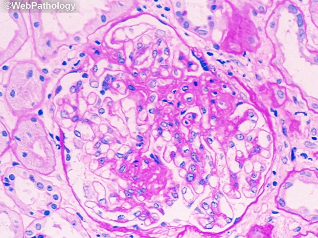
Imaging modality: light microscopy of renal tissue. A single glomerulus is shown in cross section from a renal cortical biopsy. The specimen is stained with Hematoxylin and Eosin (H&E) and viewed at high magnification. The glomerular tuft demonstrates increased cellularity within the capillary loops, consistent with endocapillary proliferation. Mesangial matrix appears expanded, and there are scattered infiltrating cells in the capillary lumens. Capillary lumina are variably narrowed, suggesting early glomerular inflammation. The basement membranes are not clearly visible as thickened in this field, and no overt segmental sclerosis or crescent formation is evident in this slice. The surrounding renal parenchyma shows mild tubulointerstitial involvement, including tubules with pale cytoplasm and interstitial edema, but the primary focus is the glomerulus. The staining highlights nuclei in blue and the extracellular matrix and cytoplasm in pink, typical of routine H&E preparation. This histopathology raises suspicion for a proliferative glomerulonephritis pattern, which can be seen in diseases such as postinfectious GN, IgA nephropathy, or lupus nephritis depending on ancillary tests. Clinically, findings may include hematuria, variable proteinuria, and potential decline in renal function; biopsy helps classify severity and guide therapy. Documentation of periglomerular inflammation and capillary loop involvement supports timely treatment decisions and assessment.
Systemic Lupus Erythematosus (SLE) - Exam High-Yield Summary
1. Definition & Epidemiology
- SLE is a systemic autoimmune disease of unclear etiology characterized by autoantibody production and multi-organ involvement with a relapsing-remitting course.
- Sex ratio: F:M = 9:1 during childbearing years; 2:1 before puberty; ratio narrows after menopause.
- Peak onset: 15-45 years (childbearing age).
- Race: More prevalent and severe in Black, Hispanic, and Asian women (2-4x higher than White women).
- Prevalence: ~100-150 per 100,000; incidence ~4-6 per 100,000/year.
2. Pathogenesis (High-Yield)
- Core defect: Defective clearance of apoptotic cells → nuclear material (DNA, histones, RNA-binding proteins) persists → acts as immunostimulatory antigen.
- Type I interferon axis: A key pathogenic driver; pDCs produce excess IFN-α triggered by nucleic acid-containing immune complexes via TLR signaling.
- Autoantibodies + immune complexes: Deposit in kidneys (glomerulus), skin, joints, blood vessels → complement activation → inflammation.
- Key autoimmune "triad": Loss of tolerance → autoantibody production → immune complex deposition → tissue injury.
- Genetics: HLA-DR2, HLA-DR3; complement deficiencies (C1q, C2, C4 - especially C1q is the strongest genetic risk factor).
- Environmental triggers: UV light, EBV infection, silica dust, estrogens, certain drugs.
3. Clinical Features (by System)
| System | Manifestation | Frequency |
|---|---|---|
| Constitutional | Fatigue, fever, weight loss | 90-95% |
| Mucocutaneous | Malar (butterfly) rash, discoid rash, photosensitivity, oral ulcers, alopecia | 70-80% |
| Musculoskeletal | Non-erosive arthritis/arthralgia | 75-90% |
| Renal | Lupus nephritis (proteinuria, hematuria, casts) | 50-60% |
| Hematologic | Hemolytic anemia, leukopenia, lymphopenia, thrombocytopenia | 50-65% |
| Serositis | Pleuritis, pericarditis | 40-60% |
| Neuropsychiatric | Seizures, psychosis, cognitive dysfunction, headache | 25-50% |
| Vascular | Raynaud's phenomenon, thrombosis (if aPL+) | 20-30% |
| Cardiac | Libman-Sacks endocarditis (non-infective, sterile verrucae on both sides of mitral valve) | 10-15% |
Mnemonic for ACR Criteria - "SOAP BRAIN MD"
Serositis, Oral ulcers, Arthritis, Photosensitivity, Blood disorders, Renal disorder, ANA, Immunologic (anti-dsDNA/anti-Sm/aPL), Neurologic, Malar rash, Discoid rash
4. Classification Criteria
1997 ACR Criteria (11 criteria; need ≥4 of 11 serially or simultaneously)
| # | Criterion | Key Detail |
|---|---|---|
| 1 | Malar rash | Fixed erythema over malar eminences, spares nasolabial folds |
| 2 | Discoid rash | Erythematous patches, keratotic scale, follicular plugging; may scar |
| 3 | Photosensitivity | Skin rash from unusual reaction to sunlight |
| 4 | Oral ulcers | Usually painless; oral or nasopharyngeal |
| 5 | Arthritis | Non-erosive, ≥2 peripheral joints |
| 6 | Serositis | Pleuritis or pericarditis |
| 7 | Renal disorder | Proteinuria >0.5 g/day or cellular casts |
| 8 | Neurologic disorder | Seizures or psychosis (without other cause) |
| 9 | Hematologic disorder | Hemolytic anemia, leukopenia <4000, lymphopenia <1000, or thrombocytopenia <100,000 |
| 10 | Immunologic | Anti-dsDNA, anti-Sm, or antiphospholipid antibodies |
| 11 | ANA | Positive ANA by immunofluorescence |
2012 SLICC Criteria
- 11 clinical + 6 immunologic criteria; classified if ≥4 criteria (≥1 clinical + ≥1 immunologic), OR biopsy-proven lupus nephritis + ANA/anti-dsDNA.
- More sensitive (97% vs 83%) but less specific (84% vs 96%) than 1997 ACR.
2019 ACR/EULAR Criteria
- Requires ANA ≥1:80 as entry criterion (mandatory gate).
- Weighted scoring across 7 clinical + 3 immunologic domains; score ≥10 = SLE.
- Best balance of sensitivity and specificity for early SLE.
5. Autoantibodies (Most Exam-Tested)
| Antibody | Frequency | Clinical Significance |
|---|---|---|
| ANA | 95-99% | Highly sensitive; screening test (negative ANA essentially rules out SLE) |
| Anti-dsDNA | 70% | High specificity for SLE; levels correlate with disease activity (especially nephritis) |
| Anti-Sm (Smith) | 25-38% | Most specific for SLE (low sensitivity, high specificity) |
| Anti-Ro/SSA | 49% | Neonatal lupus, congenital heart block, subacute cutaneous lupus (SCLE) |
| Anti-La/SSB | 35% | Seen with anti-Ro; protective against nephritis |
| Anti-histone | ~70% | Drug-induced lupus (but not exclusive) |
| Antiphospholipid (aPL) | 21% | Thrombosis, recurrent miscarriages (antiphospholipid syndrome) |
| Anti-ribosomal P | 10% | Neuropsychiatric lupus (psychosis) |
Key mnemonics: Anti-dsDNA = disease activity; Anti-Sm = specific; Anti-Ro = neonatal/SCLE; Anti-histone = drug-induced.
6. Lupus Nephritis - ISN/RPS Classification
| Class | Name | Light Microscopy | Notes |
|---|---|---|---|
| I | Minimal mesangial LN | Normal | Mesangial immune deposits on IF only |
| II | Mesangial proliferative LN | Mesangial hypercellularity | Good prognosis; no specific Rx needed |
| III | Focal LN | <50% glomeruli involved | Active (A), chronic (C), or mixed |
| IV | Diffuse LN | ≥50% glomeruli involved | Most severe; worst prognosis; "wire-loop" lesions classic |
| V | Membranous LN | Subepithelial deposits | Heavy proteinuria/nephrotic syndrome |
| VI | Advanced sclerosing LN | ≥90% global sclerosis | End-stage; irreversible |
7. Lab Findings
- ANA: Positive >95%; gold standard = HEp-2 cell indirect immunofluorescence.
- Anti-dsDNA: Rises with disease flares; correlates with nephritis activity.
- Complement (C3, C4, CH50): Low during active disease (consumed by immune complexes) - classic exam finding.
- CBC: Leukopenia, lymphopenia, thrombocytopenia, hemolytic anemia (Coombs positive).
- Urinalysis: Proteinuria, hematuria, red cell casts (nephritis).
- False-positive VDRL/RPR: Due to antiphospholipid antibodies (lupus anticoagulant).
8. Drug-Induced Lupus (DIL)
- Common culprits (mnemonic "SHIPP"): Hydralazine, Procainamide, Isoniazid, Phenytoin, Minocycline, quinidine, methyldopa.
- Key features: Anti-histone antibodies (>95%); ANA positive; anti-dsDNA usually negative; renal/CNS involvement rare; resolves on stopping drug.
9. Neonatal Lupus
- Due to maternal anti-Ro (SSA) and anti-La (SSB) antibody transfer.
- Features: Transient skin rash, cytopenias, liver disease.
- Most serious: Congenital complete heart block (3rd degree AV block) - permanent, requires pacemaker.
- Hydroxychloroquine may reduce risk of recurrent heart block.
10. SLE in Pregnancy
- Increased risk of: preeclampsia, preterm birth, IUGR, fetal loss, neonatal lupus.
- Disease may flare during pregnancy and postpartum.
- Active lupus nephritis = contraindication to pregnancy.
- Hydroxychloroquine: Safe in pregnancy; continued throughout (reduces flares).
- Avoid: Cyclophosphamide, mycophenolate (teratogenic), methotrexate.
11. Treatment
Step-up approach:
| Severity | Treatment |
|---|---|
| All patients | Hydroxychloroquine (HCQ) 200-400 mg/day - reduces flares, prevents damage, cardiovascular protection; mandatory for all SLE patients without contraindication |
| Mild disease | HCQ ± NSAIDs ± low-dose glucocorticoids |
| Moderate disease | Glucocorticoids (0.5 mg/kg/day) + immunosuppressive (azathioprine, methotrexate) |
| Severe/organ-threatening | High-dose GC ± IV cyclophosphamide (CYC) or mycophenolate mofetil (MMF) |
| Refractory | Rituximab, belimumab, voclosporin, anifrolumab |
Biologic therapy:
- Belimumab (anti-BLyS/BAFF): First biologic approved for SLE; used in moderate-to-active disease on standard therapy.
- Anifrolumab (anti-IFN-α receptor): Approved as add-on for moderate-to-severe SLE (targets the key Type I IFN pathway).
- Voclosporin + MMF: Approved for active lupus nephritis (2021).
Lupus Nephritis Treatment:
- Class I/II: No specific immunosuppression; manage as primary disease.
- Class III/IV (proliferative): High-dose GC + MMF (preferred) or IV CYC (Euro-Lupus low-dose regimen).
- Class V (membranous): MMF or calcineurin inhibitors.
- All LN patients: Hydroxychloroquine + RAAS blockade (ACEi/ARB) for renoprotection.
12. Monitoring Disease Activity
- SLEDAI (SLE Disease Activity Index) - standard scoring tool.
- Rising anti-dsDNA + falling complement = impending flare.
- Regular urinalysis, CBC, renal function, complement levels.
Classic Exam Vignette Clues
| Clue | Think |
|---|---|
| Young woman + butterfly rash + arthritis + proteinuria | SLE |
| ANA positive + anti-dsDNA positive + low C3/C4 | Active SLE |
| Painless oral ulcers + photosensitivity | SLE |
| Positive ANA + anti-histone + no renal/CNS disease | Drug-induced lupus |
| Recurrent miscarriages + thrombocytopenia + prolonged aPTT | Antiphospholipid syndrome (secondary to SLE) |
| Neonatal complete heart block | Maternal anti-Ro antibody |
| "Wire-loop" lesion on renal biopsy | Class IV diffuse lupus nephritis |
| Libman-Sacks endocarditis | SLE (both sides of mitral valve, sterile verrucae) |
| False positive VDRL | Antiphospholipid antibodies in SLE |