
Medicine 💊 for the treatment of the folliculitis
folliculitis treatment
folliculitis clinical presentation skin
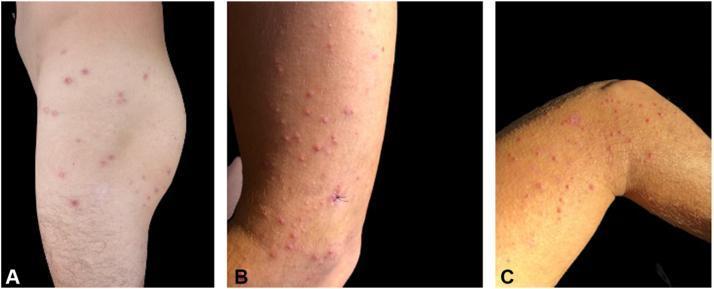
Clinical photographs of the trunk and lower extremities (thigh, knee, and calf) demonstrating a disseminated eruption characterized by multifocal skin lesions. The images show a polymorphous presentation including erythematous papules, yellow-reddish pustules, and shallow, punched-out ulcers. The pustules exhibit a follicular distribution, suggesting an underlying abscessing folliculitis. The ulcers are noted for their depressed centers and thick, red-brown crusting (eschar-like appearance), each surrounded by a distinct halo of erythema. The lesions are scattered across the integument with some areas showing higher density near the knee and thigh. A surgical suture is visible on the calf in Panel B, indicating a biopsy site. This visual presentation is consistent with a generalized infectious folliculitis, specifically Pantoea dispersa-associated folliculitis in the context of environmental exposure.
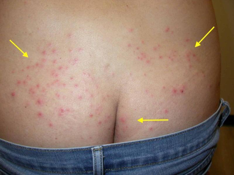
This clinical photograph shows a posterior view of the lower back and upper buttocks of a patient, demonstrating a widespread inflammatory skin eruption. The primary lesions are small, erythematous papules and pustules, some of which exhibit a central punctum or follicular orientation. The distribution is bilateral but asymmetrical, with significant clustering along the lower back and superior gluteal regions. Yellow arrows highlight representative clusters of these lesions. The distribution pattern corresponds to areas typically occluded by clothing, such as swimwear or the waistband of the denim jeans visible in the frame. The morphology and clinical presentation are highly suggestive of folliculitis, specifically 'hot tub folliculitis' (Pseudomonas folliculitis) given the context of occlusion and characteristic inflammatory papulopustular appearance. This visual serves as an educational example of bacterial folliculitis and the importance of lesion distribution in dermatological diagnosis.
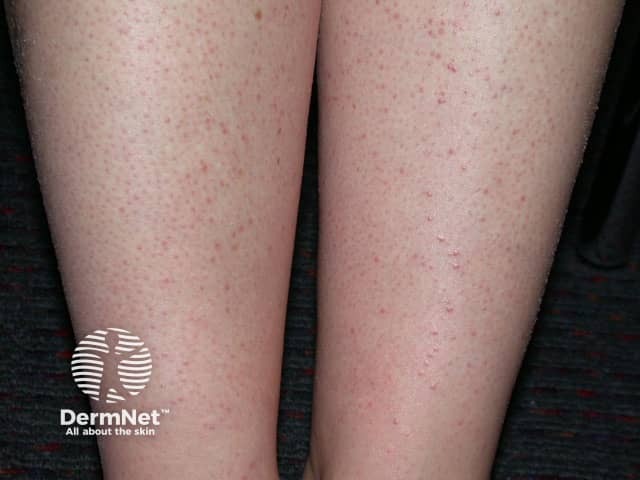
Clinical photography of exposed lower legs demonstrates a diffuse eruption consistent with folliculitis secondary to hair removal. The images depict bilateral shins with numerous small, erythematous papules and scattered pustules centered on hair follicles, surrounded by mild perifollicular erythema. The lesions are superficial and discrete, without obvious abscess formation or systemically abnormal findings. Hair shafts appear present; irritation and microtrauma from shaving or waxing likely initiated the perifollicular inflammation. The pattern is predominantly follicular, with a uniform distribution along anterior leg surfaces, suggestive of irritant contact folliculitis rather than a deep infectious process. The skin surface shows no crusting beyond minor scale in some papules, consistent with mild inflammatory response. In the clinical context, this presentation aligns with post-hair-removal folliculitis and may resolve with conservative management or topical antiseptics, exfoliation, and avoidance of aggressive hair removal. Secondary bacterial infection should be considered if pustules enlarge or purulence increases, warranting antibiotic therapy. The image serves as an educational reference for recognizing common dermatologic sequelae of depilation techniques and for differentiating folliculitis from keratosis pilaris, acneiform eruptions, or contact dermatitis. It supports patient counseling on shaving methods, skin prep, and timely treatment.
Treatment of Folliculitis
Clinical Images
1. Bacterial Folliculitis (Staphylococcus aureus - most common)
Mild / Superficial cases
| Drug | Route | Details |
|---|---|---|
| Mupirocin ointment | Topical | Antistaphylococcal; also applied to anterior nares (daily x 5 days) to reduce carriage and prevent recurrence |
| Clindamycin solution | Topical | Twice daily for at least 2 weeks |
| Bacitracin ointment | Topical | Applied to individual pustules |
| Retapamulin ointment | Topical | Pleuromutilin antibiotic; effective against S. aureus |
| Benzoyl peroxide washes | Topical wash | Antimicrobial; helps decolonize skin surface |
| Chlorhexidine washes | Topical wash | Antiseptic; use daily to reduce carriage |
| Dilute sodium hypochlorite baths | Topical bath | 1/2 cup bleach in 40-gallon tub; decolonizes skin |
Moderate-to-severe / Extensive cases
| Drug | Dose | Duration |
|---|---|---|
| Cephalexin | 500 mg twice daily | 14 days |
| Dicloxacillin | Standard oral dose | 10-14 days |
| Azithromycin | Standard dose | 5-7 days (for penicillin allergy) |
Note: Culture of pustule contents is recommended for extensive infection to guide antibiotic selection, especially if MRSA is suspected. - Goldman-Cecil Medicine, p. 4298
2. Pseudomonas Folliculitis ("Hot Tub Folliculitis")
- Usually self-limited in healthy individuals; resolves with antibacterial soap and stopping exposure.
- Twice-daily cleansing with mild hand soap + warm compresses suffice for most cases.
- If lesions persist beyond 5 days or are extensive:
| Drug | Dose | Duration |
|---|---|---|
| Ciprofloxacin | 500 mg twice daily | 10-14 days |
The hot tub must also be drained and cleaned. - Textbook of Family Medicine 9e, p. 937
3. Pityrosporum (Malassezia) Folliculitis - Fungal
| Drug | Dose | Duration |
|---|---|---|
| Topical antifungal cream (ketoconazole, clotrimazole) | Topical | 2-4 weeks |
| Selenium sulfide shampoo | Daily application | 1 month |
| Itraconazole | 200 mg daily | 1 week |
| Fluconazole | 100-200 mg daily, OR 300 mg once weekly | 1-4 weeks OR 1-2 months |
- Goldman-Cecil Medicine, p. 4298
4. Eosinophilic Pustular Folliculitis (HIV-associated)
| Drug | Dose | Notes |
|---|---|---|
| Antihistamines | Standard doses | First-line symptomatic relief |
| Topical corticosteroids | Apply to lesions | Anti-inflammatory |
| Topical tacrolimus | Apply to lesions | Calcineurin inhibitor |
| Indomethacin | 50 mg/day | Systemic NSAID option |
| Minocycline | 100 mg twice daily | Systemic antibiotic with anti-inflammatory effect |
| Dapsone | 100-200 mg/day | For refractory cases |
| Colchicine | 0.6 mg twice daily | Anti-inflammatory |
| Systemic corticosteroids | Variable | Severe/refractory |
| Narrowband UVB phototherapy | - | Useful adjunct |
- Goldman-Cecil Medicine, p. 4298
5. Sycosis Barbae / Beard Folliculitis
- Antistaphylococcal topical antibiotics (mupirocin, clindamycin)
- Systemic antibiotics for persistent/deep disease (cephalexin, dicloxacillin)
- Avoidance of shaving in the affected area until resolved
Summary Table: Drug Choice by Type
| Folliculitis Type | First-Line Treatment |
|---|---|
| Staph (mild) | Topical mupirocin, clindamycin, or benzoyl peroxide wash |
| Staph (moderate/severe) | Cephalexin 500 mg BD x 14 days |
| Pseudomonas (hot tub) | Self-limited; ciprofloxacin 500 mg BD if persistent |
| Malassezia (fungal) | Topical antifungal or oral fluconazole/itraconazole |
| Eosinophilic (HIV) | Antihistamines + topical steroids; systemic options for severe cases |
General Supportive Measures (All Types)
- Warm compresses applied several times daily to reduce inflammation
- Avoid shaving in involved areas
- Antibacterial soap for daily cleansing
- Treat underlying predisposing factors (obesity, diabetes, immunosuppression)
- Mupirocin to anterior nares daily x 5 days + bleach baths to prevent recurrence
- Andrews' Diseases of the Skin, Clinical Dermatology (Elsevier), pp. 4698-4700
- Goldman-Cecil Medicine, International Edition, pp. 4298-4299
- Tintinalli's Emergency Medicine, p. 207
- Textbook of Family Medicine 9e, p. 937
- Recent systematic review: Management of Folliculitis Decalvans (2025, Sideris et al.) - PMID 41146582 - found antibiotics and corticosteroids remain mainstay for the scarring subtype