
Helminthic disease
helminthic disease parasitic worms lifecycle
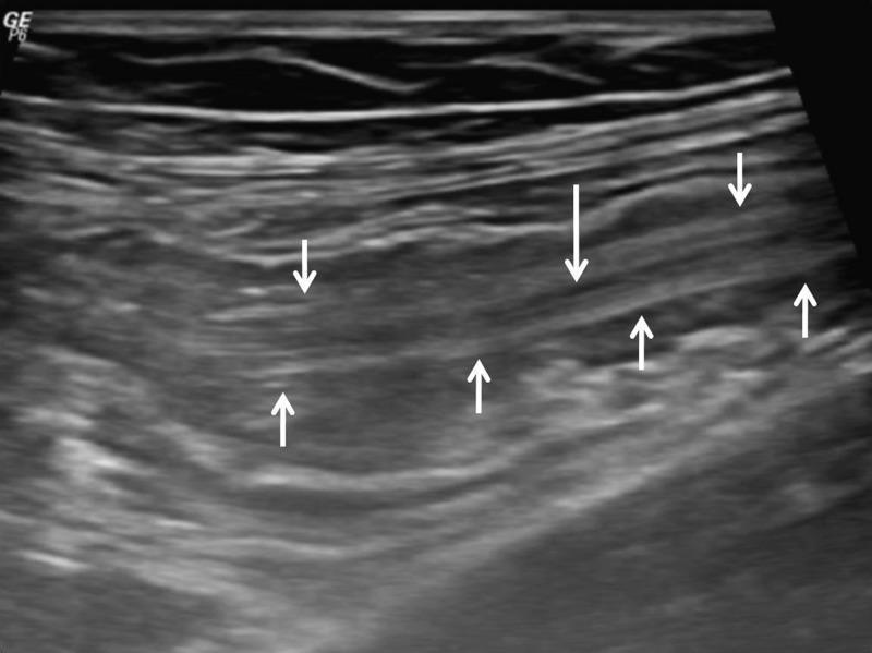
This diagnostic image is an abdominal ultrasound scan in longitudinal view, demonstrating classic sonographic signs of intestinal helminthic infestation, specifically Ascaris lumbricoides. The image reveals multiple tubular, echogenic structures within the intestinal lumen. These structures exhibit a characteristic 'railway tract' appearance, defined by parallel echogenic lines representing the worm's outer wall. A central, longitudinal anechoic line is visible between these walls (indicated by the long white arrow), representing the parasite's digestive tract. Notably, there is an absence of posterior acoustic shadowing, which helps differentiate these soft-bodied parasites from calcified structures or gallstones. The small white arrows highlight the distinct parallel margins of the worms. This imaging finding is pathognomonic for ascariasis in the appropriate clinical context and is essential for diagnosing parasitic causes of bowel obstruction or intussusception. The content is suitable for medical students and radiology residents studying gastrointestinal pathology and tropical medicine.
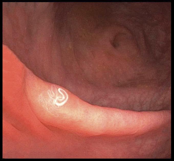
This diagnostic clinical photograph captures an endoscopic view of the colon during a colonoscopy, specifically at the terminal ileum and cecum. The image demonstrates a parasitic infection within the gastrointestinal tract. Visible on the healthy, pinkish-red mucosal surface are multiple thread-like worms, identified in clinical context as Hymenolepis nana. These parasites appear as bright white, slender, and translucent organisms exhibiting both linear and tightly coiled configurations. The underlying intestinal mucosa shows no overt signs of secondary pathology such as severe inflammation, ulceration, or mucosal bleeding. This image illustrates a case of helminthic infestation, which in this clinical setting relates to opportunistic reactivation in an immunocompromised patient. The visual is of high educational value for gastroenterology and infectious disease specialties, providing a clear demonstration of macroscopically visible intestinal parasites during direct visualization.
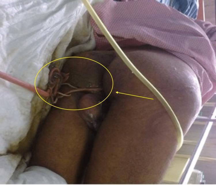
Clinical photograph of a patient in a medical setting demonstrating a severe helminthic infestation. The image captures the perineal and gluteal region, where several live, light-brown, tubular parasites, identified as Ascaris lumbricoides, are seen emerging from the anal opening. The worms exhibit a cylindrical, elongated morphology and appear coiled or intertwined. The primary area of focus is demarcated by a yellow circle and indicated by a yellow arrow. Surrounding medical context includes clinical draping, a pinkish flexible tube (possibly a urinary catheter or drainage line), and a thicker yellow tube running across the patient's right thigh. This visual demonstrates the clinical manifestation of ascariasis, specifically the spontaneous migration of adult nematodes from the gastrointestinal tract during systemic illness. The finding is relevant for infectious disease and gastroenterology specialties, illustrating complications of heavy worm burden and intestinal migration.
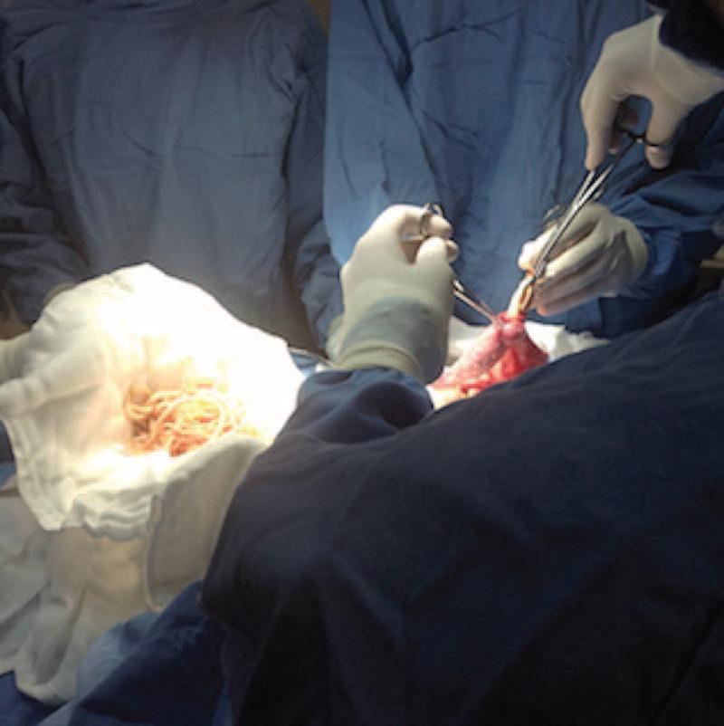
This clinical photograph captures an intraoperative scene during a surgical procedure for intestinal obstruction caused by Ascariasis. The image shows a surgeon and surgical team in sterile blue gowns and gloves performing a laparotomy. The primary focus is on a segment of small intestine (jejunum/ileum) being manipulated with surgical forceps. Adjacent to the surgical field, a large cluster of extracted helminths (roundworms, likely Ascaris lumbricoides) is visible on a white surgical gauze. These worms are numerous, long, cylindrical, and off-white to light tan in color, appearing as a tangled mass. The image demonstrates the surgical technique of milking or manually extracting helminths from the bowel lumen to resolve a mechanical obstruction. This visual serves as a critical educational example of parasitic infestation presenting as an acute surgical abdomen, illustrating the severity of helminthic load that can occur in pediatric patients in endemic regions.
schistosomiasis blood fluke lifecycle eggs granuloma
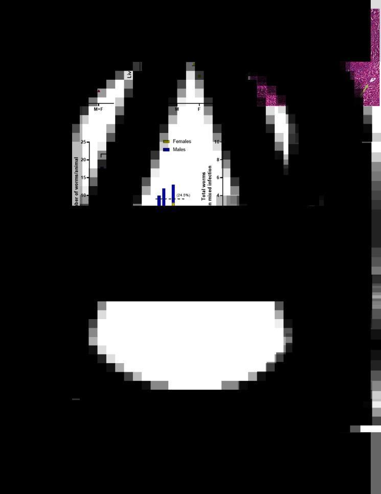
This composite educational image illustrates the pathophysiology and quantitative data associated with Schistosoma mansoni infection in a mouse model. (A-B) Scatter plots show mouse body weight and liver weight across control, male-only (M), female-only (F), and mixed infection (M+F) groups, highlighting significant hepatomegaly in the mixed infection group. (C) A high-magnification (scale bar 25 μm) histopathological micrograph of liver tissue with H&E staining reveals a classic granulomatous reaction. Arrows denote trapped S. mansoni eggs within the central area of the granuloma, surrounded by inflammatory cell infiltration. (D) A bar chart quantifies the number of adult worms recovered per animal, showing higher recovery rates in the male and mixed groups. (E) A scatter plot displays the correlation between the number of recovered male (blue triangles) and female (yellow triangles) worms and the burden of eggs per gram of liver tissue. This visual material is used to teach parasitic disease manifestations, host inflammatory response, and the experimental methodology of schistosomiasis research.
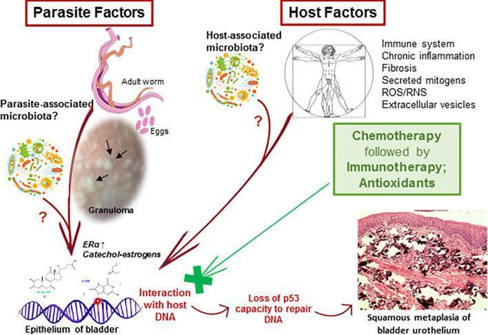
A pathophysiology diagram and clinical imaging composite illustrating the carcinogenesis pathway of urogenital schistosomiasis-induced bladder cancer. The visual is divided into 'Parasite Factors' and 'Host Factors'. Parasite factors include an illustration of an adult Schistosoma worm, pink oval eggs, a parasite-associated microbiota, and a clinical photograph of a granuloma with internal calcifications (indicated by black arrows). Host factors list immune system status, chronic inflammation, fibrosis, ROS/RNS, and host-associated microbiota. The central mechanism depicts parasite-derived catechol estrogens and mitogens interacting with host DNA within the bladder epithelium. This leads to the 'loss of p53 capacity to repair DNA', eventually progressing to the 'Squamous metaplasia of bladder urothelium', shown in a bottom-right histopathology image. The histopathology displays typical features of metaplastic transformation with a dense, pink-stained keratinized surface and irregular cellular infiltration. A green box highlights therapeutic interventions including chemotherapy, immunotherapy, and antioxidants, symbolized by a green cross blocking the DNA-interaction pathway.
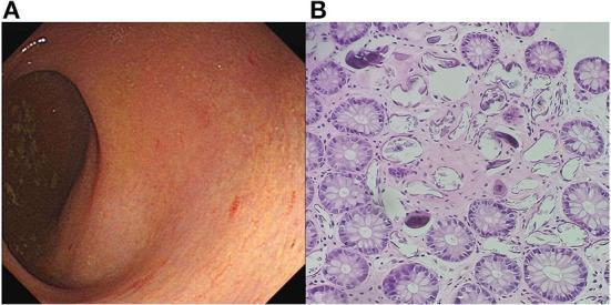
The image consists of two panels illustrating clinical and pathological findings of intestinal schistosomiasis. Panel A is a colonoscopy image showing the enteric mucosa of the rectum. It displays characteristic yellow-white plaque formations and nodules beneath the mucosal surface, accompanied by a blurred or obscured submucosal blood vessel network, indicating a chronic inflammatory response. Panel B is a high-power photomicrograph (200x) of a histological section stained with hematoxylin and eosin (H&E). It reveals numerous transverse and longitudinal sections of colonic crypts and glands. Within the surrounding lamina propria, there are multiple calcified eggs of Schistosoma japonicum, visible as distinct, darkly stained, rounded, or oval bodies. These findings demonstrate the typical pathological manifestation of schistosomiasis in the gastrointestinal tract, where egg deposition triggers localized tissue reactions.
hookworm skin penetration larva migrans
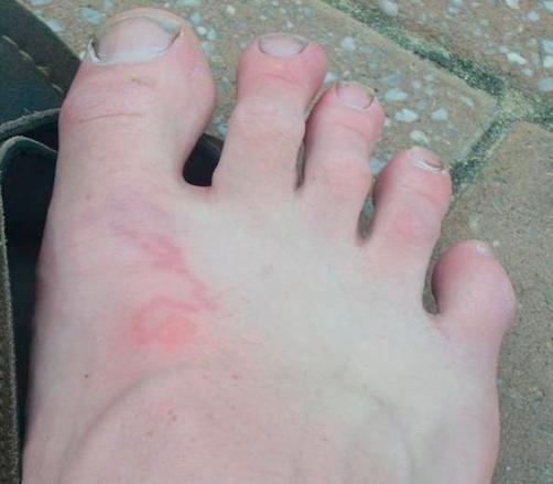
This clinical photograph shows the dorsum of a right foot of a fair-skinned individual, illustrating a classic presentation of Cutaneous Larva Migrans. The primary lesion is a thin, erythematous, and raised serpiginous (snake-like) track that winds across the mid-dorsal surface. The track is approximately 2-3 mm wide and exhibits a migratory, linear, and wavy morphology characteristic of a creeping eruption caused by hookworm larvae. At one end of the track, there is a more concentrated circular erythematous patch, possibly representing the initial site of larval penetration or a localized inflammatory response. The surrounding skin appears normal without secondary infection signs like purulence or significant edema. The toes, nails, and general skin texture are visible and unremarkable except for the parasitic track. This image is an educational example of tropical dermatology, highlighting a diagnostic visual sign for a parasitic infestation typically acquired through contact with contaminated sand or soil.
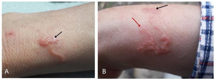
This composite clinical photograph displays cutaneous larva migrans (CLM) on the dorsum of the right forearm. Image A depicts the initial presentation of a characteristic serpiginous (snake-like) skin lesion. The tract is erythematous, elevated, and shows evidence of vesiculobullous formation with small vesicles along the inflammatory path. A black arrow indicates the presumed site of larval penetration where the inflammation is comparatively less intense. Image B illustrates the lesion's progression and response to ivermectin treatment. It shows the continued serpiginous path with varying degrees of erythema and edema. A red arrow marks the advancing, more inflamed endpoint of the burrowing larva, while the black arrow points toward the older, resolving portion of the tract. The visual findings are pathognomonic for hookworm-related cutaneous larva migrans (Hr-CLM), often caused by Ancylostoma species. This educational material is suitable for dermatology and infectious disease studies, demonstrating the clinical morphology of parasitic skin infestations and their migratory patterns.
Helminthic Diseases - A Comprehensive Overview
Classification
| Group | Shape | Examples |
|---|---|---|
| Nematodes | Nonsegmented roundworms | Ascaris, hookworm, Strongyloides, Trichuris, Enterobius, filariae |
| Cestodes | Segmented flatworms (tapeworms) | Taenia solium, T. saginata, Echinococcus, Hymenolepis |
| Trematodes | Unsegmented flatworms (flukes) | Schistosoma, Fasciola, Clonorchis, Paragonimus |
I. NEMATODE (ROUNDWORM) INFECTIONS
1. Ascariasis (Ascaris lumbricoides)
- Life cycle: Eggs ingested → gastric acid dissolves shell → larvae invade mucosa → portal circulation → lung migration (alveolar penetration → bronchial tree ascent → swallowed) → small intestine adults. Females produce >200,000 eggs/day. Adults live 1-2 years.
- Epidemiology: Tropical/subtropical; fecally contaminated soil; children most affected.
- Clinical features:
- Pulmonary phase (Löffler syndrome): transient eosinophilic pneumonitis - cough, wheeze, fever, dyspnea; larvae in sputum
- Intestinal phase: usually asymptomatic; heavy loads → abdominal pain, obstruction, biliary/pancreatic migration (cholangitis, pancreatitis)
- Diagnosis: Stool microscopy for characteristic eggs (65×45 μm); adult worm passage; ultrasound/ERCP for biliary worms
- Treatment: Albendazole 400 mg single dose; mebendazole 500 mg single dose; ivermectin 150-200 μg/kg; pyrantel pamoate 11 mg/kg. Intestinal obstruction → nasogastric piperazine; surgical for complete obstruction.
2. Hookworm (Necator americanus, Ancylostoma duodenale, A. ceylanicum)
- Life cycle: Filariform larvae penetrate bare skin → bloodstream → lung migration (same as Ascaris) → small bowel attachment. Buccal teeth (Ancylostoma) or cutting plates (Necator) attach to mucosa, ingest blood. Eggs shed in stool → soil → rhabditiform → filariform larvae.
- Pathogenesis of disease: Combination of heavy worm burden + prolonged infection + inadequate dietary iron → iron-deficiency anemia + hypoproteinemia
- Clinical features:
- Ground itch: pruritic, maculopapular rash at site of skin penetration
- Löffler syndrome: pulmonary eosinophilia during migration
- Intestinal disease: fatigue, pallor, dyspnea, edema; children suffer cognitive impairment
- Cutaneous larva migrans (CLM): Animal hookworm larvae (A. braziliense) in humans - characteristic serpiginous, creeping eruption
- Diagnosis: Stool microscopy for eggs (60×40 μm, thin-shelled); eosinophilia; low Hb/serum ferritin
- Treatment: Albendazole 400 mg single dose or mebendazole 500 mg; iron supplementation. CLM: ivermectin or albendazole
3. Strongyloides stercoralis (Threadworm)
- Life cycle: Filariform larvae penetrate skin → lung migration → intestinal adult females (parthenogenetic) → produce rhabditiform larvae in stool. Internal autoinfection: rhabditiform → filariform in the colon → reinfect the host.
- Hyperinfection/disseminated strongyloidiasis: In immunosuppressed patients (corticosteroids, HTLV-1, HIV), massive larval migration → bacteremia (gram-negative bacteria carried on larvae through gut wall), meningitis, multi-organ failure. Potentially fatal.
- Clinical features: Larva currens (rapidly migrating urticarial rash), eosinophilia, watery diarrhea, malabsorption
- Diagnosis: Stool O&P (low sensitivity); serology (ELISA); Baermann technique; duodenal aspirate
- Treatment: Ivermectin 200 μg/kg/day × 2 days (drug of choice); albendazole 400 mg bid × 7 days. Hyperinfection: prolonged ivermectin until stool/sputum clear.
4. Trichuris trichiura (Whipworm)
- Anterior whip-like portion embeds in colonic mucosa; posterior thicker "handle" free in lumen
- Heavy infection → trichuris dysentery syndrome: colitis, rectal prolapse, growth retardation
- Diagnosis: stool for barrel-shaped eggs with polar plugs (50×22 μm)
- Treatment: Mebendazole 100 mg bid × 3 days or albendazole 400 mg × 3 days
5. Enterobius vermicularis (Pinworm)
- Most common helminthic infection in developed countries; schoolchildren
- Female migrates to perianal area at night to lay eggs → nocturnal perianal pruritus
- Can cause ectopic sites: appendix, female genitalia
- Diagnosis: Scotch tape test (perianal swab, morning)
- Treatment: Albendazole 400 mg or mebendazole 100 mg once, repeated at 2 weeks (eggs and larvae not killed). Treat entire household.
6. Filarial Infections
| Species | Vector | Disease | Distribution |
|---|---|---|---|
| Wuchereria bancrofti | Culex mosquito | Lymphatic filariasis | Africa, Asia, Americas |
| Brugia malayi/timori | Mansonia mosquito | Lymphatic filariasis | Southeast Asia |
| Onchocerca volvulus | Simulium (blackfly) | River blindness (onchocerciasis) | Sub-Saharan Africa |
| Loa loa | Chrysops (deerfly) | Loiasis | Central/West Africa |
| Dracunculus medinensis | Cyclops (copepod) in water | Guinea worm disease | Sub-Saharan Africa |
- Microfilariae circulate in blood (nocturnal periodicity for W. bancrofti); adult worms in lymphatics
- Chronic: lymphedema, elephantiasis, hydrocele, chyluria
- Tropical pulmonary eosinophilia: hyperresponsiveness to filarial antigens → nocturnal wheeze, high IgE
- Diagnosis: night blood smear for microfilariae; circulating filarial antigen assay; ultrasound (dancing worm sign)
- Treatment: Diethylcarbamazine (DEC) 6 mg/kg/day × 12 days (drug of choice); albendazole; doxycycline (targets Wolbachia endosymbionts); mass drug programs use single-dose albendazole + DEC or ivermectin
- O. volvulus lives in subcutaneous nodules (onchocercoma); microfilariae migrate to eye
- Ocular manifestations: punctate keratitis → sclerosing keratitis → blindness; also anterior and posterior uveitis
- Treatment: Ivermectin 150 μg/kg single dose every 6-12 months (kills microfilariae but not adult worms); doxycycline 6 weeks (kills adult worms via Wolbachia)
II. CESTODE (TAPEWORM) INFECTIONS
1. Taenia saginata (Beef Tapeworm)
- Humans = definitive host; cattle = intermediate host
- Scolex attaches to small intestine; strobila up to 8 m; 1000-2000 proglottids
- Clinical: usually mild - perianal discomfort, proglottid passage, mild abdominal pain
- Diagnosis: stool for gravid proglottids/eggs; 15-30 uterine branches per gravid proglottid (vs 8-12 for T. solium)
- Treatment: Praziquantel 5-10 mg/kg single dose
2. Taenia solium (Pork Tapeworm / Neurocysticercosis)
- Humans can be BOTH definitive host (adult tapeworm from eating undercooked pork) AND intermediate host (cysticercosis from ingesting T. solium eggs - via fecal-oral route, even without eating pork)
- Cysticercosis: Oncospheres from ingested eggs penetrate gut → bloodstream → lodge in brain, muscle, eye, subcutaneous tissue
- Neurocysticercosis: Leading cause of acquired epilepsy in endemic regions. Cysts in brain parenchyma, ventricles, subarachnoid space → seizures, hydrocephalus, headache, focal neurological deficits
- Diagnosis: CT/MRI brain (ring-enhancing lesions, calcifications, racemose cysts); serology (EITB); stool for tapeworm
- Treatment: Albendazole (15 mg/kg/day) or praziquantel + dexamethasone (to reduce inflammatory response as cysts die); antiepileptics; VP shunt for hydrocephalus; surgical excision for ocular/spinal cysts
3. Echinococcosis (Hydatid Disease)
- Life cycle: Adult tapeworm in dog intestine → eggs in feces → humans ingest contaminated food/water → oncospheres released → bloodstream → liver (65%), lungs (25%), other organs → develop into hydatid cysts containing protoscolices
- Clinical features: Slow-growing cysts (years); hepatomegaly, RUQ pain, palpable mass. Cyst rupture → anaphylaxis, peritoneal/pleural seeding, urticaria, eosinophilia. Biliary rupture → cholangitis, obstructive jaundice.
- Diagnosis: Ultrasound/CT - large cyst with "hydatid sand" (floating protoscolices), daughter cysts, peripheral calcification. Serology (ELISA/Weinberg reaction - can be falsely negative in 38%).
- Treatment: PAIR procedure (Puncture-Aspiration-Injection-Reaspiration) with hypertonic saline + albendazole cover; surgical excision; albendazole 400 mg bid × 28-day cycles (medical management only)
III. TREMATODE (FLUKE) INFECTIONS
1. Schistosomiasis (Blood Flukes)
| Species | Disease | Intermediate Host | Region |
|---|---|---|---|
| S. mansoni | Intestinal/hepatosplenic | Biomphalaria snails | Africa, Brazil, Caribbean |
| S. japonicum | Intestinal/hepatosplenic | Oncomelania snails | China, Philippines, Indonesia |
| S. haematobium | Urogenital | Bulinus snails | Africa, Middle East |
| S. mekongi | Intestinal | Neotricula snails | Cambodia, Lao PDR |
- Life cycle: Cercariae released from snails → penetrate human skin → schistosomulae → portal circulation → mature into adult male/female worms → pair and migrate to venules (S. mansoni/japonicum: mesenteric; S. haematobium: vesical plexus) → eggs laid → egg-induced granuloma is the primary pathology
- Clinical phases:
- Cercarial dermatitis (swimmer's itch): pruritic papules 1-3 days post-exposure
- Katayama fever (acute): 4-8 weeks; fever, urticaria, eosinophilia, hepatosplenomegaly - immune complex disease
- Chronic intestinal (S. mansoni/japonicum): portal hypertension, hepatosplenic schistosomiasis, esophageal varices, "Symmer's pipe-stem fibrosis"
- Urogenital (S. haematobium): hematuria (terminal), dysuria, ureteral obstruction; chronic infection → squamous cell carcinoma of bladder
- Diagnosis: Stool/urine for eggs; Kato-Katz technique; rectal snip biopsy; serology; PCR
- Treatment: Praziquantel 40 mg/kg (S. mansoni, haematobium) or 60 mg/kg in divided doses (S. japonicum) - single-day regimen
2. Liver Flukes
| Species | Transmission | Disease |
|---|---|---|
| Clonorchis sinensis | Raw freshwater fish | Biliary obstruction, cholangiocarcinoma |
| Opisthorchis viverrini | Raw freshwater fish | Biliary disease, cholangiocarcinoma (IARC Group 1 carcinogen) |
| Fasciola hepatica | Aquatic plants (watercress) | Fascioliasis (liver rot) |
- Clonorchiasis/Opisthorchiasis: Adult flukes in bile ducts → biliary epithelial hyperplasia → recurrent cholangitis, cholelithiasis, cholangiocarcinoma. Diagnosis: stool for operculate eggs; treatment: praziquantel 75 mg/kg/day in 3 doses × 2 days.
- Fascioliasis: Unique - praziquantel often not effective; treatment is triclabendazole 10 mg/kg (single or 2 doses). Acute phase: fever, RUQ pain, eosinophilia (larval migration through liver parenchyma). Chronic: biliary obstruction.
3. Lung Fluke (Paragonimus westermani)
- Transmission: ingestion of metacercariae in raw freshwater crabs/crayfish
- Adults encyst in lung tissue → hemoptysis, chronic cough, chest pain; may mimic tuberculosis
- Can disseminate to brain, spinal cord, abdomen
- Diagnosis: eggs in sputum/stool; serology; CT (ring-enhancing lesions)
- Treatment: Praziquantel 75 mg/kg/day × 2 days
IV. IMMUNOLOGY OF HELMINTHIC INFECTIONS
- Elevated IgE (total and parasite-specific)
- Eosinophilia (blood and tissue)
- Mast cell/basophil activation
- IL-4, IL-5, IL-13 cytokine profile
- IgE-mediated mast cell degranulation contributes to parasite expulsion from gut
- Paradoxically, helminths may protect against allergic/autoimmune disease (hygiene hypothesis) by skewing toward Th2 and promoting regulatory T cell responses, and by altering gut microbiome composition - Sleisenger and Fordtran's GI & Liver Disease
V. ANTHELMINTIC DRUGS - SUMMARY
- Sherris & Ryan's Medical Microbiology, 8th Ed, Ch. 6
| Drug | Class | Mechanism | Key Indications |
|---|---|---|---|
| Albendazole | Benzimidazole | Inhibits tubulin polymerization → impairs glucose uptake; blocks cytoskeletal function | Broad spectrum: Ascaris, hookworm, Trichuris, Strongyloides, cysticercosis, echinococcosis |
| Mebendazole | Benzimidazole | Same as albendazole | Ascaris, hookworm, Trichuris, Enterobius |
| Ivermectin | Avermectin | Glutamate-gated Cl⁻ channel agonist → hyperpolarization/paralysis of parasite neuromuscular junction | Strongyloides (drug of choice), onchocerciasis, filariasis, scabies, CLM |
| Praziquantel | Isoquinoline | Increases Ca²⁺ permeability → spastic paralysis + tegument disruption | All cestodes, schistosomes, most trematodes (NOT Fasciola) |
| Diethylcarbamazine (DEC) | Piperazine derivative | Immobilizes microfilariae; promotes immune destruction | Lymphatic filariasis (drug of choice), loiasis, tropical pulmonary eosinophilia |
| Triclabendazole | Benzimidazole-like | Unique: disrupts microtubules and energy metabolism in immature/adult flukes | Fasciola (drug of choice); Paragonimus |
| Pyrantel pamoate | Depolarizing NMJ blocker | Cholinergic agonist → spastic paralysis | Ascaris, hookworm, Enterobius |
- For GI helminths, full eradication is not always necessary - reducing worm burden below disease threshold is often sufficient
- Worm burden follows a negative binomial distribution: most people have few worms; a minority harbor very heavy loads and suffer the bulk of morbidity
- Tissue-invasive helminths often require full cure, but dying worms may release antigens causing dangerous inflammatory reactions
- Drug resistance in helminths is less of a problem than in bacteria/protozoa, partly because the reproductive cycle occurs outside the human host
VI. DIAGNOSIS - GENERAL APPROACH
| Test | Use |
|---|---|
| Stool O&P (ova and parasite exam) | Most intestinal helminths; requires 3 samples on different days |
| Kato-Katz thick smear | Quantitative stool egg count for soil-transmitted helminths |
| Scotch tape (cellophane) test | Enterobius eggs (perianal) |
| Blood smear (night) | Microfilariae of W. bancrofti, Brugia spp. |
| Serology (ELISA, EITB) | Strongyloides, Echinococcus, neurocysticercosis, schistosomiasis, toxocariasis |
| Ultrasound/CT/MRI | Echinococcal cysts; neurocysticercosis; hepatosplenic schistosomiasis |
| Skin biopsy/snip | Onchocerca microfilariae |
| PCR | Increasingly used for Ascaris, Strongyloides, schistosomiasis |
| Eosinophilia + IgE | Hallmark of helminthic infection, especially with tissue migration |
VII. KEY CLINICAL ASSOCIATIONS (High-Yield)
| Helminth | Classic Clinical Clue |
|---|---|
| Ascaris | Worm expelled from mouth/nose; biliary obstruction; Löffler syndrome |
| Hookworm | Iron-deficiency anemia; ground itch; CLM (serpiginous track) |
| Strongyloides | Autoinfection; hyperinfection in immunosuppressed; larva currens rash |
| Trichuris | Rectal prolapse in children; barrel-shaped eggs with polar plugs |
| Enterobius | Nocturnal perianal itch; Scotch tape test; schoolchildren |
| W. bancrofti | Elephantiasis; hydrocele; nocturnal microfilaremia; tropical pulmonary eosinophilia |
| O. volvulus | River blindness; Simulium blackfly; subcutaneous nodules; "snowflake" corneal opacities |
| T. solium | Neurocysticercosis; seizures; ring-enhancing lesions on CT/MRI |
| Echinococcus | Hydatid cyst; anaphylaxis on cyst rupture; "daughter cysts" on CT; sheep-raising areas |
| S. haematobium | Terminal hematuria; bladder cancer (SCC); Bulinus snails; Africa/Middle East |
| S. mansoni | Pipe-stem (Symmer's) fibrosis; portal hypertension; Biomphalaria snail; Africa/Brazil |
| Opisthorchis/Clonorchis | Cholangiocarcinoma; raw freshwater fish; Asia |
| Fasciola | Eosinophilia + RUQ pain; watercress ingestion; praziquantel-resistant; treat with triclabendazole |
Define helminthic disease, classification, explain each classification with simple definition, shape, body gravity, digestive tract, sex. What are the common helminthic disease mode of transmission, risk factors, etiology, treatment, prevention and diagram of each worms.
Ascaris lumbricoides roundworm anatomy diagram
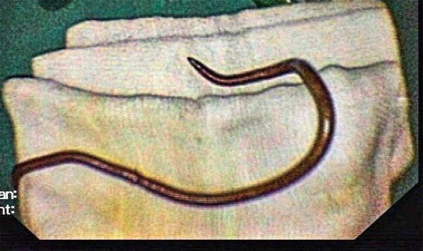
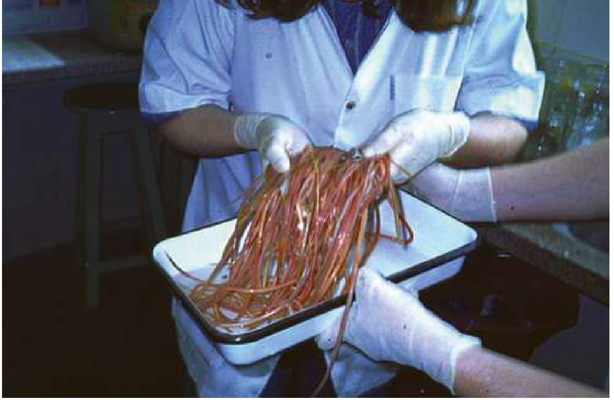
This clinical photograph displays a specimen of Ascaris lumbricoides, a human parasitic roundworm, following extraction via Endoscopic Retrograde Cholangio-Pancreaticography (ERCP). The specimen is a single, elongated, cylindrical nematode with a smooth, tan-to-brownish exterior. One visible extremity tapers to a sharp, curved point, which is characteristic of the adult worm's morphology. The organism is placed on sterile white surgical gauze, illustrating the outcome of a procedural intervention for biliary ascariasis. This image demonstrates the clinical manifestation of hepatobiliary parasitism where a nematode has migrated into the common bile duct, potentially causing obstructive jaundice or biliary colic. The educational focus is on the macroscopic identification of common helminthic infections and the visual confirmation of parasitic extraction in gastroenterology and tropical medicine.
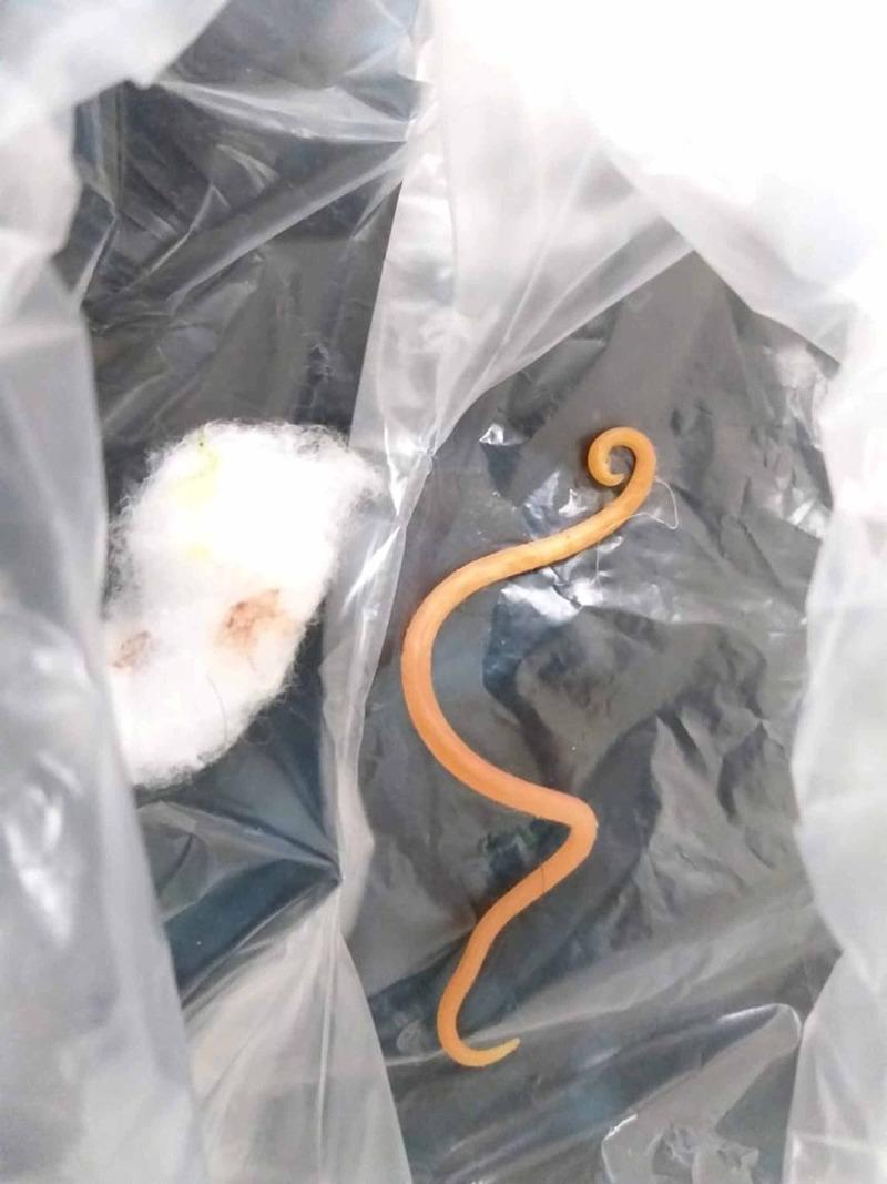
A clinical photograph of a gross pathology specimen featuring an adult nematode (roundworm), identified contextually as Ascaris lumbricoides. The specimen is displayed inside a clear specimen bag alongside a piece of white cotton wool for scale and handling. The roundworm exhibits characteristic morphology: a cylindrical, elongated, and unsegmented body with a smooth, light-orange to tan cuticle. One end demonstrates a distinctive curved or hooked appearance, while the other is tapered. The specimen measures approximately 15 cm in length. This visual provides evidence of ascariasis, a common parasitic infection where adult worms may be expelled via hematemesis or vomiting if they migrate from the small intestine to the stomach. The image is highly relevant for tropical medicine, gastroenterology, and infectious disease education, illustrating the gross appearance of a parasitic agent that can cause biliary or pancreatic complications.
tapeworm cestode anatomy scolex proglottid diagram
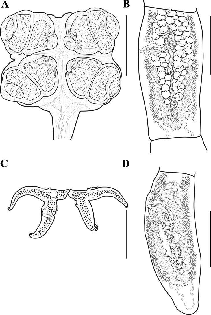
This scientific diagram illustrates the detailed morphology of a parasitic helminth, specifically the tapeworm species Acanthobothrium pulidofloresae. The composite image features four labeled panels: (A) The scolex (head), demonstrating a petaloid or 'clover-leaf' shape with four prominent bothridia. Each bothridium is divided into three loculi by transverse septa and possesses an anterior muscular pad equipped with hooks. (B) A mature proglottid (body segment), showing internal reproductive organs including oval testes arranged in two layers, a follicular ovary with symmetrical arms, and a developing sacciform uterus. (C) A high-magnification view of a pair of bipronged hooks, showing the handle and the differentiation between axial and abaxial prongs used for host attachment. (D) A terminal mature proglottid, highlighting the morphological transition of the ovary into an inverted A- or V-shape and a more prominent uterus. Scale bars are provided for each panel (75 μm to 500 μm). This material is essential for diagnostic parasitology and the study of cestode anatomy.
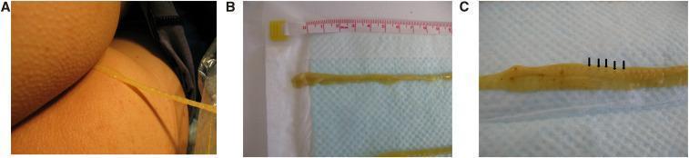
A series of clinical photographs documenting the extraction and gross morphology of a tapeworm (Cestoda). Figure A shows a long, yellowish, ribbon-like structure being carefully extracted from a patient's anal region into a clear plastic bag to maintain specimen integrity. Figure B displays the parasite post-extraction against a measuring tape and blue absorbent pad, highlighting its significant length, flat morphology, and a spoon-shaped scolex (head). Figure C provides a close-up view of the parasite's strobila, revealing distinct proglottids (segments). Black arrows point to central dark spots within the segments, which represent the gravid uterus. This image serves as a clinical reference for parasitic helminth infections, specifically diphyllobothriasis associated with the consumption of raw fish. Key educational features include the visualization of the scolex, proglottid segmentation, and macroscopic diagnostic characteristics used in identifying cestode species.
trematode fluke Schistosoma anatomy diagram
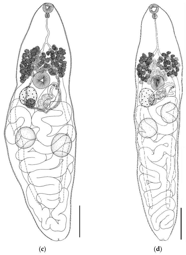
This biological diagram illustrates the internal anatomy of two trematode specimens, specifically Parabascus semisquamosus, categorized under medical helminthology and parasitology. The illustrations provide a comparative view of morphology based on different hosts. Both specimens feature a characteristic elongated body with an anterior oral sucker, followed by a muscular pharynx and a long esophagus bifurcating into intestinal ceca. In the mid-body, the ventral sucker (acetabulum) is centrally located. Key reproductive structures are visible, including granular vitelline follicles in the forebody, a prominent cirrus sac, a stippled ovary, and paired testes. The posterior two-thirds of the body are largely occupied by sinuous, winding uterine loops. Specimen (c) demonstrates a broader, more robust body shape with more convoluted digestive and reproductive tracts, while specimen (d) displays a slender, more attenuated morphology. Such diagrams are essential for identifying parasitic flatworms that may have zoonotic potential or clinical relevance in comparative pathology.
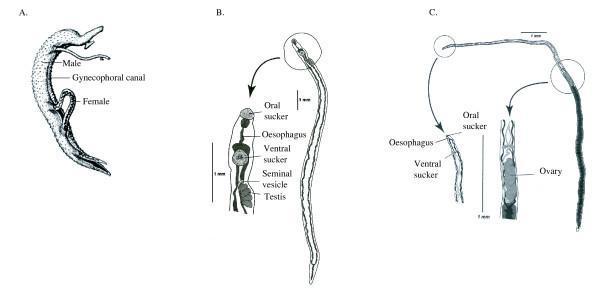
Educational anatomical diagram illustrating the sexual dimorphism and morphological features of Schistosoma species. The visual is divided into three sections: (A) A Schistosome pair during mating, highlighting the male's gynecophoral canal where the slender female resides. (B) A detailed view of a male schistosome, featuring a stout body and labeled internal structures including the oral sucker, oesophagus, ventral sucker, seminal vesicle, and multiple testes. (C) A detailed view of a female schistosome, showing its significantly longer and thinner morphology. Enlarged insets for the female specify the anterior attachment organs (oral sucker, oesophagus, and ventral sucker) and the reproductive system focusing on the ovary. The diagrams include 1 mm scale bars and use precise medical labeling to differentiate the male (stouter, with a ventral groove) from the female (longer, filiform). This content serves as a diagnostic reference for parasitology, specifically for identifying the morphological characteristics of blood flukes involved in human schistosomiasis.
pinworm Enterobius vermicularis perianal eggs microscopy
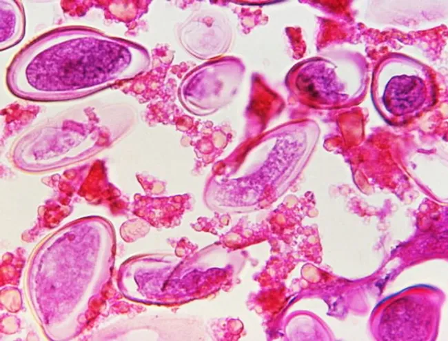
This is a bright-field light microscopy image of Enterobius vermicularis eggs (pinworm) obtained from a fecal or perianal specimen. The eggs appear as elongated, ovoid ova with a characteristic flattened side, stained pink-purple against a pale background. Measurements within the field confirm the diagnostic morphology: eggs approximately 50–60 μm long by 20–32 μm wide, consistent with standard parasitology references. The shells are thick and smooth, with a distinct taper and a subtly curved outline in many specimens. A perianal sediment of particulate material and host cells is visible, illustrating typical background in stool or tape-prep preparations. Clinically, the detection of these eggs is highly specific for Enterobius vermicularis infection and supports a diagnosis in patients with nocturnal perianal itching or observed scratching, especially in children with close-contact exposure. Differential considerations include eggs of other helminths such as Ascaris lumbricoides or Trichuris trichiura, which differ in size, shape, and shell ornamentation. The image’s diagnostic significance lies in enabling rapid identification and differentiation from artifacts in educational and clinical laboratory settings. Its CDC provenance (Dr. George R. Healy) makes it a trusted reference for teaching parasitology, epidemiology, and diagnostic parasitology workflows. This image supports education, proficiency testing, and reference material for parasitology laboratories.
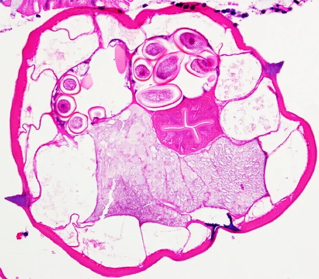
This cross-sectional light microscopy image depicts a female Enterobius vermicularis (pinworm) in transverse section within gastrointestinal tissue, stained with Hematoxylin and Eosin (H&E). The specimen shows a circular host cross-section containing a coiled nematode with pronounced lateral alae (alae) along the cuticle, a hallmark feature for identification. In the worm body, clear internal organs are visible, including a central digestive tract and reproductive structures; within the uterus numerous eggs are evident as operculated or oval, densely packed entities. The surrounding mucosa and submucosa reflect intestinal tissue with wavy smooth muscle layers and extracellular matrix; the thick, pink-stained cuticle of the nematode contrasts with the eosinophilic cytoplasm of the worm's parenchyma and the surrounding host tissue. The eggs within the worm body are ovoid, often deeply basophilic with visible shells in some sections. Notable features include the characteristic lateral alae (alae) extending along the dorsal and ventral surface and the presence of numerous eggs in the uterus, indicative of female pinworms producing eggs. Clinically, this image supports diagnosis of enterobiasis; pathologists correlate with perianal itching history and stool or cellophane tape test results. This slide is a valuable teaching reference for parasitology, histopathology and tropical medicine laboratories.
Trichuris trichiura whipworm morphology stool
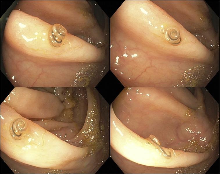
This diagnostic image is a four-panel colonoscopy collage showing a live helminth, identified as Trichuris trichiura (whipworm), located on the ileocecal valve. The organism is brown and exhibits a characteristic morphology: a thin, thread-like anterior end partially embedded in the colonic mucosa and a thicker, coiled posterior end resembling a watch spring or spiral. The surrounding intestinal lining appears healthy, characterized by a pinkish-tan hue, visible mucosal folds, and a glistening surface with normal vascular patterns. Minor amounts of yellow fecal debris and clear intestinal fluid are present. The image demonstrates the clinical manifestation of trichuriasis in a symptomatic patient presenting with weight loss and eosinophilia. It serves as an educational example of how parasitic infections can be directly visualized during lower gastrointestinal endoscopy and highlights the specific appearance of whipworms within the human gastrointestinal tract for gastroenterology and infectious disease training.
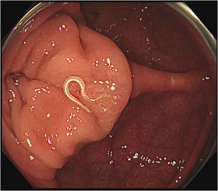
This endoscopic clinical photograph depicts a gastrointestinal parasite within the large intestine, specifically identified as a white whipworm (Trichuris trichiura) located in the cecum. The worm is characterized by a slender, elongated, and curved white body, exhibiting a characteristic whip-like morphology. It is situated on the surface of the colonic mucosa, which appears reddish-pink and healthy, though slightly nodular in texture with visible light reflections from the endoscope. The worm is positioned near the ileocecal valve area. The image demonstrates a classic presentation of trichuriasis, an intestinal nematode infection. The clinical significance of this visual is to illustrate the endoscopic detection of live helminths during colonoscopy in a patient presenting with symptoms such as chronic diarrhea and peripheral eosinophilia. The image is a valuable educational resource for gastroenterology and infectious disease specialties for the identification of parasitic infections of the human cecum and ascending colon.
lymphatic filariasis elephantiasis lymphedema
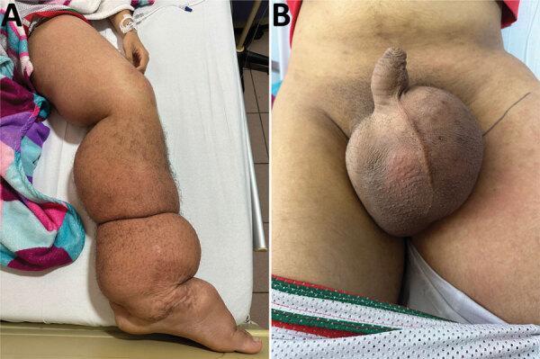
Two-panel clinical photograph illustrating the manifestations of lymphatic filariasis (elephantiasis) caused by Wuchereria bancrofti. Image A displays a side profile of the left lower limb exhibiting severe, generalized lymphedema from the foot to the groin. There is significant swelling and skin folds, erythematous patches on the thigh, and trophic skin changes including perimaleolar hypocromia and hypercromia. Image B provides a frontal view of the pelvic region, showing massive, bulbous scrotal edema (hydrocele) and induration. The skin of the scrotum appears thickened and tense. These findings are characteristic of chronic lymphatic obstruction leading to massive tissue enlargement and secondary dermatological changes. The images are highly relevant for tropical medicine and infectious disease specialties, demonstrating the late-stage physical sequelae of nematode-induced lymphatic damage.
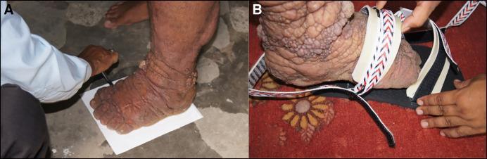
A two-part clinical photograph illustrating the management of advanced stage lymphatic filariasis (elephantiasis) and associated lymphedema. Image A displays a patient's lower leg and foot, demonstrating severe non-pitting edema with marked skin thickening (pachyderma) and deep transverse folds. The skin surface is rough and hyperkeratotic, featuring multiple nodular and verrucous (wart-like) lesions characteristic of mossy foot or podoconiosis. A healthcare provider is shown tracing the foot's enlarged outline for custom footwear. Image B shows the application of a specialized, adaptable shoe. The shoe features a thick, reinforced sole and wide, adjustable Velcro straps with a chevron pattern designed to accommodate extreme swelling and anatomical distortion while providing protection. These images illustrate key components of morbidity management and disability prevention (MMDP) in filariasis-endemic regions, emphasizing the importance of tailored footwear to prevent secondary bacterial and fungal infections and improve patient mobility.
neurocysticercosis brain CT scan cyst Taenia solium
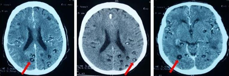
This diagnostic image consists of three axial CT scan slices of the brain demonstrating neurocysticercosis. The images show multiple small, well-defined, infra-centimetric hypodense cystic lesions scattered throughout the cerebral parenchyma, involving both the gray-white matter junction and deeper cortical structures. A key pathognomonic feature is the presence of a hyperdense mural nodule within many of the cysts, representing the scolex of the Taenia solium larva in the vesicular stage. There is a lack of significant surrounding vasogenic edema or mass effect in these specific slices, which is characteristic of the early viable stage of the parasite. This imaging presentation is highly suggestive of disseminated neurocysticercosis, illustrating the relationship between the parasitic cyst and the host brain tissue.
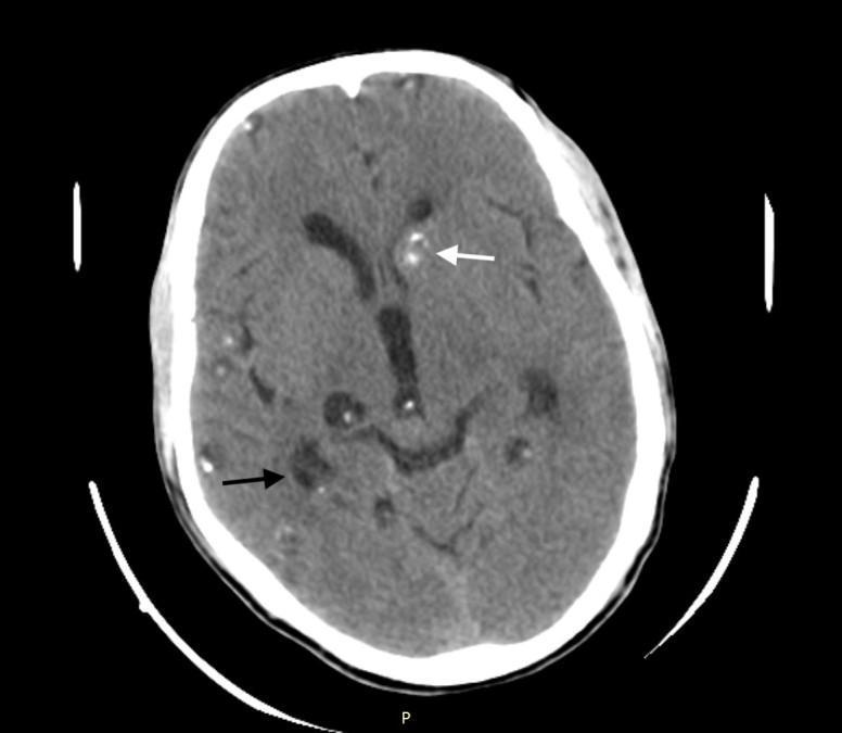
This diagnostic image is an axial non-contrast computed tomography (CT) scan of the brain demonstrating neurocysticercosis in multiple evolutionary stages. The scan shows numerous parenchymal cystic lesions distributed throughout both cerebral hemispheres. Two distinct stages are highlighted: the vesicular stage, marked by a black arrow, presents as a well-defined, low-attenuation (hypodense) fluid-filled cyst; and the nodular calcific stage, indicated by a white arrow, appearing as a high-attenuation (hyperdense) calcified lesion located in the left cerebral hemisphere near the basal ganglia. These lesions are characteristic of Taenia solium infection within the central nervous system. Anatomical landmarks including the cerebral cortex, lateral ventricles, and subcortical gray matter are visible, providing context for the multifocal distribution of the pathology. This image serves as a clinical example of the varied radiographic presentations of parasitic brain infections and is relevant for neurology and radiology education regarding seizure etiology.
hookworm Necator americanus adult worm intestine
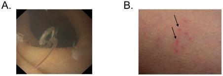
This composite clinical figure illustrates different stages and manifestations of a human hookworm infection. Panel A presents an endoscopic image of the upper gastrointestinal tract, demonstrating a mature adult hookworm attached to the intestinal mucosa. The worm is characterized by an elongated, curved, and semi-translucent body morphology. Panel B is a clinical photograph showing the percutaneous inoculation site on light-pigmented skin. It features several small, erythematous, inflammatory papules (indicated by black arrows) representing the localized cutaneous reaction to the penetration of hookworm larvae (L3 stage). The images serve to illustrate the visual findings associated with parasitic helminth infection, from initial larval entry to adult colonization of the intestine. This content is relevant for gastroenterology and infectious disease training, focusing on the diagnosis of hookworm (Ancylostoma or Necator species) through endoscopy and physical examination of skin lesions (ground itch).
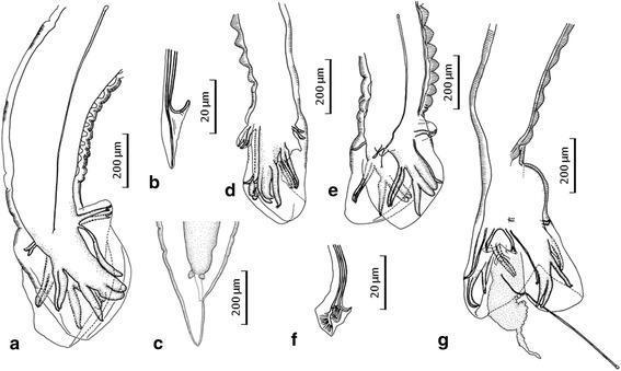
This set of line drawings illustrates the diagnostic morphological features of the posterior extremities and spicules of hookworm species, specifically comparing Necator americanus and Necator gorillae. Figures a and b display the male N. americanus, highlighting a wrinkled dorsal cuticle in the prebursal region and a characteristic recurved, barb-shaped distal end of the spicule. Figure c shows the conical, pointed tail of a female N. gorillae. Figures d through g focus on the male N. gorillae, emphasizing key taxonomic differentiators: distinctive transverse cuticular thickenings in the dorsal prebursal region and relatively thin bursal rays. High-magnification details (b, f) compare the morphology of the spicule ends, while figures d, e, and g illustrate various orientations of the copulatory bursa and spicule extrusion. These illustrations serve as a comparative tool in medical parasitology for identifying zoonotic hookworm infections and distinguishing between human-typical and great ape-associated Necator species based on micro-anatomical structures.
Strongyloides stercoralis larvae skin autoinfection rash
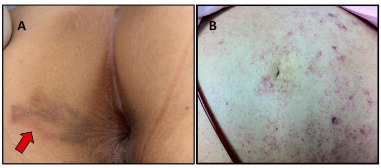
This composite clinical photograph displays two distinct dermatological manifestations of Strongyloides stercoralis infection. Image A shows larva currens in the perianal region, characterized by a serpiginous, erythematous, and slightly raised linear track (indicated by a red arrow). This pathognomonic lesion represents the rapid intradermal migration of filariform larvae. Image B displays a periumbilical 'thumbprint' purpura in a patient with disseminated strongyloidiasis. The abdominal skin exhibits extensive, non-blanching purpuric macules and petechiae, appearing as clusters of reddish-purple spots that are most concentrated in the periumbilical area. These lesions result from larvae migrating through the vessel walls into the dermis. The educational focus is on distinguishing the localized cutaneous signs of chronic infection from the systemic vascular signs associated with severe, disseminated hyperinfection syndrome in immunocompromised hosts.
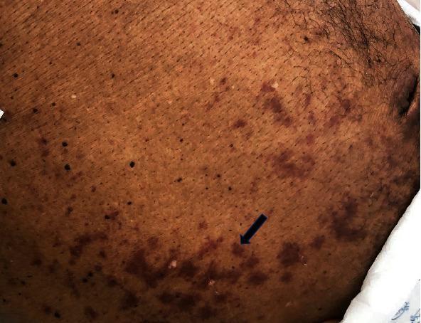
Clinical photograph of the abdomen on a dark-skinned patient demonstrating a purpuric rash. The primary visual finding is a periumbilical distribution of reddish-purple macules and patches. These lesions are both discrete and confluent, extending laterally toward the flanks. A black arrow points to a representative cluster of purpuric spots. In the clinical context of Strongyloides stercoralis hyperinfection or dissemination (as suggested by the associated metadata), this visual manifestation is classically known as the 'thumbprint sign' or 'larva currens' when presenting as migrating serpiginous tracks, though here it appears more as stationary petechiae and ecchymoses. The skin shows normal hair distribution and no overt signs of ulceration. This image is an essential educational example of cutaneous manifestations related to parasitic dissemination in immunocompromised individuals, illustrating the dermatological signs of severe systemic infection.
HELMINTHIC DISEASE - Complete Reference Guide
PART 1: DEFINITION
-
Peripheral blood eosinophilia (especially during tissue migration phases)
-
Elevated serum IgE (total and parasite-specific)
-
Chronic, often silent morbidity: anemia, malnutrition, impaired development, organ damage
-
Over 1 billion people infected worldwide, predominantly in tropical/subtropical low-income settings
-
Goldman-Cecil Medicine, International Edition
PART 2: CLASSIFICATION OF HELMINTHS
Kingdom: Animalia
  └─ Subkingdom: Metazoa
       ├─ Phylum: Platyhelminthes (Flatworms)
       │    ├─ Class: Trematoda (Flukes)
       │    └─ Class: Cestoidea (Tapeworms)
       ├─ Phylum: Nemathelminthes (Roundworms / Nematodes)
       └─ Phylum: Acanthocephala (Thorny-headed worms - rare)
- Sherris & Ryan's Medical Microbiology, 8th Ed, Table 48-3
PART 3: EACH CLASS - SIMPLE DEFINITION, SHAPE, BODY CAVITY, DIGESTIVE TRACT, AND SEX
CLASS 1: NEMATODA (Roundworms)
| Feature | Details |
|---|---|
| Simple Definition | Cylindrical, unsegmented worms that are the most common human parasites; they live mostly in the intestine but some invade tissues |
| Shape | Cylindrical and fusiform (tapered at both ends, like a spindle or thread) |
| Body Cavity | Pseudocoelom - a fluid-filled body cavity that is NOT lined by true mesoderm; acts as a hydrostatic skeleton |
| Digestive Tract | Complete - a true tubular alimentary canal running from mouth (anterior) to anus (posterior); they actively ingest host intestinal contents and blood |
| Sex | Separate sexes (dioecious); male worm is typically smaller than the female; male sperms are amoeboid (not flagellated - unique feature); females can produce up to 200,000 eggs per day |
| Body Wall | Protected by a tough cuticle that is shed (moulted) 4 times as larvae mature into adults |
| Muscles | Longitudinal muscles only - they flex but cannot shorten; movement is sinusoidal/whip-like |
| Feeding | By ingestion through the mouth |
| Reproduction | Oviparity (egg-laying), ovoviviparity (larvae born from eggs retained in utero), or parthenogenesis (Strongyloides) |
| Key Examples | Ascaris, hookworms, Strongyloides, Trichuris, Enterobius, Wuchereria, Onchocerca |
CLASS 2: TREMATODA (Flukes)
| Feature | Details |
|---|---|
| Simple Definition | Leaf-shaped or tongue-shaped flatworms that use suckers to attach to host organs; they infect blood vessels, liver, lungs, and intestines |
| Shape | Flat, leaf-like (dorso-ventrally flattened); usually oval or elongated; schistosomes are an exception - they are tubular (cylindrical) |
| Body Cavity | Acoelomate - no true body cavity; body is filled solid with parenchymal (spongy) tissue |
| Digestive Tract | Incomplete - a simple, branched gut (cecae) with a mouth but no anus; wastes are expelled back through the mouth; nutrients are also absorbed across the tegument |
| Sex | Most trematodes are hermaphroditic (contain both male and female reproductive organs in the same worm); EXCEPTION: Schistosomes are dioecious (separate sexes) - the thin female lives in the gynaecophoral canal (groove) of the stout male |
| Body Wall | Covered by a syncytial tegument (living, metabolically active outer layer); used for both nutrient absorption and waste excretion |
| Suckers | Two suckers: an oral sucker (around mouth) and a ventral sucker/acetabulum (for attachment) |
| Feeding | By absorption through the tegument AND ingestion |
| Life Cycle | Complex; require snails as first intermediate host (for larval multiplication); second intermediate hosts (fish, crabs, aquatic plants) for most; schistosomes penetrate skin directly as cercariae |
| Key Examples | Schistosoma, Fasciola, Clonorchis, Opisthorchis, Paragonimus, Fasciolopsis |
CLASS 3: CESTOIDEA (Tapeworms)
| Feature | Details |
|---|---|
| Simple Definition | Long, flat, ribbon-like worms made of many segments; they live in the intestine as adults and in tissues as larvae (cysts); they have NO mouth or gut at all |
| Shape | Flat, ribbon-like (dorso-ventrally flattened); composed of a chain of segments called proglottids forming the strobila |
| Body Cavity | Acoelomate - no body cavity; filled with parenchymal tissue and reproductive organs |
| Digestive Tract | NONE - tapeworms have no digestive system whatsoever; they absorb all nutrients directly across their tegument from the host's intestinal contents |
| Sex | Hermaphroditic - each individual proglottid contains both male and female reproductive organs; self-fertilization is possible; gravid proglottids are filled with eggs |
| Body Plan | Three regions: Scolex (head with suckers ± hooks for attachment) → narrow Neck (growth zone, continuously produces new proglottids) → Strobila (chain of immature → mature → gravid proglottids) |
| Feeding | By absorption only - through syncytial tegument; no mouth, no gut |
| Size | From a few mm (Hymenolepis nana) to over 10 meters (Taenia saginata, Diphyllobothrium latum) |
| Key Examples | Taenia solium, T. saginata, Echinococcus granulosus, Hymenolepis nana, Diphyllobothrium latum |
COMPARISON TABLE: Three Classes at a Glance
| Feature | Nematode (Roundworm) | Trematode (Fluke) | Cestode (Tapeworm) |
|---|---|---|---|
| Shape | Cylindrical, round | Flat, leaf-shaped | Flat, ribbon-like, segmented |
| Body cavity | Pseudocoelom (fluid-filled) | Acoelomate (solid) | Acoelomate (solid) |
| Digestive tract | Complete (mouth to anus) | Incomplete (mouth, no anus) | ABSENT (none at all) |
| Feeding method | Ingestion | Absorption + some ingestion | Absorption only |
| Sex | Separate (M and F worms) | Hermaphrodite (except Schistosoma) | Hermaphrodite (each segment) |
| Body wall | Cuticle (non-living) | Syncytial tegument (living) | Syncytial tegument (living) |
| Size range | 1 mm - 1 m | Few mm - several cm | Few mm - 10+ m |
| Segments | None | None | Yes (proglottids) |
PART 4: COMMON HELMINTHIC DISEASES - DETAILED PROFILES
DISEASE 1: ASCARIASIS
Causative Organism: Ascaris lumbricoides (Giant Intestinal Roundworm)
| Feature | Details |
|---|---|
| Etiology | Ascaris lumbricoides - the largest intestinal nematode (15-50 cm); female produces >200,000 eggs/day; fertilized eggs have thick, mammillated outer shell |
| Mode of Transmission | Fecal-oral - ingestion of embryonated eggs from fecally contaminated soil, food, water, or hands. Eggs can survive 15 years in soil. |
| Risk Factors | Poor sanitation; use of human feces as fertilizer; lack of hand-washing; children aged 2-15 years; rural tropical/subtropical areas; Africa, Asia, Latin America |
| Pathology | Larvae migrate through liver → lungs (Löffler syndrome) → swallowed → adult worms in small intestine; heavy burdens cause mechanical obstruction |
| Clinical Features | - Pulmonary phase: Löffler syndrome (eosinophilic pneumonia) - cough, wheeze, fever, blood-tinged sputum; - Intestinal phase: mostly asymptomatic; heavy load → abdominal pain, intestinal obstruction, biliary migration (cholangitis, pancreatitis); worm passed in stool/vomit/nose |
| Diagnosis | Stool microscopy (fertilized eggs 65×45 µm with thick mammillated shell); CBC shows eosinophilia; abdominal X-ray/ultrasound for obstruction; ERCP for biliary worms |
| Treatment | Albendazole 400 mg single dose (first-line); OR Mebendazole 500 mg single dose; OR Ivermectin 150-200 µg/kg; OR Pyrantel pamoate 11 mg/kg. Intestinal obstruction: NG tube + piperazine; surgical for complete obstruction |
| Prevention | Hand washing with soap; safe disposal of human feces; sanitation improvements; not using raw human manure as fertilizer; mass drug administration (MDA) programs in schools; cook vegetables from soil |
DISEASE 2: HOOKWORM DISEASE
Causative Organisms: Necator americanus, Ancylostoma duodenale, A. ceylanicum
| Feature | Details |
|---|---|
| Etiology | N. americanus (predominant, tropics/subtropics worldwide); A. duodenale (Mediterranean, India, China); ~1 cm long; attach to small bowel mucosa using buccal cutting plates (Necator) or teeth (Ancylostoma) and ingest blood |
| Mode of Transmission | Skin penetration - filariform (L3) larvae penetrate bare skin (usually feet) from contaminated soil; less commonly, Ancylostoma spp. can be ingested orally in contaminated food/water |
| Risk Factors | Walking barefoot on contaminated soil; poor sanitation; agricultural work in endemic areas; children and farmers most at risk; warm, moist climates |
| Pathology | Larvae: skin inflammation (ground itch) → lung migration → Löffler syndrome; adults: attach to mucosa, secrete anticoagulants, ingest blood daily → iron-deficiency anemia + hypoproteinemia; >400 million infected worldwide |
| Clinical Features | "Ground itch" (pruritic papular rash at entry site); Löffler syndrome during migration; iron-deficiency anemia (fatigue, pallor, dyspnea, palpitations); hypoproteinemia → edema; cognitive impairment in children; chronic heavy infection → Plummer-Vinson syndrome |
| Cutaneous Larva Migrans (CLM) | From animal hookworm (A. braziliense) in humans - larvae cannot complete life cycle; track serpiginously in skin → intensely pruritic serpiginous rash |
| Diagnosis | Stool microscopy for thin-shelled oval eggs (60×40 µm); CBC: eosinophilia + microcytic hypochromic anemia; low serum ferritin |
| Treatment | Albendazole 400 mg single dose; OR mebendazole 500 mg once; PLUS iron supplementation. CLM: ivermectin 200 µg/kg or albendazole 400 mg/day × 3 days |
| Prevention | Wear footwear (sandals/shoes); improved sanitation; avoid walking barefoot in endemic areas; MDA programs; proper disposal of human waste; treat infected individuals |
DISEASE 3: STRONGYLOIDIASIS
Causative Organism: Strongyloides stercoralis (Threadworm)
| Feature | Details |
|---|---|
| Etiology | Strongyloides stercoralis - unique among helminths for its capacity for autoinfection (rhabditiform larvae → filariform larvae in the colon → reinfect the same host); only females (parthenogenetic) live in the intestinal mucosa of humans |
| Mode of Transmission | Skin penetration of filariform larvae from contaminated soil (same as hookworm); also autoinfection via perianal skin; rare oral transmission |
| Risk Factors | Tropical regions; poor sanitation; immunosuppression (corticosteroids, HTLV-1, HIV, organ transplant) - risk of fatal hyperinfection; institutionalized patients; veterans returning from endemic areas |
| Clinical Features | (1) Cutaneous: "Larva currens" - rapidly migrating urticarial linear rash on trunk/buttocks (pathognomonic); (2) Intestinal: diarrhea, abdominal pain, malabsorption; (3) Pulmonary: cough, wheeze; (4) Hyperinfection syndrome in immunosuppressed: massive larval migration carrying gut bacteria → gram-negative bacteremia/meningitis, multi-organ failure - potentially fatal |
| Diagnosis | Stool O&P (low sensitivity; Baermann technique preferred); serology (ELISA - most sensitive); duodenal aspirate; larvae in sputum during hyperinfection |
| Treatment | Ivermectin 200 µg/kg/day × 2 days (drug of choice); Albendazole 400 mg bid × 7 days (second-line). Hyperinfection: prolonged ivermectin until clearance confirmed |
| Prevention | Wear footwear; improved sanitation; screen all patients before immunosuppression; treat with ivermectin before starting steroids/chemotherapy in endemic-area residents |
DISEASE 4: TRICHURIASIS (Whipworm)
Causative Organism: Trichuris trichiura
| Feature | Details |
|---|---|
| Etiology | Trichuris trichiura - has a characteristic whip shape: thin anterior 2/3 (embedded in colonic mucosa) and thick posterior 1/3 (free in lumen); ~3-5 cm long; inhabits cecum and ascending colon |
| Mode of Transmission | Fecal-oral - ingestion of embryonated eggs from contaminated soil, food, or water |
| Risk Factors | Poor sanitation; tropical/subtropical regions; children 5-15 years; geophagia (eating soil); co-infection with Ascaris is very common |
| Clinical Features | Light infection: usually asymptomatic; Heavy infection: trichuris dysentery syndrome - chronic bloody diarrhea, tenesmus, rectal prolapse (especially in children), iron-deficiency anemia, growth retardation, clubbing |
| Diagnosis | Stool microscopy: barrel-shaped/lemon-shaped eggs with bipolar plugs (50×22 µm); colonoscopy shows whip-shaped worm in cecum |
| Treatment | Mebendazole 100 mg bid × 3 days; OR Albendazole 400 mg/day × 3 days |
| Prevention | Hand washing; improved sanitation; avoid eating soil; wash raw vegetables; MDA with mebendazole/albendazole in school-age children |
DISEASE 5: ENTEROBIASIS (Pinworm)
Causative Organism: Enterobius vermicularis
| Feature | Details |
|---|---|
| Etiology | Enterobius vermicularis - small white worm (~1 cm female, ~3 mm male); adult worms live in cecum/ascending colon; gravid female migrates to perianal area at night to deposit eggs |
| Mode of Transmission | Fecal-oral - ingestion of eggs; direct contact (perianal scratching → eggs on fingers → mouth); indirect via contaminated bedding, clothing, toilet seats; retroinfection (eggs hatch near anus, larvae migrate back into rectum) |
| Risk Factors | Most common helminth in developed countries; schoolchildren (5-10 years); crowded institutions (daycares, dormitories); family clusters; poor hand hygiene |
| Clinical Features | Nocturnal perianal pruritus (itching at night when females migrate); disturbed sleep; perianal scratching → excoriation; vulvovaginitis in girls; rarely appendicitis, ectopic sites (ovaries, fallopian tubes) |
| Diagnosis | Scotch tape (cellophane) test: transparent tape pressed to perianal skin on waking → microscopy shows flat-sided oval eggs (50-60×20-32 µm); NOT found on routine stool O&P |
| Treatment | Albendazole 400 mg once, repeated at 2 weeks; OR Mebendazole 100 mg once, repeated at 2 weeks; OR Pyrantel pamoate 11 mg/kg, repeated at 2 weeks. Treat ALL household members simultaneously |
| Prevention | Hand washing especially after toilet and before meals; cut and clean fingernails; wash bedding/underwear in hot water; shower/bath in morning (removes eggs deposited overnight); treat all household contacts |
DISEASE 6: LYMPHATIC FILARIASIS (Elephantiasis)
Causative Organisms: Wuchereria bancrofti, Brugia malayi, Brugia timori
| Feature | Details |
|---|---|
| Etiology | Filarial nematodes transmitted by mosquitoes; adult worms (males 4 cm, females 8 cm) live in lymphatic vessels; produce microfilariae that circulate in blood with nocturnal periodicity (W. bancrofti) |
| Mode of Transmission | Mosquito bite - infective L3 larvae deposited on skin by mosquito (Culex for W. bancrofti; Mansonia for Brugia spp.) → larvae enter lymphatics through bite wound |
| Risk Factors | Tropical/subtropical regions; Africa, South/Southeast Asia, Pacific Islands, parts of Americas; sleeping outdoors; lack of mosquito control; long-term residence in endemic areas |
| Clinical Features | Acute: fever, lymphangitis, lymphadenitis (acute dermatolymphangioadenitis/ADL); Chronic: lymphedema (leg, arm, breast), elephantiasis (massive limb enlargement, skin thickening), hydrocele (scrotal enlargement), chyluria; Tropical pulmonary eosinophilia (hyperreactive immune response) - nocturnal asthma, high IgE |
| Diagnosis | Night blood smear for microfilariae (10 pm - 2 am); circulating filarial antigen (CFA) card test; ultrasound - "filarial dance sign" (live worms moving in lymphatics); serology |
| Treatment | DEC 6 mg/kg/day × 12 days (drug of choice); + albendazole 400 mg; doxycycline × 6 weeks (kills Wolbachia endosymbionts → macrofilaricidal); Mass drug administration: single dose albendazole + DEC or ivermectin. Lymphedema management: hygiene, physiotherapy, compression |
| Prevention | Mosquito nets (especially at night); insect repellent; insecticide-treated bed nets; indoor residual spraying; MDA in endemic communities; WHO Global Programme to Eliminate Lymphatic Filariasis |
DISEASE 7: NEUROCYSTICERCOSIS / TAENIASIS (Taenia solium)
Causative Organism: Taenia solium (Pork Tapeworm)
| Feature | Details |
|---|---|
| Etiology | Taenia solium; humans are definitive host for adult tapeworm (after eating undercooked pork); humans can ALSO be intermediate host for larvae (cysticercosis) by ingesting T. solium eggs via fecal-oral route |
| Mode of Transmission | (1) Taeniasis: eating undercooked pork containing cysticerci → adult tapeworm grows in intestine; (2) Cysticercosis: ingesting T. solium eggs from fecally contaminated food/water/hands (even without eating pork); autoinfection possible |
| Risk Factors | Eating undercooked or raw pork; poor sanitation; open defecation; endemic regions: Latin America, sub-Saharan Africa, South and Southeast Asia, Eastern Europe |
| Clinical Features | Taeniasis (adult worm): mostly asymptomatic; mild abdominal pain, proglottid passage; Neurocysticercosis (larval cysts in brain): seizures (most common cause of acquired epilepsy in endemic areas), headache, hydrocephalus, focal neurological deficits, altered consciousness |
| Diagnosis | Taeniasis: stool for eggs/proglottids (8-12 uterine branches in T. solium vs 15-30 in T. saginata); NCC: CT/MRI brain showing ring-enhancing cystic lesions with scolex (pathognomonic), calcified lesions; serology (EITB immunoblot - most specific) |
| Treatment | Taeniasis: Praziquantel 5-10 mg/kg single dose or niclosamide; NCC: Albendazole 15 mg/kg/day (preferred) or praziquantel + dexamethasone (mandatory to reduce inflammation as cysts die); antiepileptics; VP shunt for hydrocephalus; surgical/endoscopic for ocular or ventricular cysts |
| Prevention | Cook pork thoroughly (>65°C internal temperature); improved sanitation and open-defecation-free communities; hand washing; inspect meat; pig vaccination programs; treat taeniasis carriers |
DISEASE 8: SCHISTOSOMIASIS (Bilharzia)
Causative Organisms: Schistosoma mansoni, S. haematobium, S. japonicum
| Feature | Details |
|---|---|
| Etiology | Blood flukes; adult male and female worms live paired in mesenteric venules (S. mansoni, S. japonicum) or vesical venous plexus (S. haematobium); eggs trapped in tissues cause granulomatous inflammation - the primary pathology |
| Mode of Transmission | Cercarial skin penetration - free-swimming cercariae released from freshwater snails penetrate exposed human skin during contact with contaminated freshwater (swimming, wading, bathing, farming) |
| Risk Factors | Freshwater exposure in endemic areas; Africa (S. haematobium - most common; S. mansoni); Brazil/Caribbean (S. mansoni); China/Philippines (S. japonicum); agricultural workers; fishermen; children playing in water; poor sanitation facilitating snail habitats |
| Clinical Features | (1) Cercarial dermatitis ("swimmer's itch"): pruritic urticarial rash 1-3 days post-exposure; (2) Katayama fever (acute): 4-8 weeks later - fever, urticaria, hepatosplenomegaly, diarrhea, eosinophilia (immune complex disease); (3) Chronic intestinal (S. mansoni/japonicum): portal hypertension, Symmer's pipe-stem fibrosis, esophageal varices, ascites; (4) Urogenital (S. haematobium): terminal hematuria, dysuria, obstructive uropathy, bladder squamous cell carcinoma (IARC Group 1 carcinogen) |
| Diagnosis | Stool (S. mansoni/japonicum) or urine (S. haematobium) for eggs; Kato-Katz smear; rectal snip biopsy; serology; urine dipstick for hematuria; liver ultrasound (periportal fibrosis) |
| Treatment | Praziquantel: 40 mg/kg in 1-2 doses (S. mansoni, haematobium); 60 mg/kg in 3 doses over 1 day (S. japonicum) |
| Prevention | Avoid freshwater contact in endemic areas; use protective clothing/boots; safe water supply; snail control (molluscicides - niclosamide); MDA with praziquantel; health education; improve sanitation to reduce fecal contamination of water |
DISEASE 9: ECHINOCOCCOSIS (Hydatid Disease)
Causative Organism: Echinococcus granulosus
| Feature | Details |
|---|---|
| Etiology | Cestode (tapeworm) larvae; dogs = definitive host; sheep/cattle = normal intermediate hosts; humans = accidental intermediate hosts; larvae form fluid-filled hydatid cysts in liver (65%), lungs (25%), or other organs |
| Mode of Transmission | Fecal-oral - ingestion of eggs from food/water contaminated with dog feces; direct contact with infected dogs (especially sheepdogs); eggs are ingested → oncospheres hatch → travel via blood → lodge and form cysts |
| Risk Factors | Sheep-raising communities; close contact with dogs; drinking contaminated water; areas endemic for the disease: Mediterranean, Middle East, Africa, South America, Central Asia, Australia |
| Clinical Features | Often asymptomatic for years; enlarging liver cyst → RUQ pain, hepatomegaly, palpable mass; Cyst rupture → anaphylaxis, urticaria, peritoneal seeding of daughter cysts; biliary rupture → cholangitis, obstructive jaundice |
| Diagnosis | Ultrasound/CT: large cyst with "hydatid sand" (protoscolices), floating daughter cysts (pathognomonic), peripheral calcification; Serology: ELISA/Weinberg reaction (may be falsely negative in 38%); eosinophilia only if cyst ruptures |
| Treatment | PAIR procedure (Puncture-Aspiration-Injection-Reaspiration with hypertonic saline) + albendazole cover 400 mg bid × 28-day cycles (pre and post procedure); Surgical excision with precautions to avoid spillage; Albendazole alone for inoperable or small cysts |
| Prevention | Prevent dogs from eating sheep offal; regular deworming of dogs with praziquantel; wash hands after contact with dogs; safe water and food; sheep vaccination (EG95 vaccine); avoid contact between dogs and slaughter waste |
PART 5: SUMMARY - MODE OF TRANSMISSION BY DISEASE
| Disease | Pathogen | How It Enters the Body |
|---|---|---|
| Ascariasis | A. lumbricoides | Ingestion of eggs in contaminated food/water/soil |
| Hookworm | N. americanus / A. duodenale | Skin penetration by larvae in contaminated soil |
| Strongyloidiasis | S. stercoralis | Skin penetration + autoinfection |
| Trichuriasis | T. trichiura | Ingestion of eggs in contaminated soil/food |
| Enterobiasis | E. vermicularis | Ingestion of eggs; direct contact; retroinfection |
| Lymphatic Filariasis | W. bancrofti / Brugia spp. | Mosquito bite (larval injection) |
| Onchocerciasis | O. volvulus | Blackfly (Simulium) bite |
| Taeniasis/NCC | T. solium | Eating raw/undercooked pork; fecal-oral (for cysticercosis) |
| Schistosomiasis | Schistosoma spp. | Cercarial penetration through skin in freshwater |
| Echinococcosis | E. granulosus | Ingestion of eggs from dog feces |
PART 6: ANTHELMINTIC DRUG SUMMARY (Treatment at a Glance)
| Drug | How It Works (Simply) | Main Diseases Treated |
|---|---|---|
| Albendazole | Blocks worm's skeleton proteins → worm starves and is paralyzed | Broad spectrum: Ascaris, hookworm, Trichuris, cysticercosis, echinococcosis |
| Mebendazole | Same as albendazole | Ascaris, hookworm, Trichuris, Enterobius |
| Ivermectin | Paralyzes worm's nerves and muscles by opening chloride channels | Strongyloides (1st choice), onchocerciasis, filariasis |
| Praziquantel | Increases calcium permeability → worm spasms and dies; disrupts surface membrane | All tapeworms, schistosomiasis, most flukes |
| DEC (diethylcarbamazine) | Immobilizes microfilariae; promotes immune system destruction of worms | Lymphatic filariasis (1st choice), loiasis |
| Triclabendazole | Disrupts microtubules of immature and adult flukes | Fasciola (drug of choice) |
| Pyrantel pamoate | Causes rigid paralysis of worm muscles | Ascaris, hookworm, Enterobius |
PART 7: GENERAL PREVENTION PRINCIPLES FOR ALL HELMINTHIC DISEASES
- Sanitation and hygiene - safe disposal of human feces; access to toilets; open-defecation-free communities
- Hand hygiene - wash hands with soap before eating and after using the toilet
- Safe food practices - cook meat thoroughly; wash vegetables; safe water supply
- Footwear - wearing shoes prevents skin-penetrating larvae (hookworm, Strongyloides)
- Vector control - insecticide-treated nets, indoor spraying (for mosquito/blackfly-borne filariae)
- Avoid freshwater exposure in endemic areas (schistosomiasis)
- Proper cooking of meat - pork (T. solium), beef (T. saginata), freshwater fish (Clonorchis)
- Animal deworming - dogs (Echinococcus), cats and dogs (Toxocara)
- Mass drug administration (MDA) - WHO-supported programs using albendazole/mebendazole/ivermectin/DEC in endemic communities
- Health education - teach communities about transmission and prevention
Search PubMed for latest WHO guidelines on helminthiasis elimination programs
"neglected tropical diseases"[MeSH Terms] AND "helminthiasis"[MeSH Terms] AND "mass drug administration"
"soil-transmitted helminths"[MeSH Terms] AND "elimination" AND "WHO"
WHO 2025 2026 helminthiasis elimination guidelines neglected tropical diseases roadmap
"soil-transmitted helminths" AND "elimination" AND "mass drug administration"
"schistosomiasis"[MeSH Terms] AND "elimination" AND "WHO" AND "guideline"
"lymphatic filariasis"[MeSH Terms] AND "elimination" AND "global program"
https://www.who.int/teams/control-of-neglected-tropical-dise…
PMID: 40752908
PMID: 35594896
"lymphatic filariasis" AND "elimination" AND "preventive chemotherapy"
"neglected tropical diseases" AND "2030 targets" AND "WHO roadmap"
PMID: 36549443
PMID: 34928944
https://apps.who.int/gb/ebwha/pdf_files/EB158/B158_9-en.pdf
WHO Guidelines on Helminthiasis Elimination Programs: Latest Evidence (2021-2026)
1. THE WHO NTD ROADMAP 2021-2030: THE MASTER FRAMEWORK
- Accelerate programmatic action (MDA coverage, treatment scale-up)
- Intensify cross-cutting approaches (WASH integration, vector control, surveillance)
- Change operating models and culture (country ownership, sustainability, innovation)
- Reduce by 90% the number of people requiring interventions against NTDs (from 2010 baseline)
- Reduce disability-adjusted life years (DALYs) from NTDs by 75%
- Achieve zero countries with NTDs as public health problems in which the disease had been eliminated
- At least 100 countries achieving elimination of at least one NTD by 2030
- In 2024, 1.4 billion people still required NTD interventions - a 36% reduction from 2010
- Disease burden fell from 17.2 to 14.1 million DALYs between 2015 and 2021; mortality fell from 139,000 to 119,000
- Over 880 million people treated in 2024, 99% through mass drug administration (MDA)
- As of early 2026, 58 countries have eliminated at least one NTD - progress toward the target of 100 by 2030
2. DISEASE-SPECIFIC WHO TARGETS AND GUIDELINE UPDATES
A. Soil-Transmitted Helminthiases (STH)
- WHO published new guidelines on strongyloidiasis (2023-2025 period) - a previously under-addressed STH
- A new monitoring and evaluation framework for STH (and schistosomiasis) was released in this period
- MDA with albendazole or mebendazole remains the cornerstone, now with biannual treatment recommended in high-prevalence settings
- MDA for STH reached 502 million people in 2024 - one of the world's largest public health interventions
- WHO acknowledged six countries for eliminating at least one NTD by the end of 2024
B. Schistosomiasis
Review of 2022 WHO Guidelines on Control and Elimination of Schistosomiasis - Lo NC et al., Lancet Infect Dis 2022. PMID: 35594896
| Recommendation | Previous (2006) | New (2022) |
|---|---|---|
| Target population | School-aged children (SAC) predominantly | All ages ≥2 years in endemic communities |
| Prevalence threshold for annual MDA | ≥50% in SAC | Lowered: treat annually at lower prevalence thresholds |
| MDA frequency | Annual in high settings | Increased frequency based on infection intensity |
| Elimination goal | Morbidity control | Explicit transmission interruption goal added |
| Diagnostic tools | Kato-Katz stool microscopy | Includes serology and molecular diagnostics |
| Praziquantel supply | Limited | Expanded donated praziquantel supply framework |
C. Lymphatic Filariasis (LF)
- Albendazole + DEC (outside Africa, where onchocerciasis is co-endemic)
- Albendazole + ivermectin (in Africa, where DEC is contraindicated due to onchocerciasis)
- Triple therapy (IDA): Ivermectin + DEC + Albendazole (single dose) - newer regimen endorsed by WHO; shown to sustain microfilarial clearance for at least 2 years
- Since 2023, global MDA coverage for LF returned to pre-COVID-19 levels
- In 2024, record MDA coverage of 74.7% was achieved for LF
- Several countries progressively stopping MDA for onchocerciasis and transitioning to post-treatment surveillance
- In both 2023 and 2024, over 170 million people treated with ivermectin each year - the highest annual figures to date
D. Onchocerciasis (River Blindness)
- The Africa Programme for Onchocerciasis Control (APOC) achieved major successes
- Ivermectin MDA remains the cornerstone; moxidectin 8 mg (for ages >12 years) has been added as an alternative
- Several countries now stopping MDA and transitioning to post-treatment surveillance (2023-2024)
- New WHO guidelines on onchocerciasis were published in this cycle
E. Echinococcosis
- Tiered management approach: PAIR (Puncture-Aspiration-Injection-Reaspiration), surgery, medical therapy with albendazole
- Dog deworming with praziquantel in endemic areas as primary prevention
- WHO classifies echinococcosis under diseases targeted for control (not elimination), given its zoonotic nature and complexity
3. KEY RECENT CLINICAL EVIDENCE FROM PUBMED
[RCT - Tier 3 - 2025] DeWorm3 Trial - Can STH Transmission Actually Be Interrupted?
- Design: Open-label community cluster-RCT; 120 clusters (357,716 individuals); 3 countries; 3 years
- Intervention: Community-wide biannual MDA with albendazole 400 mg (all ages) vs. school-based deworming only
- Key findings:
- Community-wide MDA significantly reduced hookworm (N. americanus) prevalence vs. school-based deworming in all 3 countries (prevalence ratio 0.40-0.44)
- Transmission interruption was achieved in 55% of intervention clusters in Benin (vs. 30% control), but only in 5% in India and 0% in Malawi
- Conclusion: Community-wide MDA is feasible and superior to school-based treatment alone, but transmission interruption is difficult to sustain consistently across settings - challenges remain especially in high-transmission environments
Policy significance: This trial directly informs the ongoing WHO debate about whether school-based MDA alone is sufficient, or whether community-wide treatment is needed to reach the 2030 elimination target. Results suggest community-wide MDA is necessary but not always sufficient.
[Review - Tier 7 - 2022] 2022 WHO Schistosomiasis Guidelines - Lancet Review
- Written by the 2018-2022 Schistosomiasis Guidelines Development Group
- Approximately 250 million people infected globally; praziquantel is the cornerstone drug
- The 2022 guidelines mark a major strategic shift: from school-age-child-focused morbidity control to community-wide elimination as the goal
- Expansion of treatment eligibility from SAC to all ages ≥2 years is the single biggest operational change
[Systematic Review - Tier 1 - 2021] Preventive Chemotherapy Impact - Philippines
- Finding: Following preventive chemotherapy, moderate-heavy intensity Ascaris prevalence dropped from 23.6% to 6.6%; Trichuris from 12.2% to 2.7%; schistosomiasis in SAC from 30.5% to 1%
- Only 2 of 12 provinces still had LF prevalence >1% after MDA
- Conclusion: Preventive chemotherapy works, but community-wide expansion + WASH is needed for full elimination
[Journal Article - 2023] Helminth Infections: Enabling the WHO Roadmap
- Identifies critical gaps blocking 2030 targets: lack of new anthelmintic drugs, inadequate diagnostics for low-intensity infections, and disease siloing
- Advocates for cross-disciplinary helminth elimination platforms and new drug pipelines
- Points out that the current benzimidazoles (albendazole, mebendazole) have suboptimal efficacy against hookworm, threatening hookworm elimination goals
[Review - Tier 7 - 2023] STH Co-infections and Elimination
- Co-infections with multiple STH species are common; they interact to worsen morbidity
- Integrated MDA programs addressing multiple NTDs simultaneously are more cost-effective
- WASH improvements are essential complements to MDA - MDA alone cannot achieve elimination without improved sanitation
4. CURRENT CHALLENGES TO ACHIEVING 2030 TARGETS
| Challenge | Details |
|---|---|
| Funding gap | Deep cuts in official development assistance (ODA) for NTDs in 2025-2026 threaten program continuity |
| COVID-19 recovery | MDA programs disrupted 2020-2022; recovery achieved for LF (74.7% coverage in 2024) but gaps remain for others |
| Drug efficacy | Suboptimal efficacy of albendazole/mebendazole against hookworm (N. americanus); no new anthelmintic drugs licensed |
| Transmission rebound | As DeWorm3 showed, even high-coverage community MDA may not consistently interrupt transmission |
| Last-mile coverage | Hard-to-reach communities, migrants, and displaced populations remain undertreated |
| Diagnostics | Low-sensitivity tools for low-intensity infections hinder surveillance as prevalence falls |
| Data gaps | Incomplete country reporting; lack of gender-disaggregated data |
| Climate change | Expanding geographic range of vector-borne NTDs; altered snail habitats for schistosomiasis |
| WHO restructuring | In 2025, WHO merged the Global NTD Programme with the Global Malaria Programme - new department: Malaria and Neglected Tropical Diseases |
5. SUMMARY TABLE: WHO 2030 TARGETS FOR HELMINTHIC NTDs
| Disease | WHO 2030 Target | Key Drug(s) | 2024 Coverage |
|---|---|---|---|
| Soil-transmitted helminthiases | Elimination as a public health problem | Albendazole / Mebendazole | 502 million people reached with MDA |
| Schistosomiasis | Elimination as a public health problem | Praziquantel | >100 million treated; 253 million requiring treatment |
| Lymphatic filariasis | Elimination as a public health problem | Albendazole + DEC or ivermectin | 74.7% coverage (record high) |
| Onchocerciasis | Elimination of transmission (selected areas) | Ivermectin / Moxidectin | >170 million treated per year |
| Echinococcosis | Control | Albendazole + PAIR/surgery | Zoonotic; focus on dog deworming |
6. LATEST WHO GUIDELINE DOCUMENTS (2023-2025)
- Guidelines on echinococcosis (new)
- Guidelines on strongyloidiasis (new - previously had no WHO-specific guideline)
- Guidelines on arboviral diseases (vector control)
- Guidelines on human African trypanosomiasis
- Guidelines on tungiasis
- Monitoring and evaluation framework for schistosomiasis and soil-transmitted helminthiases
- Operational guidance to eliminate human fascioliasis as a public health problem in the Americas