
Laq Syphillus Microbiology
syphilis Treponema pallidum stages chancre rash
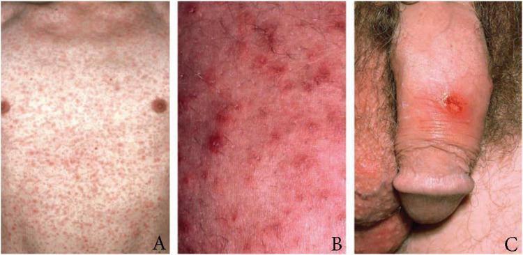
This composite of three clinical photographs illustrates the dermatological and urogenital manifestations of early syphilis (Treponema pallidum infection). Panel A shows a wide-field view of the anterior torso featuring a dense, generalized maculopapular rash, typical of secondary syphilis. The lesions are symmetric, non-confluent, and reddish-pink. Panel B provides a high-magnification view of the same rash, highlighting individual erythematous papules with distinct borders and underlying skin inflammation. Panel C depicts a primary syphilis lesion: a solitary, well-demarcated chancre on the dorsal penile shaft. The chancre presents as a round, reddish-brown ulcer with indurated edges, a clean base, and associated localized erythema. This educational visual is designed to assist in the clinical identification of primary versus secondary stages of syphilis, focusing on the transition from a localized inoculation site (chancre) to systemic cutaneous dissemination (maculopapular rash). It is intended for medical education in infectious diseases, dermatology, and sexual health.
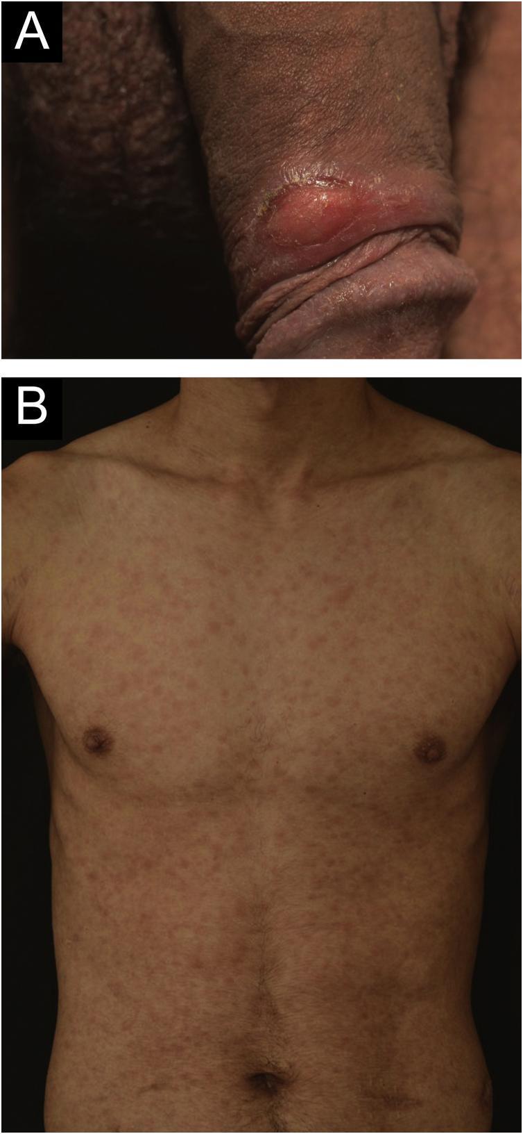
Two-panel clinical photograph illustrating manifestations of syphilis in a 24-year-old male. Panel A (top) is a close-up clinical photograph of the male genitalia showing a solitary, well-demarcated genital ulcer (chancre) located on the foreskin. The lesion exhibits clean, well-defined edges and an erythematous, indurated base, characteristic of primary syphilis. Panel B (bottom) is an anterior clinical photograph of the patient's trunk demonstrating a diffuse, symmetrical cutaneous eruption. The rash consists of multiple, discrete, erythematous maculopapular lesions distributed across the chest and abdomen. These findings represent secondary syphilis occurring concurrently with the resolving primary lesion. The images serve as an educational resource for identifying the clinical progression of Treponema pallidum infection, particularly in immunocompromised patients such as kidney transplant recipients.
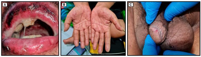
A series of three clinical photographs demonstrating multisystem manifestations of a systemic infection, likely secondary syphilis with concurrent secondary manifestations. (A) Close-up of the oral cavity showing significant inflammation, characterized by edematous, erythematous, and crusted lips. Thick, white pseudomembranous plaques are visible on the hard and soft palates, accompanied by viscous, purulent hypersalivation. (B) Bilateral palmar view showing a disseminated maculopapular rash. The lesions are reddish-copper in color, discrete, and non-confluent, typical of a secondary syphilitic eruption. (C) Genital examination revealing a single, well-demarcated, painless indurated ulcer (chancre) located on the penile shaft adjacent to the glans, consistent with primary syphilis. These visuals illustrate the potential overlap of primary and secondary syphilis stages in an immunocompromised or complex clinical presentation. Educational focus includes dermatologic and mucosal signs of Treponema pallidum infection, clinical morphology of genital ulcers, and secondary palmar syphilids.
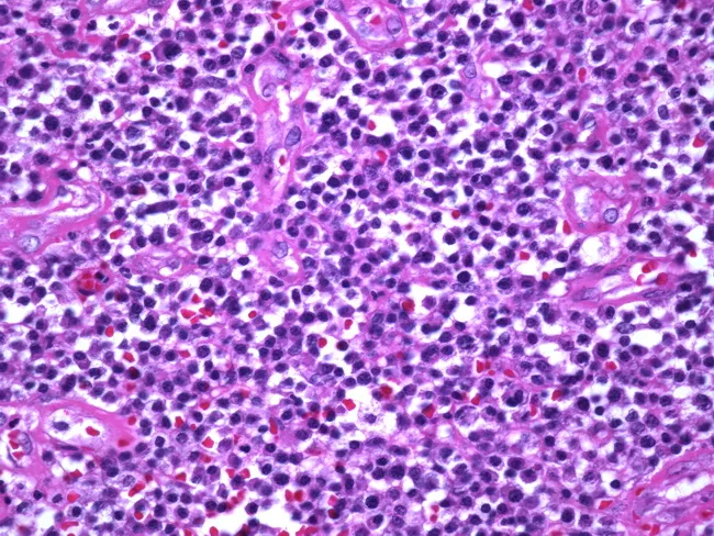
Histopathology image from a cutaneous syphilitic chancre after Hematoxylin and Eosin staining demonstrates a dense inflammatory infiltrate in the dermis with a perivascular pattern. The cellular milieu is dominated by plasma cells, accompanied by lymphocytes and histiocytes. Endothelial swelling and small-vessel endarteritis are evident, forming the classic histologic triad associated with Treponema pallidum infection. The epidermis may be relatively preserved with only mild spongiosis; necrosis is minimal. Although non-specific in isolation, the combination of perivascular plasma cell–rich infiltrate and endarteritis strongly points toward primary syphilis in the appropriate clinical context. Special stains or immunohistochemistry can reveal spirochetes, though they may be absent in this field. Clinically relevant for differentiating chronic inflammatory dermatoses and serving as an educational exemplar of syphilitic histopathology. Diagnostic significance lies in supporting early syphilis when integrated with serology (RPR/VDRL, FTA-ABS/TP-PA) and clinical findings; informs treatment decisions and epidemiologic interventions; useful for medical education, dermatopathology reviews, and infectious disease reference repositories.
Syphilis - Microbiology
The Pathogen
- T. pallidum subsp. pertenue - causes yaws
- T. pallidum subsp. endemicum - causes endemic syphilis (bejel)
- Treponema carateum - causes pinta
Morphology & Structure
- Thin, helical (corkscrew-shaped) spirochete
- 0.15 μm wide, 6-15 μm long
- Has 6 to 14 spirals, tapered at both ends
- Cannot be seen by ordinary Gram stain (too thin)
- Visualized by:
- Darkfield microscopy (in wet mounts from lesions)
- Silver stain (Warthin-Starry stain)
- Fluorescent antibody methods (DFA-TP)
Unique Structural Features
- Outer membrane is composed predominantly of phospholipids with very few surface-exposed proteins (unlike most bacteria which have protein-rich outer membranes) - this helps the organism evade immune detection
- Six axial fibrils (endoflagella): three attached at each end, overlapping at the center - structurally similar to flagella and responsible for characteristic motility
- Three-axis motility: forward/backward along long axis + rotation on long axis + side-to-side bending
Genome & Cultivation
- Genome contains ~1 million base pairs with only ~1,000 genes - about 1/4 the number of most bacteria
- Lacks significant metabolic capacity (cannot synthesize most amino acids, lipids, nucleotides)
- Cannot be cultured in vitro (obligate intracellular pathogen-like dependence) - lacks the enzymes needed for growth outside a host
- Survives only briefly outside the host
- Sensitive to temperature, drying, soap, and common disinfectants
- All strains are antigenically similar and remain universally susceptible to penicillin
Transmission & Epidemiology
- Acquired through direct contact with infectious lesions (chancres, mucous patches, condylomata lata) during sexual intercourse (vaginal, anal, oral)
- Rarely via: contaminated needles, organ transplantation, or occupational exposure
- Congenital syphilis: transplacental transmission to the fetus
- Risk of acquisition from a single sexual contact with an infected partner: 16-30%
- Fomite transmission is extremely uncommon
- Natural hosts: humans and certain primates only
Clinical Stages
Primary Syphilis
- Appears 2-6 weeks after exposure (incubation 10-90 days)
- Chancre: painless, indurated (hard) ulcer with well-defined, raised borders and a clean base
- Usually solitary; located on genitalia, anus, rectum, lips, or oral cavity
- Associated with regional painless lymphadenopathy
- Heals spontaneously in 3-6 weeks even without treatment
Secondary Syphilis
- Develops in 60-90% of untreated primary syphilis patients
- Occurs 2-8 weeks after chancre appears; represents hematogenous dissemination
- Classic features:
- Maculopapular rash - diffuse, symmetric, copper-red; involves palms and soles (highly characteristic)
- Generalized lymphadenopathy
- Fever, malaise, headache, arthralgia, sore throat
- Condylomata lata - flat, moist, wart-like lesions in moist body areas (highly infectious)
- Mucous patches - painless, silvery-gray patches on oral/genital mucosa
- Patchy alopecia ("moth-eaten" appearance)
- Can involve CNS (meningitis), eyes (uveitis), liver, and kidneys
Latent Syphilis
- Defined by reactive serology without clinical signs
- Early latent: infection within the previous 12 months
- Late latent: infection >12 months (or of unknown duration)
- Not clinically infectious (except for vertical transmission)
Tertiary (Late) Syphilis
| Form | Timing | Features |
|---|---|---|
| Gummatous | 3-10 yrs | Granulomatous lesions (gummata) in skin, bone, liver; central necrosis |
| Cardiovascular | 10-30 yrs | Aortic root endarteritis → aortic aneurysm (especially ascending aorta), aortic regurgitation, coronary ostial stenosis |
| Neurosyphilis | Variable | See below |
Neurosyphilis
- Syphilitic meningitis: within first year; cranial nerve palsies
- Meningovascular syphilis: 5-10 years; endarteritis → cerebrovascular infarction
- General paresis: 10-20 years; chronic meningoencephalitis, dementia, personality change, seizures
- Tabes dorsalis: 20-30 years; degeneration of posterior columns and dorsal roots → progressive ataxia, loss of reflexes, Charcot joints, lancinating pains, urinary incontinence
- Argyll Robertson pupil: small, irregular pupils that accommodate but do not react to light ("prostitute's pupil" - accommodates but doesn't react) - seen in ~90% of tabes dorsalis
Congenital Syphilis
- Rhinitis ("snuffles"), maculopapular rash, hepatosplenomegaly, jaundice, periostitis, osteochondritis, anemia, lymphadenopathy
- Hutchinson's teeth - peg-shaped, notched upper central incisors
- Interstitial keratitis - corneal vascularization and scarring
- Eighth nerve deafness
Laboratory Diagnosis
Direct Detection
- Darkfield microscopy: gold standard for primary lesions - demonstrates motile spirochetes from fresh chancre exudate
- DFA-TP (Direct Fluorescent Antibody for T. pallidum): can distinguish from oral commensals (darkfield of oral lesions is unreliable)
- PCR: sensitive, especially for congenital syphilis and neurosyphilis (CSF)
Serological Tests
| Test | Use |
|---|---|
| VDRL (Venereal Disease Research Laboratory) | Screening; only validated CSF test for neurosyphilis |
| RPR (Rapid Plasma Reagin) | Screening; quantitative monitoring of treatment response |
- Both detect antibodies to cardiolipin (a non-specific antigen)
- Used for quantitative monitoring of treatment: a 4-fold decrease in titer = adequate response
- Can give false positives (Jarisch-Herxheimer reaction, autoimmune disease, pregnancy, viral infections)
- Prozone phenomenon: very high antibody titers can cause a false-negative result; diluting the serum resolves this
| Test | Use |
|---|---|
| FTA-ABS (Fluorescent Treponemal Antibody-Absorption) | Confirmatory; not for routine screening |
| TP-PA (T. pallidum Particle Agglutination) | Confirmatory; can be quantified and automated |
| EIA / CIA (Enzyme/Chemiluminescence Immunoassay) | Confirmatory; increasingly used for screening |
- Once positive, treponemal tests remain positive for life (even after successful treatment)
- Used to confirm positive nontreponemal tests
Sensitivity of Serological Tests by Stage
| Stage | VDRL (%) | FTA-ABS (%) | TP-PA (%) |
|---|---|---|---|
| Primary | 70 | 85 | 50-60 |
| Secondary | 99 | 100 | 100 |
| Latent/Late | 70 | 98 | 98 |
- Secondary syphilis is the only stage where serologic testing is nearly 100% reliable
- RPR titers are highest in secondary syphilis (usually ≥1:16)
- Most false-positives have titers <1:8
Treatment
| Stage | Drug of Choice |
|---|---|
| Primary, secondary, early latent | Benzathine penicillin G 2.4 million units IM single dose |
| Late latent / tertiary (non-CNS) | Benzathine penicillin G 2.4 million units IM weekly × 3 doses |
| Neurosyphilis | Aqueous crystalline penicillin G IV × 10-14 days |
| Penicillin allergy | Doxycycline or ceftriaxone (desensitize if pregnant) |
Clinical Images