
Cardiomyopathies detailed explanation
Your credits are exhausted. You can purchase a top-up or subscribe to a plan to continue using Orris. If you're already subscribed, your daily credits will reset tomorrow.
Cardiomyopathy in detail
Reading File
Reading File
Finding Sources
Finding Sources
Finding Sources
Searching PubMed
cardiomyopathies
Searching the Web
2023 ESC guidelines cardiomyopathies diagnosis management
Reading File
Reading File
Cardiomyopathy is a disease of the heart muscle that can impair pumping or filling, and may lead to heart failure, arrhythmias, thromboembolism, or sudden cardiac death. Major forms include dilated, hypertrophic, arrhythmogenic, restrictive, and left-ventricular noncompaction cardiomyopathy. Many are inherited, while others arise from inflammation, toxins such as alcohol, endocrine/metabolic disease, pregnancy, or persistent tachyarrhythmia. Current ESC guidance emphasizes phenotype definition, cardiac imaging, cause-directed investigation, genetic counselling/testing where appropriate, and family screening (ESC guideline overview).
A detailed study of cardiomyopathy can be organized as:
- Definition and classification
- Causes and genetics
- Dilated cardiomyopathy (DCM)
- Hypertrophic cardiomyopathy (HCM)
- Arrhythmogenic cardiomyopathy (ACM/ARVC)
- Restrictive cardiomyopathy (RCM)
- Left ventricular noncompaction and secondary forms
- Symptoms, signs, investigations, and differential diagnosis
- Treatment, devices, transplantation, and sudden-death prevention
- Family screening, genetic counselling, prognosis, and follow-up
Which section should I explain in full first?
Asking for Preferences
Choose the cardiomyopathy topic to cover in detail.
Your credits are exhausted. You can purchase a top-up or subscribe to a plan to continue using Orris. If you're already subscribed, your daily credits will reset tomorrow.Cardiomyopathy is a disease of the heart muscle that can impair pumping or filling, and may lead to heart failure, arrhythmias, thromboembolism, or sudden cardiac death. Major forms include dilated, hypertrophic, arrhythmogenic, restrictive, and left-ventricular noncompaction cardiomyopathy. Many are inherited, while others arise from inflammation, toxins such as alcohol, endocrine/metabolic disease, pregnancy, or persistent tachyarrhythmia. Current ESC guidance emphasizes phenotype definition, cardiac imaging, cause-directed investigation, genetic counselling/testing where appropriate, and family screening (ESC guideline overview).
A detailed study of cardiomyopathy can be organized as:
- Definition and classification
- Causes and genetics
- Dilated cardiomyopathy (DCM)
- Hypertrophic cardiomyopathy (HCM)
- Arrhythmogenic cardiomyopathy (ACM/ARVC)
- Restrictive cardiomyopathy (RCM)
- Left ventricular noncompaction and secondary forms
- Symptoms, signs, investigations, and differential diagnosis
- Treatment, devices, transplantation, and sudden-death prevention
- Family screening, genetic counselling, prognosis, and follow-up
Which section should I explain in full first?Cardiomyopathy is a disease of the heart muscle that causes abnormal contraction, relaxation, wall thickness, chamber size, or electrical stability. It can lead to heart failure, arrhythmias, stroke from intracardiac thrombus, and sudden cardiac death.
For a detailed study, choose one section:
-
Classification and causes
- Genetic versus acquired causes
- Dilated, hypertrophic, arrhythmogenic, restrictive, and noncompaction phenotypes
-
Dilated cardiomyopathy
- Causes, clinical features, echocardiography, heart-failure treatment, ICD/CRT, transplant
-
Hypertrophic cardiomyopathy
- Obstructive versus non-obstructive disease, murmur, diagnosis, sudden-death risk, medicines, septal reduction
-
Arrhythmogenic and restrictive cardiomyopathies
- ARVC/ACM, cardiac amyloidosis, endomyocardial fibrosis, sarcoidosis
-
Investigations and management overview
- ECG, echocardiography, cardiac MRI, genetic testing, family screening, anticoagulation, device therapy
Reply with a number, and I will explain that section in depth.
Explain cardiomyopathies by type in a comparison table
Reading File
Finding Sources
Finding Sources
Finding Sources
Searching PubMed
cardiomyopathies
Searching the Web
2023 ESC Guidelines management cardiomyopathies phenotypes dilated hypertrophic arrhythmogenic restrictive
Reading File
Reading File
Reading File
Finding Sources
Cardiomyopathies: comparison by type
Cardiomyopathies are disorders of the myocardium that cause impaired contraction, impaired relaxation, abnormal chamber size/wall thickness, or electrical instability. The traditional major patterns are dilated, hypertrophic, and restrictive cardiomyopathy. Modern classification also includes arrhythmogenic cardiomyopathy and non-dilated left-ventricular phenotypes. 2023 ESC guidance emphasizes defining both the phenotype and the underlying cause, including genetic disease.
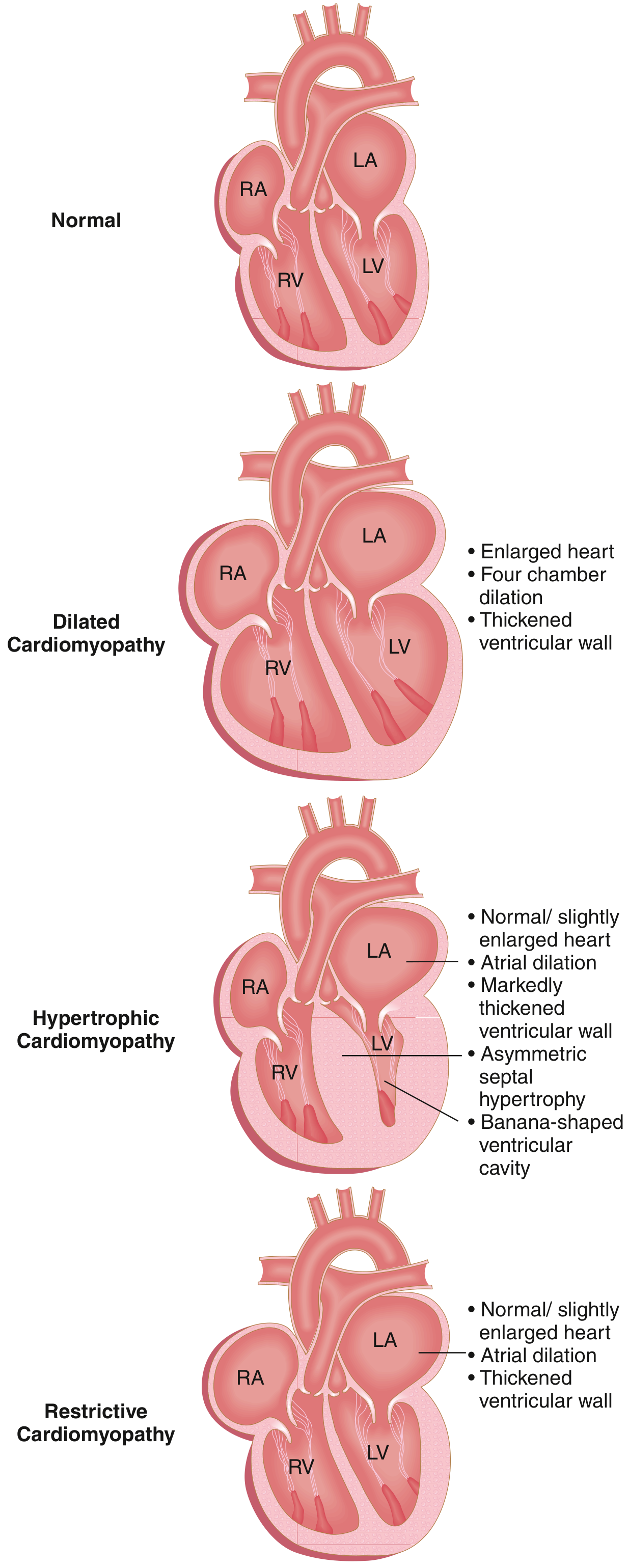
Dilated cardiomyopathy chiefly causes systolic dysfunction; hypertrophic and restrictive cardiomyopathies chiefly cause diastolic dysfunction. Robbins & Kumar Basic Pathology, p. 370.
| Type | Core structural/functional abnormality | Common causes | Typical presentation | Key investigations/findings | Major complications | Main management principles |
|---|---|---|---|---|---|---|
| Dilated cardiomyopathy (DCM) | Dilated LV, often both ventricles; reduced systolic function and reduced LVEF. Functional mitral/tricuspid regurgitation may occur. | Idiopathic/genetic; myocarditis; alcohol; anthracyclines and other cardiotoxins; tachycardia-induced disease; thyroid/metabolic disease; nutritional deficiency; peripartum disease; Chagas disease. | Progressive exertional dyspnea, fatigue, orthopnea, edema, palpitations. May first present with embolic stroke, ventricular arrhythmia, or sudden death. | Echo: enlarged LV and global hypokinesia with low EF. ECG: nonspecific ST-T changes, conduction delay, atrial/ventricular arrhythmias. Cardiac MRI helps identify scar/inflammation. Coronary imaging may exclude ischemic cardiomyopathy. | HFrEF, atrial fibrillation, ventricular tachycardia/fibrillation, LV mural thrombus and systemic embolism, sudden cardiac death. | Treat reversible cause. Guideline-directed HFrEF therapy: ARNI/ACE inhibitor/ARB, evidence-based beta-blocker, mineralocorticoid receptor antagonist, SGLT2 inhibitor, and diuretics for congestion. Anticoagulate for AF or documented LV thrombus. Consider ICD/CRT when indicated after optimized therapy; LV assist device or transplant in advanced disease. |
| Hypertrophic cardiomyopathy (HCM) | Unexplained LV hypertrophy, usually asymmetric septal hypertrophy. LV systolic function is often normal or hyperdynamic, but relaxation is impaired. Some have dynamic LV outflow tract obstruction, called obstructive HCM. | Usually autosomal-dominant sarcomeric gene variants, commonly involving beta-myosin heavy chain or myosin-binding protein C. Must distinguish from hypertension, aortic stenosis, and infiltrative/storage disease. | Often asymptomatic. Exertional dyspnea, chest discomfort, palpitations, presyncope/syncope. Sudden death may occur, especially in young people with high-risk features. | Echo/CMR: LV wall thickness usually ≥15 mm in an adult without another adequate cause; asymmetric septal hypertrophy; systolic anterior motion of mitral valve; resting or provoked LVOT gradient. ECG often shows LVH, deep T-wave inversion, or AF. CMR identifies fibrosis. | AF and embolic stroke, diastolic heart failure, LVOT obstruction, ventricular arrhythmia, sudden cardiac death, apical aneurysm in some. | Avoid dehydration and unnecessary vasodilators in obstructive disease. First-line symptom control: nonvasodilating beta-blocker; alternatives include verapamil or diltiazem in appropriate patients. Add disopyramide or a cardiac myosin inhibitor in selected obstructive HCM under specialist care. Septal myectomy or alcohol septal ablation for persistent severe obstruction and symptoms. Anticoagulate all patients with HCM plus AF unless contraindicated. ICD for previous cardiac arrest/sustained VT or selected high-risk patients. |
| Arrhythmogenic cardiomyopathy (ACM), including ARVC | Progressive myocardial loss with fibrosis or fibrofatty replacement. Classically affects the RV, but may be biventricular or LV predominant. Electrical disease can precede obvious structural change. | Usually inherited, often desmosomal gene variants. Endurance exercise can accelerate disease expression and arrhythmic risk. | Palpitations, exertional syncope, ventricular tachycardia, or sudden cardiac arrest, frequently in adolescents/young adults and athletes. | ECG: T-wave inversion in V1-V3 or beyond, epsilon waves in some, ventricular ectopy/VT often with LBBB morphology. Echo and CMR show regional RV/LV dysfunction, dilation, and scar. Diagnosis uses integrated clinical, ECG, imaging, pathology, and genetic criteria. | Sustained VT/VF, sudden death, progressive RV or biventricular failure, atrial arrhythmias. | Restrict high-intensity/endurance exercise. Beta-blockers and antiarrhythmics may reduce arrhythmia burden but do not eliminate risk. Catheter ablation for recurrent VT. ICD for secondary prevention and selected high-risk primary prevention. Screen and counsel first-degree relatives. |
| Restrictive cardiomyopathy (RCM) | Stiff, noncompliant ventricles with severe diastolic dysfunction; ventricular size is usually normal or only mildly enlarged, while both atria become markedly enlarged. EF may remain near normal until late. | Amyloidosis is a leading cause. Others: Fabry disease, hemochromatosis, sarcoidosis, radiation fibrosis, scleroderma, endomyocardial fibrosis, hypereosinophilic syndrome, carcinoid disease, and rare genetic forms. | Exercise intolerance and congestive symptoms, often with dominant right-sided failure: edema, ascites, hepatomegaly, abdominal discomfort. | Echo: biatrial enlargement with restrictive filling pattern and relatively nondilated ventricles. CMR and nuclear imaging can help identify amyloid. Serum/urine monoclonal-protein tests and genetic testing may establish cause. Differentiate from constrictive pericarditis. | Severe right and left heart failure, AF, thromboembolism, pulmonary hypertension, conduction disease. | Treat the underlying cause whenever possible: for example, disease-specific treatment for AL or transthyretin amyloidosis, iron reduction for hemochromatosis, enzyme therapy for Fabry disease. Use cautious diuresis for congestion because these patients are preload-sensitive. Manage AF and anticoagulate when appropriate. Advanced cases may need transplant evaluation. |
| Left-ventricular noncompaction cardiomyopathy (LVNC) | Prominent LV trabeculations with deep intertrabecular recesses, often at the apex/lateral wall. It may coexist with dilated, hypertrophic, or normal LV phenotype. | Genetic cardiomyopathy; may occur with neuromuscular or metabolic disorders. Sometimes detected in pregnancy or athletes and may represent physiological trabeculation rather than disease. | Variable: asymptomatic, HF symptoms, palpitations, syncope, or embolic event. | Echo and CMR demonstrate excessive trabeculation relative to compacted myocardium. Diagnosis requires phenotype and clinical context, not trabeculation alone. | HF, AF/VT, LV thrombus, embolic stroke, sudden death in affected high-risk individuals. | Treat the actual phenotype: HFrEF therapy if systolic dysfunction, antiarrhythmic/device treatment based on arrhythmic risk, anticoagulation for AF, LV thrombus, or selected high-risk cases. Consider genetic evaluation and family screening if cardiomyopathy is established. |
| Peripartum cardiomyopathy | New LV systolic dysfunction and HF occurring toward the end of pregnancy or in the months after delivery, after excluding another cause. Usually a DCM phenotype. | Multifactorial. Risk is higher with prior peripartum cardiomyopathy, hypertensive disorders of pregnancy, multifetal pregnancy, advanced maternal age, and some genetic susceptibility. | Breathlessness, orthopnea, edema, fatigue, cough, chest discomfort, or palpitations near delivery/postpartum. Can be mistaken for normal pregnancy symptoms. | Echo shows reduced LVEF, often LV dilation. Evaluate for preeclampsia, myocarditis, thrombus, ischemia, and pulmonary embolism as appropriate. | Acute HF, cardiogenic shock, LV thrombus/systemic embolism, arrhythmias, persistent LV dysfunction; recurrence with future pregnancy. | Multidisciplinary cardio-obstetric care. During pregnancy, use pregnancy-compatible HF therapy and avoid ACE inhibitors/ARBs/ARNI/MRA. After delivery, use standard HFrEF therapy when appropriate. Consider anticoagulation when EF is severely reduced or LV thrombus is present. Counsel regarding future pregnancy based on LV recovery. |
| Takotsubo syndrome or stress cardiomyopathy | Acute, usually reversible regional LV systolic dysfunction, often apical ballooning, extending beyond one coronary territory. | Physical or emotional stress; often postmenopausal women. It is not generally an inherited chronic cardiomyopathy. | Sudden chest pain, dyspnea, ECG changes, raised troponin, mimicking acute coronary syndrome. | Coronary angiography shows no culprit obstruction sufficient to explain the dysfunction. Echo/ventriculography/CMR show characteristic regional wall-motion pattern. | Acute HF, pulmonary edema, LVOT obstruction, arrhythmia, LV thrombus, rarely cardiogenic shock or death. | Initially manage as possible acute coronary syndrome until excluded. Then supportive HF care, treat triggers/complications, consider anticoagulation for severe LV dysfunction or apical thrombus. LV function commonly recovers over days to weeks. |
Quick differentiation
| Feature | DCM | HCM | ACM/ARVC | RCM |
|---|---|---|---|---|
| LV cavity | Enlarged | Small/normal | Usually normal or variable | Normal or mildly enlarged |
| Wall thickness | Normal or relatively thin | Increased, often asymmetric septum | RV/LV regional thinning and scar | Usually normal, except infiltrative causes may thicken walls |
| Dominant dysfunction | Systolic | Diastolic, sometimes dynamic obstruction | Arrhythmogenic, later systolic dysfunction | Diastolic |
| Atrial size | May enlarge | LA often enlarged | May enlarge later | Marked biatrial enlargement |
| Characteristic issue | HFrEF and thrombus | LVOT obstruction and sudden-death risk | VT/VF in young people | Infiltration/fibrosis and right-sided congestion |
| Key clinical clue | Displaced apex, S3, HF | Harsh systolic murmur that rises with Valsalva/standing | Exercise-related palpitations or syncope | Raised JVP, edema, ascites, Kussmaul sign |
General approach to every suspected cardiomyopathy
- ECG, transthoracic echocardiography, ambulatory rhythm monitoring, and cardiac MRI when needed.
- Search for reversible causes: coronary disease, alcohol/toxins, tachyarrhythmia, endocrine/metabolic disease, myocarditis, infiltrative disease.
- Obtain a three-generation family history of cardiomyopathy, heart failure, pacemaker/ICD implantation, premature sudden death, and unexplained syncope.
- Offer genetic counselling/testing and clinical screening to relatives when an inherited form is suspected.
Restrictive cardiomyopathy is characterized by noncompliant ventricles, high filling pressures, marked atrial enlargement, and often relatively prominent right-sided symptoms. Amyloidosis is the commonest infiltrative cause. Harrison’s Principles of Internal Medicine, 22nd ed., p. 270.
Seek urgent assessment for syncope, new sustained palpitations, chest pain, severe breathlessness at rest, pulmonary edema, or a family history of sudden cardiac death.
This is a shared conversation. Sign in to Orris to start your own chat.