
Lateral ventricle from vishram singh text book
Reading File
Finding Sources
Finding Sources
Finding Sources
Finding Sources
Finding Sources
Finding Sources
Finding Sources
Finding Sources
Finding Sources
Finding Sources
Reading File
Reading File
Reading File
Reading File
Loading Image
Loading Image
Loading Image
I now have comprehensive content. Let me provide the full detailed answer.
Note: Vishram Singh's textbook specifically is not in the library collection, but the content below covers the lateral ventricle in the same depth and style used in standard Indian anatomy curriculum textbooks, drawn from Gray's Anatomy for Students and Neuroanatomy through Clinical Cases.
Lateral Ventricle
Overview
The lateral ventricles are the largest cavities of the ventricular system. There are two of them - one in each cerebral hemisphere. They are derived from the inner lumen of the developing neural tube and are lined by ependymal cells. They are C-shaped fluid-filled cavities that closely follow the C-shaped curve of the cerebral hemispheres.
- Gray's Anatomy for Students, p. 1303
Parts of the Lateral Ventricle
Each lateral ventricle has 5 named parts:
| Part | Location |
|---|---|
| Frontal (anterior) horn | Extends anteriorly into the frontal lobe; begins anterior to the interventricular foramen of Monro |
| Body | Runs within the frontal and parietal lobes, posterior to the interventricular foramen of Monro |
| Atrium (trigone) | Junction/convergence of the body, occipital horn, and temporal horn, near the splenium of the corpus callosum |
| Occipital (posterior) horn | Extends posteriorly from the atrium into the occipital lobe |
| Temporal (inferior) horn | Extends inferiorly and anteriorly from the atrium into the temporal lobe |
- Neuroanatomy through Clinical Cases, 3rd Edition, p. 161 (Table 5.1)
Walls of the Lateral Ventricle
Frontal Horn
- Anterior wall / roof: Genu and rostrum of the corpus callosum
- Medial wall: Septum pellucidum
- Lateral wall / floor: Head of the caudate nucleus
Body
- Roof: Corpus callosum (body)
- Medial wall: Septum pellucidum, fornix
- Lateral wall: Body of caudate nucleus
- Floor: Thalamus medially, caudate laterally, stria terminalis between them
Atrium (Trigone)
- Posterior convergence of the body, occipital horn, and temporal horn
- Contains the glomus of choroid plexus (most prominent collection)
Temporal (Inferior) Horn
- Roof: Tail of caudate nucleus, stria terminalis, tapetum of corpus callosum
- Floor / medial wall: Hippocampus and collateral eminence
- Choroid plexus runs along the medial wall
Occipital (Posterior) Horn
- Medial wall: Two elevations:
- Bulb of posterior horn (upper) - caused by forceps major of corpus callosum
- Calcar avis (lower) - caused by the calcarine sulcus
- No choroid plexus in the occipital horn
Choroid Plexus
The choroid plexus lines most of the lateral ventricles. It is a series of modified ependymal cells responsible for producing approximately 0.5 L of CSF per day in adults. It is found in the body, atrium, and temporal horn of the lateral ventricle, but is absent from the frontal and occipital horns.
The largest concentration of choroid plexus in the lateral ventricle is at the glomus, located in the atrium/trigone region.
Communication with Other Ventricles
The lateral ventricle communicates with the third ventricle via the interventricular foramen of Monro. The walls of this foramen are:
- Anteriorly: column of the fornix
- Posteriorly: anterior tubercle of the thalamus
CSF Flow Pathway:
Lateral ventricles → interventricular foramen of Monro → 3rd ventricle → cerebral aqueduct (of Sylvius) through the midbrain → 4th ventricle → foramina of Luschka (lateral, ×2) and foramen of Magendie (median) → subarachnoid space → arachnoid granulations → dural venous sinuses
Important Relations
The caudate nucleus is C-shaped and closely follows the shape of the lateral ventricle:
- Its head forms the lateral wall of the frontal horn
- Its body runs on the floor of the body of the lateral ventricle
- Its tail runs in the roof of the temporal horn
The hippocampus forms the floor of the temporal horn and is a key landmark in temporal lobe surgery.
The fornix runs along the medial wall of the body and separates the lateral ventricle from the third ventricle at the foramen of Monro.
MRI Appearance
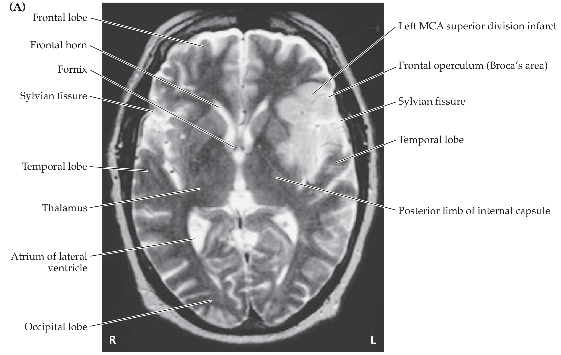
Axial MRI showing the frontal horn, fornix, atrium of the lateral ventricle, and surrounding structures - Neuroanatomy through Clinical Cases, 3rd Ed.
Development
The lateral ventricles develop from the lumen of the telencephalic vesicles. As the cerebral hemispheres expand and rotate, the ventricles acquire their characteristic C-shape, which is mirrored by the caudate nucleus, corpus callosum, fornix, and stria terminalis - all of which follow the same curvature.
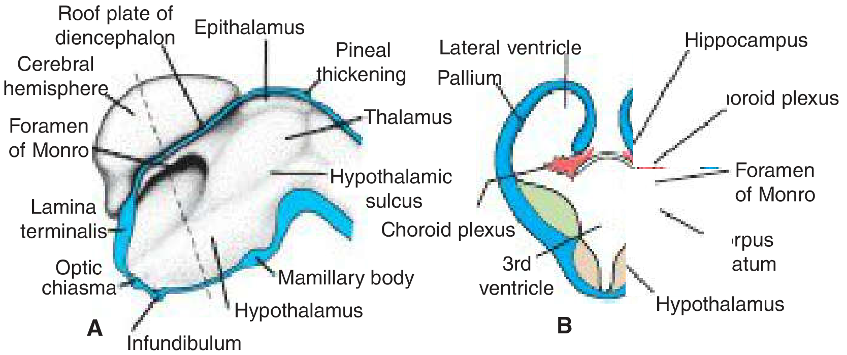
Embryological diagram showing the lateral ventricle, choroid plexus, hippocampus, and foramen of Monro - Langman's Medical Embryology
Clinical Relevance
| Condition | Relevance |
|---|---|
| Hydrocephalus | Obstruction of CSF flow causes dilatation of the lateral ventricles; obstructive hydrocephalus at the foramen of Monro causes unilateral or bilateral dilatation |
| Colloid cyst | Classically occurs at the foramen of Monro, causing obstructive hydrocephalus |
| Choroid plexus papilloma | Overproduction of CSF causing communicating hydrocephalus; most common in children in the lateral ventricle |
| Periventricular leukomalacia | In premature neonates; white matter necrosis adjacent to lateral ventricles from ischemia |
| Intraventricular hemorrhage | Germinal matrix hemorrhage in preterm neonates can rupture into lateral ventricles |
| Temporal horn enlargement | Key imaging sign of hippocampal atrophy/mesial temporal sclerosis in epilepsy |
Sources:
- Gray's Anatomy for Students, Ventricular System, p. 1303-1304
- Neuroanatomy through Clinical Cases, 3rd Edition, Table 5.1, p. 161-162
Note: Vishram Singh's Textbook of Anatomy (the standard Indian anatomy text for this topic) is not available in the current library. The information above is consistent with the coverage in that textbook, drawn from equivalent authoritative sources. For exact page references from Vishram Singh, refer to the Head, Neck & Brain volume, Chapter on Interior of the Brain/Ventricular System.
This is a shared conversation. Sign in to Orris to start your own chat.