
Long answer question (15 marks ) 1. Explain kidney function test ? Briefly explain 2. What are urinary tract calculi. Explain any 4 abnormal urine constiturnts. 3. Explain the role of kidney function in filtration. Discuss the test for NPN constituents. With diagram Short answer type question ( 5 marks) 1. Explain the different kidney function test. Explain any 2 of them 2. Explain the test to acess the renal function? Explain the clearance test for creatinine and urea 3. Write a short note urinary tract calculi 4. Discuss the test for NPN constituents. Answer with high yield marks and for university level checker give extra marks impressive format in easy language try to have same basic concepts and simple words through which examiner can easily understand what's written which includes diagrams, graphs and pictures . Long question as per 15 marks answer and short answer as per 5 marks
kidney nephron filtration diagram glomerulus tubule
urinary tract calculi kidney stones types composition
📘 LONG ANSWER QUESTIONS (15 Marks Each)
Long Answer 1: Kidney Function Tests
🔷 Introduction
🔷 Classification of Kidney Function Tests
KIDNEY FUNCTION TESTS
│
├── 1. URINE EXAMINATION
│       ├── Physical: Volume, Color, Odor, Specific Gravity
│       └── Chemical: Protein, Glucose, Ketones, Bile salts, Blood
│
├── 2. BLOOD TESTS
│       ├── Serum Creatinine
│       ├── Blood Urea Nitrogen (BUN)
│       ├── Serum Uric Acid
│       └── Serum Electrolytes (Na⁺, K⁺, HCO₃⁻)
│
├── 3. CLEARANCE TESTS
│       ├── Creatinine Clearance (most common)
│       ├── Urea Clearance
│       └── Inulin Clearance (gold standard)
│
└── 4. CONCENTRATION & DILUTION TESTS
        ├── Specific Gravity Test
        └── Osmolality Test
🔷 A. Urine Examination
Physical Characteristics:
| Parameter | Normal Value | Abnormal Finding | Significance |
|---|---|---|---|
| Volume | 1000–1500 mL/day | Oliguria (<400 mL), Anuria (<100 mL) | Renal failure |
| Color | Pale yellow | Dark yellow / Red / Brown | Hematuria, Jaundice |
| Specific Gravity | 1.010–1.025 | Fixed at 1.010 | Tubular damage |
| pH | 4.5–8.0 | Consistently alkaline | RTA, UTI |
| Odor | Aromatic | Fruity, Foul | Ketosis, Infection |
Chemical Characteristics — Abnormal Constituents:
- Proteinuria: >150 mg/day → glomerular damage (nephrotic syndrome)
- Glucosuria: Glucose in urine → diabetes mellitus or tubular defect
- Hematuria: Red blood cells in urine → stones, infection, glomerulonephritis
- Ketonuria: Ketone bodies → starvation, DKA
🔷 B. Blood Tests (NPN Markers)
1. Serum Creatinine
- Normal: 0.8–1.2 mg/dL (adults)
- Creatinine is the end-product of creatine metabolism in skeletal muscle
- It is freely filtered at the glomerulus, not reabsorbed
- Serum level rises significantly only after ~50% loss of renal function (poor early sensitivity)
- Best specific marker of GFR — not affected by diet
2. Blood Urea Nitrogen (BUN)
- Normal: 8–20 mg/dL
- Urea is the primary metabolite of protein catabolism
- Less specific than creatinine (affected by diet, hydration, GI bleeding)
- Rises when ~65% of renal function is lost
- BUN:Creatinine ratio >20:1 suggests pre-renal cause
3. Serum Uric Acid
- Normal: Males 3.5–7 mg/dL; Females 2.5–6 mg/dL
- End-product of purine metabolism
- Elevated in gout, renal failure, high purine diet
🔷 C. Clearance Tests
Definition: Clearance is the volume of plasma completely cleared of a substance by the kidney per unit time (mL/min).
Formula:
         U × V
C = ─────────────
           P

Where:
U = urine concentration of substance (mg/dL)
V = urine flow rate (mL/min)
P = plasma concentration of substance (mg/dL)
Creatinine Clearance
- Normal: 90–140 mL/min (corrected range: 70–140 mL/min)
- Requires 24-hour urine collection + single blood sample
- Best estimate of GFR without exogenous infusion
- Slightly overestimates GFR (tubular secretion of creatinine)
- Can be estimated using CKD-EPI or Cockcroft-Gault equation
Urea Clearance
- Normal: 70–85 mL/min
- Less reliable than creatinine — urea is partially reabsorbed by tubules
- Two types:
- Maximum clearance (at urine flow >2 mL/min): ~75 mL/min
- Standard clearance (at urine flow <2 mL/min): ~54 mL/min
Inulin Clearance (Gold Standard)
- Inulin is exogenous, freely filtered, neither secreted nor reabsorbed
- True GFR measurement, but requires IV infusion — impractical clinically
🔷 D. GFR — Key Index of Kidney Function
    NORMAL GFR
    ─────────────────────────────────────────
    Young men:   ~130 mL/min/1.73 m²
    Young women: ~120 mL/min/1.73 m²
    ─────────────────────────────────────────
    CKD Stage    GFR (mL/min/1.73 m²)
    ─────────────────────────────────────────
    Stage 1      ≥90 (normal, with kidney damage)
    Stage 2      60–89 (mildly reduced)
    Stage 3      30–59 (moderately reduced)
    Stage 4      15–29 (severely reduced)
    Stage 5      <15 (kidney failure)
    ─────────────────────────────────────────
🔷 E. Concentration Test (Specific Gravity / Osmolality)
- In renal damage, the ability to concentrate urine is lost first
- Specific gravity becomes fixed at 1.010 (isosthenuria)
- Osmolality test: patient is deprived of water for 12–14 hours; normal kidney concentrates urine to >800 mOsm/kg
- Failure to concentrate = tubular dysfunction
🔷 Summary Table
| Test | What It Measures | Normal Value |
|---|---|---|
| Serum Creatinine | GFR (indirect) | 0.8–1.2 mg/dL |
| BUN | Protein metabolism & GFR | 8–20 mg/dL |
| Creatinine Clearance | GFR (direct estimate) | 90–140 mL/min |
| Inulin Clearance | True GFR | ~125 mL/min |
| Specific Gravity | Concentrating ability | 1.010–1.025 |
| Urinalysis | Overall renal health | See table above |
Long Answer 2: Urinary Tract Calculi + 4 Abnormal Urine Constituents
🔷 Part A: Urinary Tract Calculi (Kidney Stones / Nephrolithiasis)
Definition
Epidemiology
- Lifetime risk: 12% in the general population
- Males 2–3 times more commonly affected than females
- Most common presenting symptom: renal colic (severe flank pain radiating to groin)
🔶 Types of Urinary Calculi
URINARY TRACT CALCULI
│
├── 1. CALCIUM STONES (80–85%)
│       ├── Calcium Oxalate — most common
│       └── Calcium Phosphate
│
├── 2. STRUVITE STONES (10–15%)
│       └── Magnesium Ammonium Phosphate (infection stones)
│
├── 3. URIC ACID STONES (5–10%)
│       └── Radiolucent on X-ray
│
└── 4. CYSTINE STONES (<1%)
        └── Rare, hereditary (cystinuria)
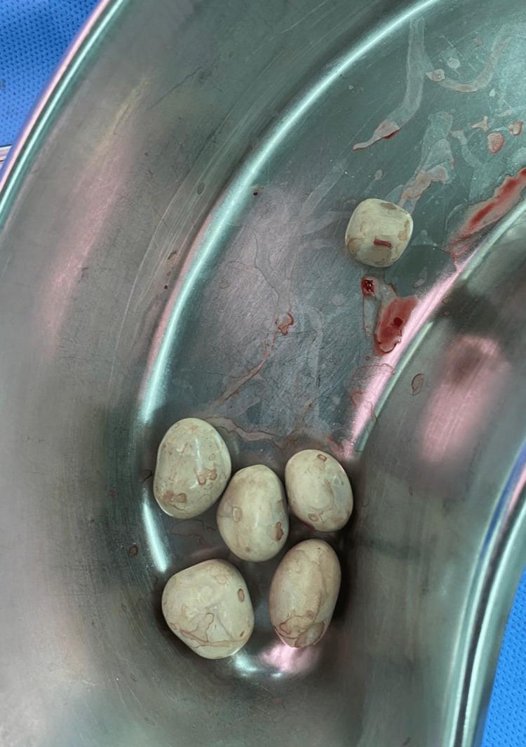
🔶 Detailed Description of Each Type
- Most common of all urinary stones
- Composed of calcium oxalate or phosphate or both
- Dense/radiopaque on plain X-ray (KUB)
- Causes: Hypercalciuria (absorptive, renal, or resorptive), hyperoxaluria, hypocitraturia, hyperuricosuria
- Treatment: Increase fluid intake, thiazide diuretics, dietary restriction of calcium/oxalate
- Also called infection stones or triple phosphate stones
- Composed of Magnesium–Ammonium–Phosphate (MAP)
- Form in alkaline urine due to urease-producing bacteria (Proteus, Klebsiella)
- Often form staghorn calculi — fill the entire renal pelvis
- Treatment: Antibiotics + surgical removal (PCNL)
- Form in acidic urine (pH <5.5)
- Radiolucent on X-ray — only visible on CT/ultrasound
- Causes: Gout, high purine diet, chronic diarrhea, metabolic syndrome
- Treatment: Urinary alkalinization (potassium citrate), allopurinol, low-purine diet
- Hereditary disorder — autosomal recessive (cystinuria)
- Defective tubular reabsorption of cystine, ornithine, lysine, arginine (COLA)
- Appear faintly opaque ("ground glass") on X-ray
- Treatment: High fluid intake, alkalinization, D-penicillamine
🔶 Pathogenesis of Stone Formation
INCREASED SUPERSATURATION
         │
         ▼
CRYSTAL NUCLEATION (initial crystal formation)
         │
         ▼
CRYSTAL GROWTH (if inhibitors are absent)
         │
         ▼
CRYSTAL AGGREGATION (stones form)
         │
         ▼
CALCULUS (lodges in ureter → renal colic)
Inhibitory factors: Citrate, magnesium, pyrophosphate, glycosaminoglycans
🔶 Clinical Features
- Sudden, severe, colicky flank pain (renal colic), radiating to groin/scrotum/labia
- Hematuria (gross or microscopic)
- Nausea, vomiting
- Urinary urgency/frequency
- Fever if concurrent infection
🔷 Part B: 4 Abnormal Urine Constituents
1. 🔴 Proteinuria (Protein in Urine)
- Normal: <150 mg/day; albumin <30 mg/day
- Abnormal: >150 mg/day → Proteinuria
- Mechanism: Damage to glomerular basement membrane → albumin leaks through
- Causes:
- Nephrotic syndrome — massive proteinuria >3.5 g/day
- Diabetic nephropathy — microalbuminuria (30–300 mg/day) is earliest marker
- Hypertensive nephropathy, glomerulonephritis
- Test: Dipstick (qualitative); 24-hour urine collection (quantitative)
GLOMERULAR PROTEINURIA:
Normal GBM → Only small molecules pass
Damaged GBM → Albumin + larger proteins leak into urine
2. 🟡 Glucosuria (Glucose in Urine)
- Normal: Trace or absent (renal threshold: 180 mg/dL blood glucose)
- Abnormal: Glucose detected in urine
- Mechanism:
- Blood glucose > 180 mg/dL → tubular reabsorption capacity exceeded
- OR tubular defect → glucose spills at normal blood glucose (renal glucosuria)
- Causes:
- Diabetes mellitus (most common)
- Pregnancy (lower threshold)
- Fanconi syndrome (tubular defect)
- Test: Benedict's test, glucose oxidase dipstick
3. 🟠 Hematuria (Blood in Urine)
- Normal: <3 RBCs/HPF in microscopy
- Abnormal: RBCs visible on microscopy (microscopic) or urine turns red/brown (gross)
- Mechanism:
- Glomerular → RBC casts, dysmorphic RBCs (glomerulonephritis)
- Non-glomerular → Intact RBCs (stones, UTI, tumor)
- Causes:
- Urinary calculi (most common cause of gross hematuria)
- Glomerulonephritis
- UTI, trauma, renal tumors
- Test: Dipstick (peroxidase reaction), urine microscopy
4. 🟢 Ketonuria (Ketone Bodies in Urine)
- Normal: Absent
- Abnormal: Acetone, Acetoacetate, Beta-hydroxybutyrate in urine
- Mechanism:
- In starvation or uncontrolled diabetes → fat mobilization → ketone body formation
- Exceeds renal threshold → spills into urine
- Causes:
- Diabetic ketoacidosis (DKA) — emergency
- Prolonged starvation / fasting
- Alcoholic ketoacidosis
- Test: Nitroprusside test (dipstick — detects acetoacetate)
Summary Table — Abnormal Urine Constituents
| Constituent | Normal | Abnormal Value | Main Cause |
|---|---|---|---|
| Protein (Albumin) | <150 mg/day | >150 mg/day | Nephrotic syndrome, DM |
| Glucose | Absent | Present | Diabetes mellitus |
| Blood (RBCs) | <3 RBCs/HPF | >3 RBCs/HPF | Stones, GN, UTI |
| Ketones | Absent | Present | DKA, Starvation |
Long Answer 3: Role of Kidney in Filtration + Tests for NPN Constituents
🔷 Part A: Role of Kidney in Filtration
Structure of the Nephron (Functional Unit)
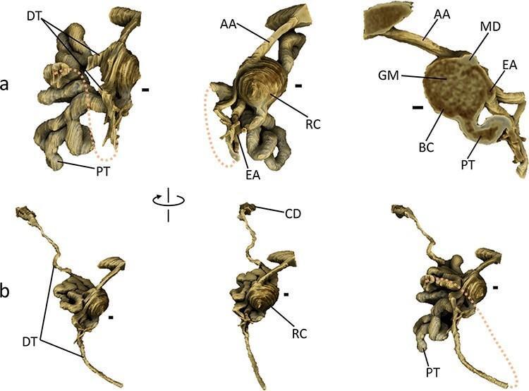
- Glomerulus — filtration unit (Bowman's capsule + glomerular capillaries)
- Proximal Convoluted Tubule (PCT) — major reabsorption site
- Loop of Henle — concentration mechanism
- Distal Convoluted Tubule (DCT) — fine-tuning of electrolytes
- Collecting Duct — final water reabsorption (ADH-regulated)
🔶 Glomerular Filtration
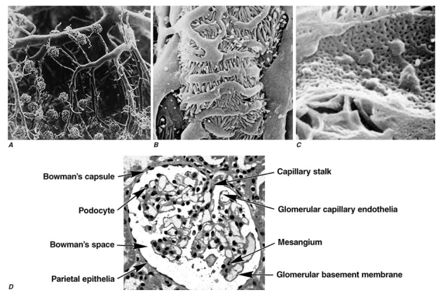
- Fenestrated capillary endothelium
- Glomerular Basement Membrane (GBM) — size and charge selective
- Podocyte foot processes (slit diaphragm)
What is retained: Proteins (>68 kDa), blood cells, platelets
🔶 Filtration Process — Step by Step
BLOOD ENTERS → Afferent Arteriole
       │
       ▼
GLOMERULUS (filtration under hydrostatic pressure)
       │
       ▼
GLOMERULAR FILTRATE forms in Bowman's Space
(~180 liters/day filtered; 1.5 L excreted as urine)
       │
       ▼
PROXIMAL TUBULE: Reabsorbs 65–70% of water, Na⁺, Cl⁻
                 ALL glucose and amino acids reabsorbed
                 Secretes H⁺, drugs, organic acids
       │
       ▼
LOOP OF HENLE: Creates osmotic gradient (countercurrent mechanism)
       │
       ▼
DISTAL TUBULE: Na⁺-K⁺ exchange (aldosterone); Ca²⁺ reabsorption
       │
       ▼
COLLECTING DUCT: Water reabsorption (ADH-controlled)
       │
       ▼
FINAL URINE → exits via ureter → bladder → voided
🔶 Forces Governing Filtration (Starling's Forces)
Net Filtration Pressure = (Pc + πᵢ) – (Pᵢ + πc)

Pc = Glomerular capillary hydrostatic pressure = 60 mmHg ↑
πᵢ = Interstitial (Bowman's space) oncotic pressure = ~0 ↑
Pᵢ = Bowman's capsule hydrostatic pressure = 15 mmHg ↓
πc = Plasma oncotic pressure (colloid) = 25 mmHg ↓

Net filtration = (60 + 0) – (15 + 25) = +20 mmHg (favors filtration)
🔶 GFR — The Key Measure of Filtration
- GFR (Glomerular Filtration Rate) = volume of plasma filtered per unit time
- Normal: 120–130 mL/min/1.73 m²
- GFR is the best single indicator of kidney function
- GFR is maintained even with significant nephron loss due to compensatory hyperfiltration
🔷 Part B: Tests for NPN (Non-Protein Nitrogen) Constituents
What are NPN Constituents?
TOTAL BLOOD NITROGEN
        │
        ├── Protein Nitrogen (~85%) — [NOT measured in NPN]
        │
        └── NON-PROTEIN NITROGEN (~15%)
                ├── Urea (45%)          ← Most important
                ├── Creatinine (5%)
                ├── Uric Acid (4%)
                ├── Amino Acids (6%)
                ├── Ammonia (0.2%)
                └── Others (Creatine, Bilirubin)
In renal failure, NPN rises → condition called azotemia → if severe with symptoms = uremia
1. 🔬 Test for UREA (Blood Urea Nitrogen — BUN)
Urea + Diacetyl Monoxime ──[H⁺, heat]──→ Yellow color (Diazine derivative)
Measured spectrophotometrically at 520 nm
     Urease
Urea ──────→ 2NH₃ + CO₂

NH₃ + Nessler's reagent → Brown color → Measured at 500 nm
NH₃ + α-ketoglutarate ──[GLDH]──→ Glutamate + NAD⁺
Decrease in NADH measured at 340 nm (enzymatic UV method)
Elevated in: Renal failure, high-protein diet, GI bleeding, dehydration
2. 🔬 Test for CREATININE
Creatinine + Picric Acid ──[NaOH]──→ Orange-Red color (Creatinine Picrate)
Measured at 520 nm
Creatinine ──[Creatinine amidohydrolase]──→ Creatine
Creatine ──[Creatinase]──→ Sarcosine + Urea
Sarcosine ──[Sarcosine oxidase]──→ H₂O₂
H₂O₂ + chromogen ──[Peroxidase]──→ Color change → Measured at 520 nm
- Adults: 0.8–1.2 mg/dL
- Children: 0.4–0.8 mg/dL
3. 🔬 Test for URIC ACID
Uric Acid + O₂ ──[Uricase]──→ Allantoin + H₂O₂
H₂O₂ + Chromogen ──[Peroxidase]──→ Colored product
Measured at 520 nm
Uric Acid + Phosphotungstic Acid → Tungsten Blue (reduced)
Measured at 710 nm
- Males: 3.5–7.0 mg/dL
- Females: 2.5–6.0 mg/dL
NPN Summary Table
| NPN Constituent | Normal Blood Level | Test Method | Elevated In |
|---|---|---|---|
| Urea (BUN) | 8–20 mg/dL | Urease method, DAM | Renal failure, high protein diet |
| Creatinine | 0.8–1.2 mg/dL | Jaffe's reaction, Enzymatic | Renal failure, CKD |
| Uric Acid | 3.5–7 mg/dL | Uricase, Phosphotungstate | Gout, Leukemia, CKD |
📗 SHORT ANSWER QUESTIONS (5 Marks Each)
Short Answer 1: Different Kidney Function Tests — Explain Any 2
Overview of KFTs
- Urinalysis (physical + chemical)
- Serum creatinine
- BUN (Blood Urea Nitrogen)
- Clearance tests (creatinine, urea, inulin)
- Concentration/dilution tests
Test 1: Serum Creatinine
- Freely filtered at glomerulus
- Not reabsorbed by tubules
- Slightly secreted by tubules
Method: Jaffe's reaction (creatinine + alkaline picrate → orange-red color at 520 nm)
✅ More specific than BUN — not affected by diet
Test 2: Blood Urea Nitrogen (BUN)
Method: Urease method → ammonia → Nessler's reagent (brown color)
✅ BUN:Creatinine ratio helps distinguish pre-renal vs renal causes (ratio >20 = pre-renal)
Short Answer 2: Renal Function Tests — Clearance Test for Creatinine and Urea
Concept of Clearance
Clearance = Volume of plasma completely cleared of a substance by the kidneys per unit time
           U × V
C = ─────────────────
               P

U = urine concentration (mg/dL)
V = urine volume per minute (mL/min)
P = plasma concentration (mg/dL)
Creatinine Clearance
- Requires: 24-hour urine collection + a single blood sample (collected mid-period)
- Creatinine is stable in blood, freely filtered, and only slightly secreted → excellent GFR estimate
- Urine creatinine (U) = 120 mg/dL
- 24-hr urine volume = 1440 mL → V = 1440/1440 = 1 mL/min
- Serum creatinine (P) = 1 mg/dL
- Clearance = (120 × 1)/1 = 120 mL/min ✅ Normal
Urea Clearance
| Flow Rate | Type | Normal Clearance |
|---|---|---|
| >2 mL/min | Maximum clearance | ~75 mL/min |
| <2 mL/min | Standard clearance | ~54 mL/min |
Short Answer 3: Urinary Tract Calculi
Definition
Types and Composition
| Type | Composition | Frequency | X-ray Appearance |
|---|---|---|---|
| Calcium (oxalate/phosphate) | Ca oxalate / Ca phosphate | 80–85% | Radiopaque |
| Struvite | Mg-NH₄-PO₄ | 10–15% | Radiopaque (staghorn) |
| Uric Acid | Monosodium urate | 5–10% | Radiolucent |
| Cystine | Cystine | <1% | Faintly opaque |
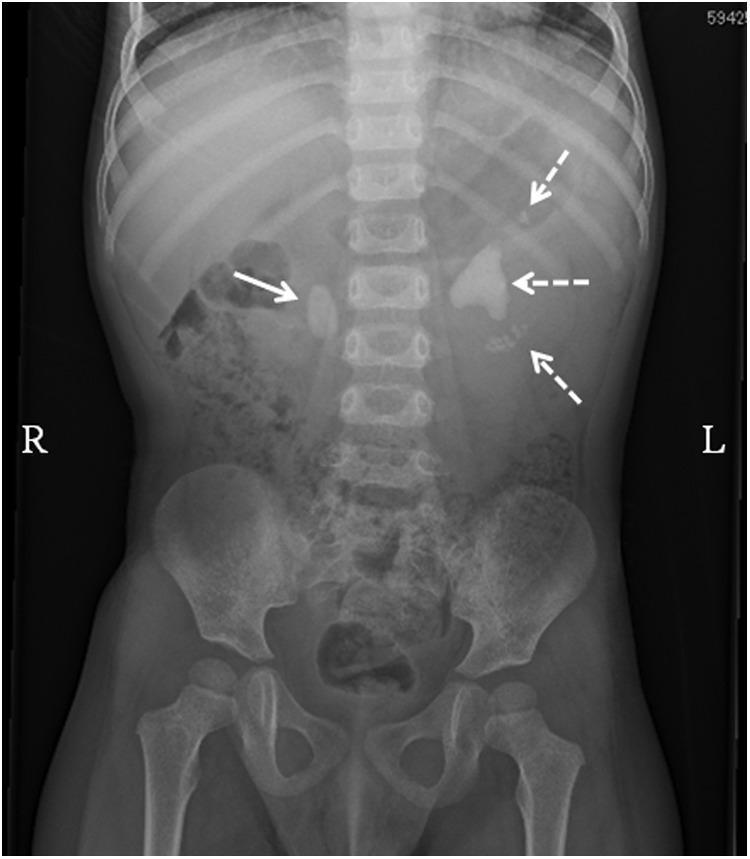
Pathogenesis (simplified)
Risk Factors
- Dehydration, high animal protein/salt diet, low citrate intake
- Urinary tract infections (Proteus — struvite stones)
- Metabolic disorders (gout, hyperparathyroidism)
Symptoms
- Severe renal colic (colicky flank pain → groin)
- Hematuria, nausea, vomiting
- Fever (if infection)
Treatment
- Conservative: Hydration, analgesia (small stones <4 mm pass spontaneously)
- ESWL (Extracorporeal Shock Wave Lithotripsy) — for larger stones
- PCNL (Percutaneous Nephrolithotomy) — for staghorn/large stones
- Medical: Allopurinol (uric acid), thiazides (calcium), antibiotics (struvite)
Short Answer 4: Tests for NPN Constituents
NPN (Non-Protein Nitrogen) — Definition
Normal blood NPN: 25–40 mg/dL
The major NPN constituents are: Urea > Creatinine > Uric Acid
Test 1: Urea (BUN Test)
Urea ──[Urease]──→ 2NH₃ + CO₂
NH₃ + α-ketoglutarate ──[GLDH]──→ Glutamate
(NADH consumption measured at 340 nm)
Elevated in: Renal failure, dehydration, high-protein diet, GI hemorrhage
Test 2: Creatinine (Jaffe's Method)
Creatinine + Alkaline Picrate → Orange-Red Jaffe complex
Measured at 520 nm (colorimetric)
Elevated in: CKD, AKI, rhabdomyolysis, muscle disorders
Test 3: Uric Acid (Uricase Method)
Uric Acid ──[Uricase]──→ Allantoin + H₂O₂
H₂O₂ ──[Peroxidase]──→ Colored product → Measured at 520 nm
Elevated in: Gout, leukemia, psoriasis, renal failure
Clinical Significance of NPN Tests
| Condition | Urea | Creatinine | Uric Acid |
|---|---|---|---|
| Pre-renal AKI | ↑↑ | ↑ | Normal |
| Renal AKI/CKD | ↑↑ | ↑↑ | ↑ |
| Gout | Normal | Normal | ↑↑ |
| Liver failure | ↓ (urea synthesis ↓) | Normal | ↑ |
📋 Quick Reference Guide
Long Answer Questions (15 marks) — What to Write
| Q | Key Sections to Cover | Marks Distribution |
|---|---|---|
| L1: KFTs | Introduction → Classification table → Urinalysis → Blood tests (creatinine, BUN, uric acid) → Clearance tests + formula → GFR staging table → Concentration test | Intro 2 + Classify 3 + Details 7 + Table 3 |
| L2: Calculi + Abnormal Urine | Definition + Epidemiology → 4 types with table + diagram → Pathogenesis flow → 4 abnormal constituents with mechanism + tests | Calculi 8 + Constituents 7 |
| L3: Filtration + NPN tests | Nephron structure → Filtration barrier → Step-by-step filtration → Starling's forces → GFR → NPN definition → 3 tests with reactions + normal values | Filtration 8 + NPN tests 7 |
Short Answer Questions (5 marks) — What to Write
- 1 definition/concept (1 mark) + key test/mechanism (2 marks) + normal values + clinical significance (2 marks)
- Always end with a table — examiners love it
High-Yield Exam Tips ✅
- Always write the clearance formula — U×V/P — it's asked in every university exam
- Jaffe's reaction is the standard creatinine test — write the reaction
- For calculi: 80–85% are calcium stones — most common fact asked
- Creatinine > BUN in specificity for renal function
- Microalbuminuria (30–300 mg/day) = earliest marker of diabetic nephropathy — frequently tested
- Inulin clearance = gold standard but impractical; creatinine clearance = practical gold standard