
I want you to help me prepare a comprehensive case presentation for a medical scenario that is both informative and engaging, ensuring that it adheres to a structured format suitable for academic or clinical review. You are: - A seasoned medical professional with expertise in case presentations - A critical thinker who applies clinical reasoning and evidence-based practices - An educator who understands how to convey complex medical information clearly You think carefully before writing, ensuring that each section of the presentation logically flows into the next while highlighting key insights. The audience: - Medical students preparing for exams - Residents or fellows looking for case presentation examples - Attending physicians seeking concise and relevant patient information Assume they: - Have a foundational understanding of medical terminology - Are looking for clarity and thoroughness in case presentations - Value logical progression and evidence-based conclusions The topic: [PLACEHOLDER: brief description of the medical case] The essential elements to cover in the presentation include: 1. History: [PLACEHOLDER: relevant patient history] 2. Salient examination findings: [PLACEHOLDER: key physical exam results] 3. Provisional and differential diagnosis: [PLACEHOLDER: initial and alternative diagnoses] 4. Investigations: [PLACEHOLDER: necessary tests and results] 5. Final diagnosis: [PLACEHOLDER: confirmed diagnosis] 6. Management: [PLACEHOLDER: treatment plan and follow-up care] Do NOT include: - Unverified claims or anecdotal evidence - Excessive jargon without explanation - Unrelated personal anecdotes or opinions - Overly complex medical terminology that may confuse the audience Structure the presentation with: - Clear headings for each section - A logical flow that connects history to management - Concise bullet points or paragraphs for clarity - Key findings or insights highlighted where appropriate Length: 600 words Tone: Professional, educational, and concise Evidence rules: - Cite relevant guidelines or studies when applicable - Avoid fabricating patient data or outcomes - Clearly distinguish between established facts and clinical judgments Now write the case presentation.
cd /home/daytona/workspace/attachments/d79aa525-6c4e-40d2-9c8e-170f323424f8 && python -m markitdown "TRENDO 2025 POSTER HHS.pptx"
cd /home/daytona/workspace/attachments/d79aa525-6c4e-40d2-9c8e-170f323424f8 && python3 -c " from pptx import Presentation prs = Presentation('TRENDO 2025 POSTER HHS.pptx') for i, slide in enumerate(prs.slides): print(f'=== SLIDE {i+1} ===') for shape in slide.shapes: if hasattr(shape, 'text') and shape.text.strip(): print(shape.text) print() "
pip install --user python-pptx markitdown 2>&1 | tail -5
cd /home/daytona/workspace/attachments/d79aa525-6c4e-40d2-9c8e-170f323424f8 && python3 -c " from pptx import Presentation prs = Presentation('TRENDO 2025 POSTER HHS.pptx') for i, slide in enumerate(prs.slides): print(f'=== SLIDE {i+1} ===') for shape in slide.shapes: if hasattr(shape, 'text') and shape.text.strip(): print(repr(shape.name), ':', shape.text[:2000]) print() "
central pontine myelinolysis hyperglycemia osmotic demyelination
Case Presentation: Central Pontine Myelinolysis Associated with Hyperglycemia
1. History
- Sudden onset difficulty in speaking (dysarthria) and swallowing (dysphagia) — 10 days duration
- Nasal regurgitation, predominantly with liquids
- Unsteadiness while walking (gait ataxia) with bilateral swaying — 10 days duration
- Type 2 diabetes mellitus — 15 years, poorly controlled (irregular medications)
- No prior neurological events documented
2. Salient Examination Findings
| Domain | Finding |
|---|---|
| Consciousness | Alert and oriented to time, place, and person |
| Memory | Intact |
| Speech | Dysarthria present |
| Cranial Nerves I & II | Normal |
| CN III/IV/VI | Full extraocular movements; bilateral gaze-evoked nystagmus; pupils equally reactive |
| CN V | Normal |
| CN VII | Bilateral eye closure weakness |
| CN IX/X | Reduced gag reflex bilaterally; reduced palatal movements bilaterally |
| CN XII | Reduced tongue movement bilaterally; spasticity noted; no fasciculations |
- Tone: Normal bilaterally | Power: 5/5 bilaterally
- Deep tendon reflexes: Sluggish bilaterally
- Plantar response: Flexor bilaterally
- Bilateral finger-nose incoordination
- Bilateral knee-heel incoordination
Clinical Insight: The combination of bilateral lower cranial nerve palsies (IX, X, XII), cerebellar signs, and dysarthria without frank limb weakness localizes the lesion to the brainstem — specifically the pons and its connections.
3. Provisional and Differential Diagnosis
| Diagnosis | Distinguishing Features |
|---|---|
| Brainstem stroke (ischemic/hemorrhagic) | Acute onset, vascular risk factors; focal deficits; no osmolar derangement |
| Brainstem encephalitis | Fever, CSF pleocytosis, inflammatory markers |
| Multiple sclerosis | Younger patient, relapsing-remitting course, oligoclonal bands |
| Wernicke's encephalopathy | Ophthalmoplegia, confusion, thiamine deficiency; responds to thiamine replacement |
| Neuromyelitis optica (NMOSD) | Optic neuritis, transverse myelitis; AQP4-IgG positive |
| Pontine glioma | Gradual onset, mass effect on MRI |
4. Investigations
- Hb: 13.2 g/dL | PCV: 37.2% | TLC: 7,700/mm³ | Platelets: 2.46 L | ESR: 10 mm/hr
| Parameter | Value | Reference |
|---|---|---|
| Random blood glucose (admission) | 538 mg/dL | 70–140 mg/dL |
| Urea | 33.9 mg/dL | Normal |
| Creatinine | 1.37 mg/dL | 0.7–1.4 mg/dL |
| Urine ketones | Negative | Negative |
| Na⁺ (30/05/23) | 133 mEq/L | 135–145 mEq/L |
| Na⁺ (06/06/23) | 138 mEq/L | 135–145 mEq/L |
| K⁺ | 3.58 mEq/L | Normal |
| Serum osmolality | 302 mOsm/L | 275–295 mOsm/L |
| LFTs | Normal | Normal |
Critical Finding: Sodium was normonatremic throughout admission (133 → 138 mEq/L), yet serum osmolality was elevated. This distinguishes this case as hyperglycemia-induced CPM — not the classical hyponatremia-correction-related ODS.
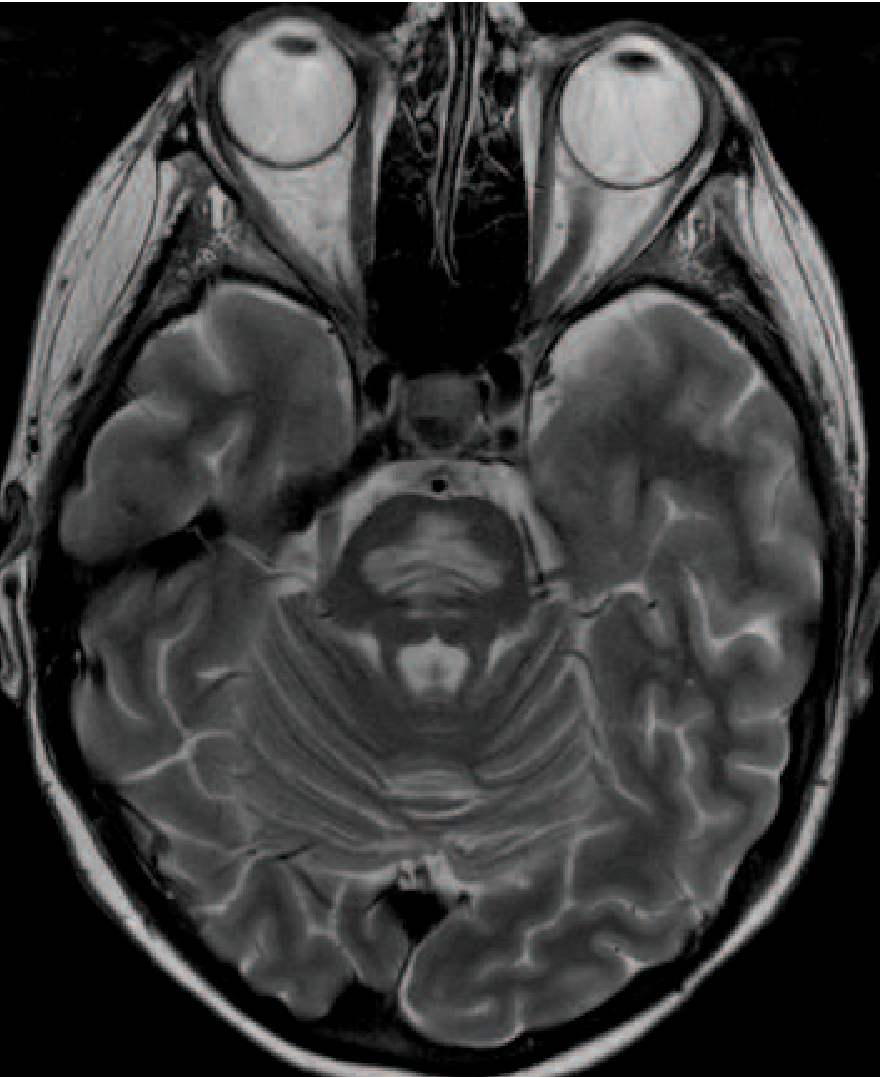
5. Final Diagnosis
- Severe hyperglycemia → hyperosmolar state → osmotic stress on the blood-brain barrier
- Endothelial cell injury → disruption of the blood-brain barrier → vasogenic edema
- Fluctuating osmotic forces → cellular edema compressing fibre tracts → demyelination
- Rapid osmolality correction during treatment (hydration + insulin) → abrupt fluid shifts → oligodendrocyte injury
6. Management
- Glycaemic control: Cautious correction of hyperglycemia — avoid precipitous drops in plasma glucose/osmolality
- Target: reduction in serum osmolality ≤3 mOsm/kg/hr
- Glucose reduction: ~50–75 mg/dL/hr (not faster)
- Fluid resuscitation: IV normal saline for volume replacement; careful monitoring of fluid balance
- Electrolyte monitoring: Frequent sodium, potassium, and glucose checks (every 2–4 hours)
- Supportive care: Nasogastric tube feeding given dysphagia; speech and language therapy referral
- Aspiration precautions: Head-of-bed elevation, thickened fluids (when oral intake resumed)
- Strict diabetic control — regular HbA1c monitoring, structured medication adherence
- Neurological follow-up: serial MRI brain at 4–6 weeks (MRI lesions may evolve or partially resolve)
- Physiotherapy for gait rehabilitation
- Patient education on the dangers of medication non-compliance in diabetes
Teaching Points
- CPM/ODS is not exclusively a complication of hyponatremia correction — hyperglycemia alone can induce osmotic demyelination through blood-brain barrier disruption and vasogenic edema.
- The clinical triad of dysarthria, dysphagia, and cerebellar ataxia in a diabetic with severe hyperglycemia should prompt MRI pons and consideration of ODS.
- Normonatremia does not exclude ODS — serum osmolality is the critical parameter.
- Management hinges on controlled, gradual correction of the metabolic derangement rather than aggressive normalisation.
- MRI brain (T2/FLAIR) remains the investigation of choice — symmetric pontine hyperintensity is pathognomonic.
- Harrison's Principles of Internal Medicine, 22nd Ed. (2025), Chapter 318 — Osmotic Demyelination Syndrome
- Robbins, Cotran & Kumar: Pathologic Basis of Disease — Demyelinating Diseases, p. 1158
- Sun WP et al. "A rare presentation of central pontine myelinolysis secondary to hyperglycaemia." BMC Endocr Disord 2023. PMID 37165361
- Jain E et al. "Osmotic Demyelination After Rapid Correction of Hyperosmolar Hyperglycemia." Cureus 2023. PMID 36874309
What was done
| Section | Notable Choice |
|---|---|
| History | Emphasized the 10-day sub-acute onset and poorly-controlled diabetes as the contextual red flag |
| Exam | Presented as a table for rapid scanning; added a localization insight for the pons |
| Differentials | Structured as a comparison table — aids exam-style recall |
| Investigations | Highlighted normonatremia explicitly, since this is the atypical/teaching point of the case |
| Final Diagnosis | Included a 4-step pathophysiology chain specific to hyperglycemia-driven ODS |
| Management | Anchored correction rate targets to Harrison's guideline figures |
| MRI Image | Included the T2 pontine signal image from Harrison's for visual reinforcement |