
Left coronary arteries
left coronary artery branches anatomy diagram LAD circumflex
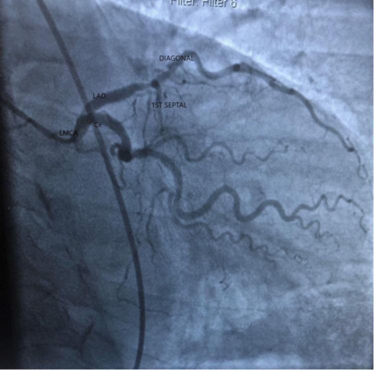
Diagnostic coronary angiogram of the left coronary system. The image shows the Left Main Coronary Artery (LMCA) bifurcating into the Left Anterior Descending (LAD) artery and the Circumflex (Cx) artery. The LAD is visualized proximally, giving off the first septal branch. Distal to the septal branch, a prominent diagonal branch is visible, exhibiting a critical high-grade stenosis (approximately 90%). The circumflex artery demonstrates significant obstructive disease with an estimated 80-90% stenosis in its mid-segment. The diagonal branch appears moderately tortuous compared to the main vessels. This image serves as a clinical demonstration of multi-vessel coronary artery disease (CAD), specifically involving major branches of the left coronary system. Key anatomical landmarks including the LMCA, LAD, Cx, and branching septal/diagonal vessels are labeled to illustrate arterial anatomy and the localization of atherosclerotic lesions for diagnostic and interventional planning.
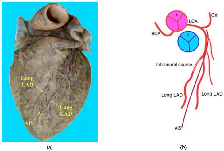
This educational image consists of a clinical photograph (a) and a schematic diagram (b) illustrating a dual left anterior descending (LAD) coronary artery variant. The photograph displays a dissected human heart specimen in the anterior view, highlighting the course of the coronary vasculature. Labeled structures include the Anterior Interventricular Sulcus (AIS) and two distinct 'Long LAD' branches descending parallel to each other. The schematic representation details the origin of these vessels from the Left Coronary Artery (LCA), alongside the Right Coronary Artery (RCA) and Circumflex Artery (CX). A critical anatomical feature demonstrated is the 'intramural course' of the right-sided LAD branch, where a portion of the vessel is embedded within the myocardium (myocardial bridge) before resurfacing. This variant is clinically significant for cardiothoracic surgery and interventional cardiology, as it represents a deviation from standard coronary anatomy (specifically resembling Pellegrini Type XIII) that can impact diagnostic interpretation and surgical planning for revascularization.
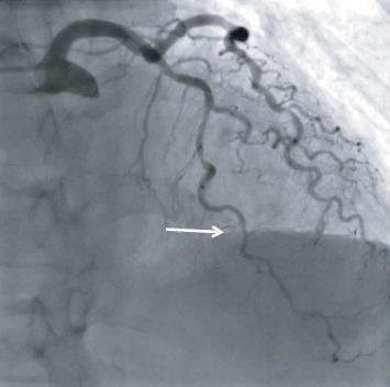
This diagnostic coronary angiogram, captured in the right anterior oblique (RAO) cranial view, illustrates the anatomy and pathology of the left coronary system. The image displays a diffusely diseased and calcified left anterior descending artery (LAD). A white arrow points to an intermediate stenosis located in the midportion of the LAD, characterized by a visible narrowing of the luminal diameter compared to the proximal and distal segments. The distal LAD and several diagonal branches show subtle tortuosity and signs of atherosclerosis. The left circumflex artery (LCx) is also visible, exhibiting mild luminal irregularities. This clinical imaging is essential for evaluating coronary artery disease and determining the functional significance of lesions, often in conjunction with physiological assessments like Fractional Flow Reserve (FFR). It serves as an educational tool for cardiology training, specifically in identifying stenotic lesions and interpreting angiographic projections during cardiac catheterization.
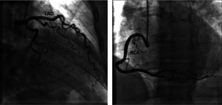
This diagnostic image is a coronary angiogram consisting of two panels demonstrating normal coronary anatomy. The left panel shows the left coronary system with labeled Left Anterior Descending (LAD) and Left Circumflex (LCx) arteries. The LAD is seen coursing anteriorly and inferiorly, giving off diagonal branches, while the LCx travels within the atrioventricular groove, exhibiting normal tapering and branching without evidence of luminal narrowing, stenosis, or occlusion. The right panel illustrates the Right Coronary Artery (RCA) in a characteristic 'C' shape, originating from the right coronary sinus and extending along the posterior interventricular groove. The RCA appears smooth with patent distal flow and visible side branches. This invasive imaging study serves as a clinical reference for angiographically normal epicardial coronary arteries, typically used in cardiology and radiology to rule out obstructive coronary artery disease (CAD) or vasospastic disorders in the setting of chest pain or ischemic ECG changes.
Left Coronary Arteries - Anatomy & Clinical Relevance
Origin
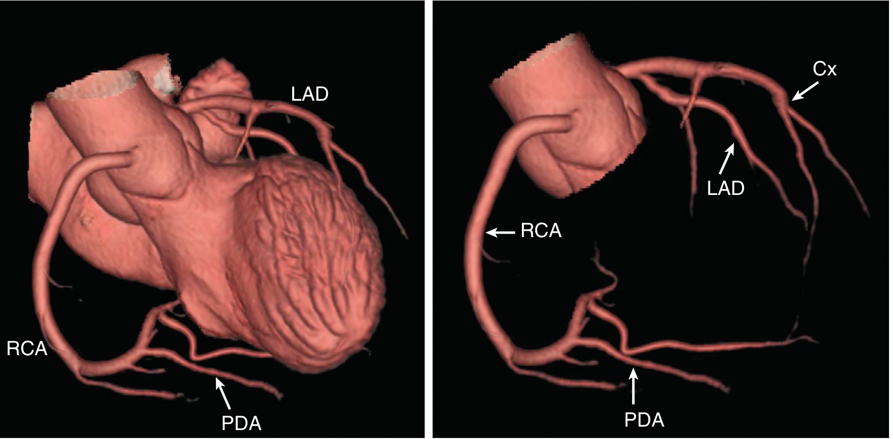
Main Branches
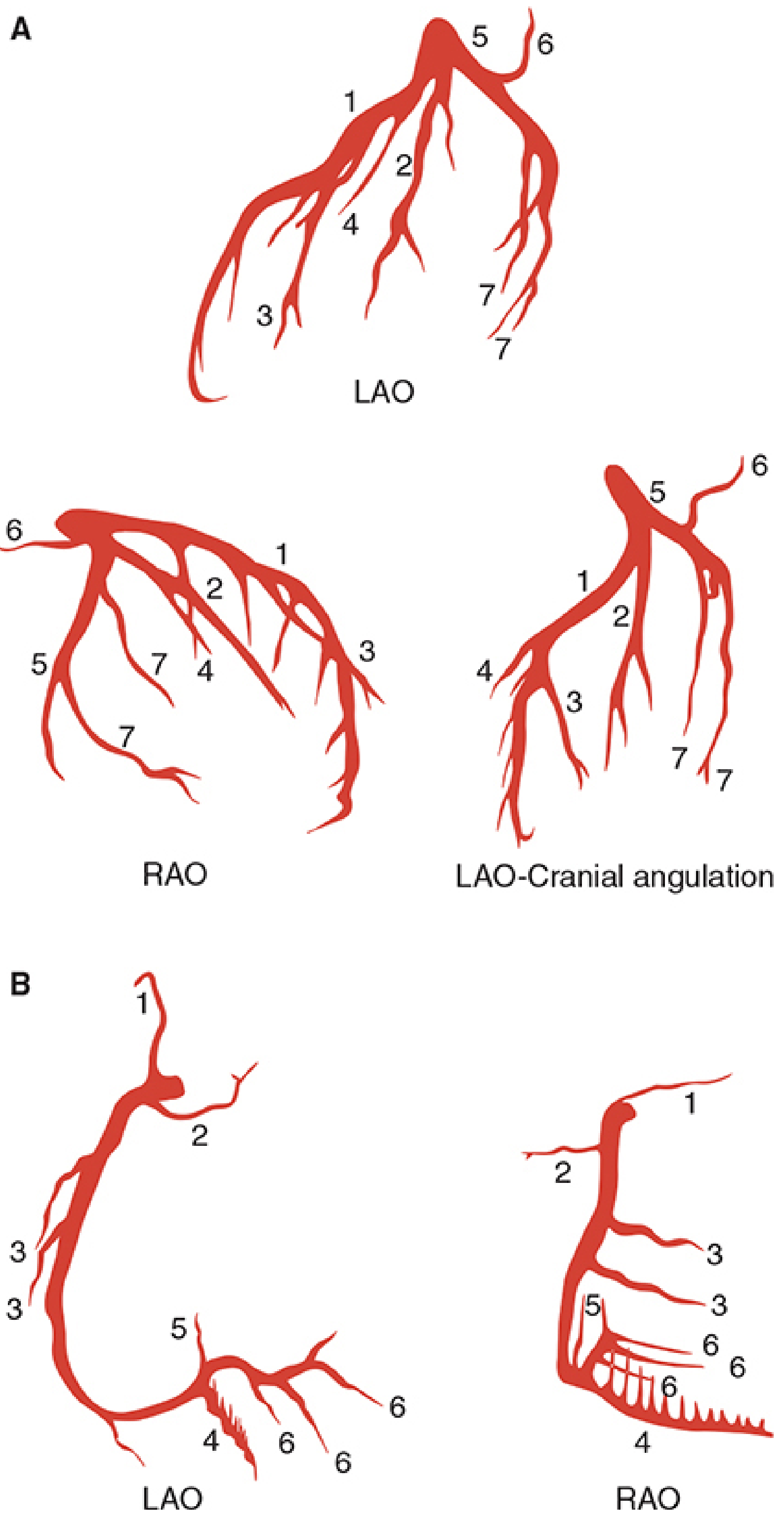
1. Left Anterior Descending Artery (LAD)
- Septal perforating branches - supply the anterior two-thirds of the interventricular septum
- Diagonal branches - supply the anterolateral wall of the left ventricle
- First septal branch - largest and most important of the septal perforators
2. Left Circumflex Artery (LCx / Cx)
- Obtuse marginal (OM) branches - supply the lateral and posterolateral LV free wall; this is the circumflex artery's main territory
- Left atrial circumflex artery - supplies the left atrium
- Posterior descending artery (PDA) - only in left-dominant systems (10-20% of patients; see Dominance section below)
3. Ramus Intermedius (Ramus Medianus) - Optional Third Branch
Territory Supplied
| Structure | Artery |
|---|---|
| Anterior LV wall | LAD (diagonal branches) |
| Anterior 2/3 of interventricular septum | LAD (septal perforators) |
| LV apex | LAD +/- PDA (variable) |
| Lateral LV wall | LCx (obtuse marginal branches) |
| Posterolateral LV wall | LCx or RCA (variable) |
| Left atrium | LCx (left atrial branch) |
| AV node | RCA (in most); LCx (in left-dominant) |
Coronary Dominance
- Right dominant (~80% of people): RCA gives rise to the PDA
- Left dominant (~10-15%): LCx gives rise to the PDA - in these cases the LCx is a much larger and more significant artery
- Co-dominant: Both RCA and LCx contribute to the PDA (~5-10%)
Anatomical Variations (from Netter's Atlas)
- Short LAD: The LAD may be very short, with the apical and inferior sternocostal surface then supplied by branches from the PDA curving around the apex
- Absent PDA: The area is supplied by small branches from the LCx and RCA
- Elongated LAD: The LAD curves around the apex to supply the inferior apical wall normally supplied by PDA
- Left-dominant circulation: PDA arises from LCx instead of RCA
Clinical Significance
- Left main stem disease: Stenosis of the LMCA carries the worst prognosis of any coronary disease without intervention. Because it supplies a large proportion of the LV, any significant stenosis is treated with revascularization (CABG preferred over PCI for most cases). The artery is inaccessible at its origin, so grafts are anastomosed to its distal branches (LAD and OM branches of LCx).
- LAD occlusion ("widow maker"): Proximal LAD occlusion causes anterior STEMI, affecting the anterior wall and septum - often a large territory infarct with high mortality.
- LCx occlusion: Causes lateral or inferolateral wall MI. In left-dominant patients, occlusion can cause inferior MI as well.