
Aquavorin
Aquaporins are a family of membrane channel proteins that facilitate the rapid, selective transport of water across cell membranes. They are found in virtually all living organisms - bacteria, plants, and animals. In humans, 13 subtypes (AQP0-AQP12) have been identified, each with distinct tissue distribution and function.
Key points:
- They form tetrameric complexes in the lipid bilayer, with each monomer containing its own water pore
- The selectivity filter (the "ar/R" region) excludes ions and protons while allowing water through
- Some aquaporins (aquaglyceroporins) also transport glycerol and other small solutes
- Clinically relevant in conditions like nephrogenic diabetes insipidus, brain edema, and neuromyelitis optica (AQP4 antibodies)
Searching sources...
Reading File
Searching the Web
aquaporin water channels structure function physiology 2025
Now let me search the medical library for aquaporin content:
Finding Sources
Searching PubMed
aquaporin water channel physiology disease
Finding Sources
Good - let me read the dedicated aquaporin section from Ganong's physiology and the kidney textbook:
Reading File
Reading File
Reading File
Reading File
Let me also get the essential role of AQP2 section and the Goodman & Gilman pharmacology context:
Reading File
Reading File
I now have rich content from multiple authoritative sources. Let me also fetch the aquaporin structure diagram:
Loading Image
Loading Image
I now have comprehensive information from multiple authoritative sources. Here is the full answer:
Aquaporins (AQPs) - Water Channel Proteins
Overview
Aquaporins are a family of small (~30 kDa) integral membrane proteins that function as selective water channels, enabling rapid water diffusion across cell membranes. The first aquaporin (AQP1) was identified in 1991 by Peter Agre, work for which he was awarded the Nobel Prize in Chemistry (2003). To date, 13 mammalian aquaporins (AQP0-AQP12) have been cloned, expressed in virtually every tissue involved in fluid transport.
Molecular Structure
The diagram from Goodman & Gilman's illustrates the architecture:
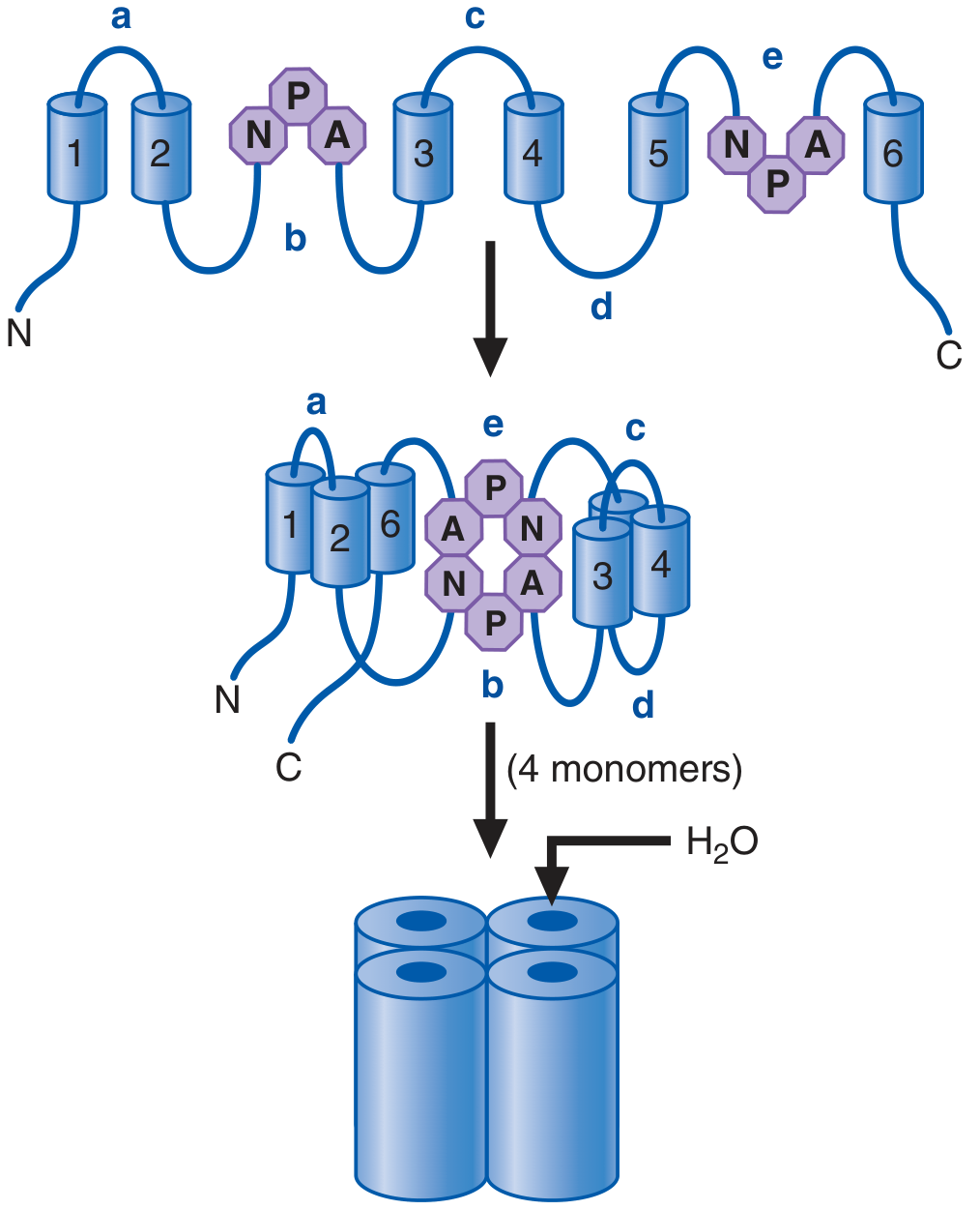
- Each monomer has 6 transmembrane (TM) helical domains with intracellular N- and C-termini
- Loops b and e each contain the signature asparagine-proline-alanine (NPA) motif - these loops dip into the membrane and their two NPA sequences align to form the actual water pore
- TM domains 1, 2, and 6 cluster together; domains 3, 4, and 5 juxtapose on the other side
- Four monomers assemble into a tetramer in the membrane, though each monomer functions as its own independent water pore
- The pore geometry excludes protons (H⁺) and ions - water molecules pass in single file, and their orientation at the NPA region prevents proton conductance (the "Grotthuss mechanism" is blocked)
Classification
| Class | Members | Permeability |
|---|---|---|
| Strict aquaporins | AQP0, AQP1, AQP2, AQP4, AQP5, AQP6 | Water only |
| Aquaglyceroporins | AQP3, AQP7, AQP8, AQP9 | Water + glycerol (+ larger solutes for AQP9) |
| Dual water/ion channels | AQP0, AQP1, AQP6 | Water + ions (controversial/context-dependent) |
Renal Aquaporins - The Key Four
At least seven aquaporins are expressed in distinct kidney segments. The four most physiologically important are:
AQP1
- Location: Apical and basolateral membranes of proximal tubule; thin descending limbs of long-loop nephrons; descending vasa recta
- Function: Allows water to follow the osmotic gradient set up by active solute transport in the proximal tubule, keeping tubular fluid isosmotic. Responsible for ~60-70% of filtered water reabsorption
- Knockout phenotype: AQP1-null mice show 80% reduction in proximal tubular water permeability, severe polyuria, and inability to concentrate urine even after water deprivation - confirming AQP1 is essential for the countercurrent multiplication mechanism
AQP2
- Location: Exclusively in principal cells of the connecting tubule and collecting duct (apical plasma membrane + intracellular vesicles)
- Function: The vasopressin-regulated water channel - the master switch for urine concentration
- Regulation by vasopressin (AVP):
- AVP binds V2 receptor → adenylyl cyclase activation → ↑cAMP → PKA activation
- PKA phosphorylates AQP2 at Ser256 - this is the critical phosphorylation site
- AQP2-containing vesicles traffic to and fuse with the apical plasma membrane (exocytosis)
- Water permeability increases dramatically; water is reabsorbed from the tubule lumen
- On AVP withdrawal, AQP2 is internalized by clathrin-mediated endocytosis and recycled back to subapical vesicles
- Chronic dehydration/AVP stimulation also upregulates AQP2 mRNA and protein expression via cAMP
The fluorescence and electron microscopy images below directly demonstrate AQP2 trafficking:
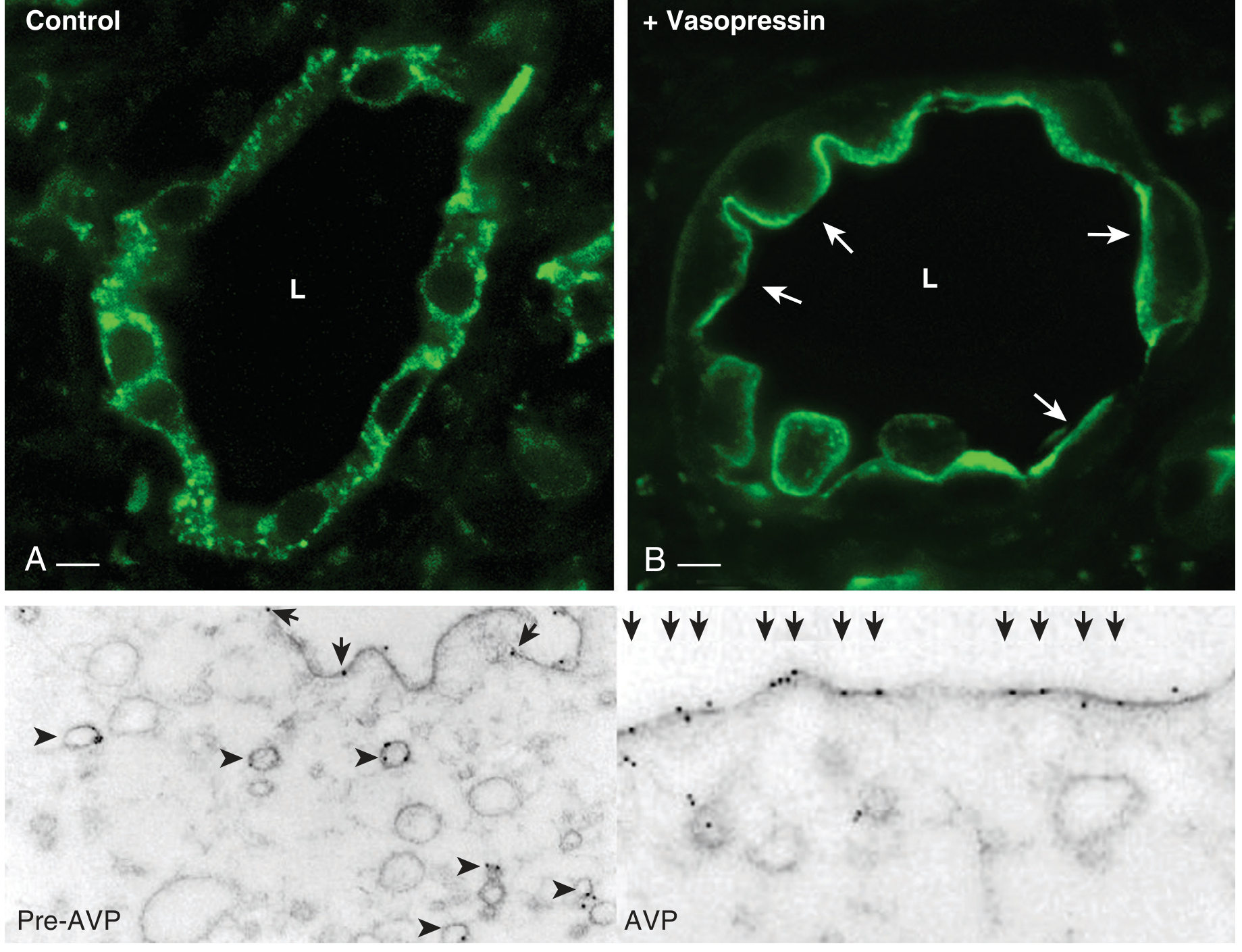
- Left (Control/Pre-AVP): AQP2 (green) is scattered in intracellular vesicles. Gold particles are on cytoplasmic vesicles with only a few at the apical membrane
- Right (+ Vasopressin/AVP): AQP2 accumulates at the apical plasma membrane (arrows); cytoplasmic vesicle labeling decreases dramatically
AQP3 and AQP4
- Location: Basolateral membranes of collecting duct principal cells (exit pathways for water reabsorbed apically by AQP2)
- AQP3 dominates in the cortical collecting duct; AQP4 in the inner medullary collecting duct
- AQP3 knockout mice: >10-fold increase in urine volume (polyuria), markedly reduced urinary osmolality
- AQP4 knockout mice: Only modestly reduced maximal urine concentrating ability (reduced AQP4 inner medullary permeability is partially compensated because most osmotic reabsorption occurs in the cortex where AQP3 is dominant)
Other Notable Aquaporins
| AQP | Key Location | Clinical / Physiological Relevance |
|---|---|---|
| AQP0 (MIP) | Lens fiber cells | Cataract formation when mutated |
| AQP4 | Brain astrocyte endfeet, spinal cord | Target of autoantibodies in neuromyelitis optica spectrum disorder (NMOSD) |
| AQP5 | Salivary/lacrimal/sweat glands, type I alveolar cells | Saliva and tear production |
| AQP7 | Adipocytes, proximal tubule apical brush border | Glycerol release from fat; glucose metabolism |
| AQP9 | Hepatocytes | Glycerol uptake for gluconeogenesis |
Pharmacological Regulation
From Goodman & Gilman's:
- NSAIDs (e.g., indomethacin): Enhance antidiuretic response to vasopressin by inhibiting prostaglandin synthesis (PGs attenuate vasopressin's effect; NSAIDs remove this brake)
- Carbamazepine, chlorpropamide: Enhance vasopressin's antidiuretic effects (mechanism incompletely understood)
- Lithium: Inhibits V2 receptor-mediated adenylyl cyclase stimulation → reduces AQP2 trafficking → causes nephrogenic diabetes insipidus (NDI)
- Demeclocycline: Attenuates antidiuretic effects of vasopressin by reducing cAMP accumulation; used historically to treat SIADH
- AVP-induced cAMP-PKA activation also directly upregulates AQP2 mRNA/protein expression with chronic stimulation
Clinical Diseases
| Disease | AQP Involved | Mechanism |
|---|---|---|
| Nephrogenic diabetes insipidus (NDI) | AQP2 | Loss-of-function mutations in AQP2, or V2R mutations; also acquired (lithium, demeclocycline) |
| Central diabetes insipidus | AQP2 | Absent AVP → no AQP2 membrane insertion |
| Neuromyelitis optica (NMOSD) | AQP4 | IgG autoantibodies target AQP4 on astrocytic endfeet → complement-mediated destruction of spinal cord/optic nerve |
| Cataract | AQP0 | Mutations in the lens major intrinsic protein |
| Brain edema | AQP4 | AQP4 facilitates cytotoxic edema; AQP4 deletion reduces edema in some models |
| Glymphatic dysfunction | AQP4 | AQP4 on astrocyte endfeet drives perivascular CSF-interstitial fluid exchange; linked to Alzheimer's disease pathology (Keil et al., Science 2025) |
Sources:
- Brenner and Rector's The Kidney, 2-Volume Set - Chapter 10 (AQP sections)
- Ganong's Review of Medical Physiology, 26th Ed. - Chapter 37 (Aquaporins)
- Goodman & Gilman's The Pharmacological Basis of Therapeutics - Chapter 29 (Vasopressin/AQP pharmacology)
- Verkman AS. Structure and function of aquaporin water channels. Am J Physiol Renal Physiol 2000
This is a shared conversation. Sign in to Orris to start your own chat.