
Teach me general anatomy. From skeletal system to lymphatic system.
Teach me general anatomy. From skeletal system to lymphatic system. Frm basics to pro
- Anatomical basics: terminology, planes, directions, body cavities, tissues
- Skeletal system: bone types and structure, axial and appendicular skeleton, joints
- Muscular system: muscle types, major muscle groups, actions, innervation
- Nervous system: CNS, PNS, autonomic system, cranial and spinal nerves
- Special senses: eye, ear, taste, smell
- Integumentary system: skin and its appendages
- Endocrine system: glands and their anatomy
- Cardiovascular system: heart, arteries, veins, circulation
- Respiratory system: airways, lungs, pleura
- Digestive system: GI tract, liver, pancreas, biliary tree
- Urinary system: kidneys, ureters, bladder, urethra
- Reproductive systems: male and female
- Lymphatic and immune system: vessels, nodes, spleen, thymus, drainage
- Basics: You learn a shared language. Standard anatomical position, planes (sagittal, coronal, transverse), and directional terms (proximal/distal, superficial/deep) let you describe any structure the same way. The four tissue types are epithelial, connective, muscle, and nervous. Every organ is built from them.
- Structure by system: The skeleton (206 bones, with joints) gives support and protection. Muscles attach to bone and move it. The nervous and endocrine systems control and coordinate the body. The cardiovascular, respiratory, digestive, and urinary systems handle transport, gas exchange, nutrient intake, and waste removal. The reproductive system handles reproduction. The lymphatic system drains tissue fluid back to the blood and supports immunity.
- Advanced level: You move to regional and clinical anatomy. That covers relations between structures, blood supply and nerve supply, surface landmarks, variations, and how injury or disease shows up (for example, a nerve injury after a fracture, or lymph node spread of cancer).
Which topic should we start with? I'll go deep on it, then we can continue in order. · What depth do you want?
- Anatomical basics: terminology, planes, directions, body cavities, tissues
- Skeletal system: bone types and structure, axial and appendicular skeleton, joints
- Muscular system: muscle types, major muscle groups, actions, innervation
- Nervous system: CNS, PNS, autonomic system, cranial and spinal nerves
- Special senses: eye, ear, taste, smell
- Integumentary system: skin and its appendages
- Endocrine system: glands and their anatomy
- Cardiovascular system: heart, arteries, veins, circulation
- Respiratory system: airways, lungs, pleura
- Digestive system: GI tract, liver, pancreas, biliary tree
- Urinary system: kidneys, ureters, bladder, urethra
- Reproductive systems: male and female
- Lymphatic and immune system: vessels, nodes, spleen, thymus, drainage
- Basics: You learn a shared language. Standard anatomical position, planes (sagittal, coronal, transverse), and directional terms (proximal/distal, superficial/deep) let you describe any structure the same way. The four tissue types are epithelial, connective, muscle, and nervous. Every organ is built from them.
- Structure by system: The skeleton (206 bones, with joints) gives support and protection. Muscles attach to bone and move it. The nervous and endocrine systems control and coordinate the body. The cardiovascular, respiratory, digestive, and urinary systems handle transport, gas exchange, nutrient intake, and waste removal. The reproductive system handles reproduction. The lymphatic system drains tissue fluid back to the blood and supports immunity.
- Advanced level: You move to regional and clinical anatomy. That covers relations between structures, blood supply and nerve supply, surface landmarks, variations, and how injury or disease shows up (for example, a nerve injury after a fracture, or lymph node spread of cancer).
anatomical planes and directional terms
human skeleton anterior view axial and appendicular
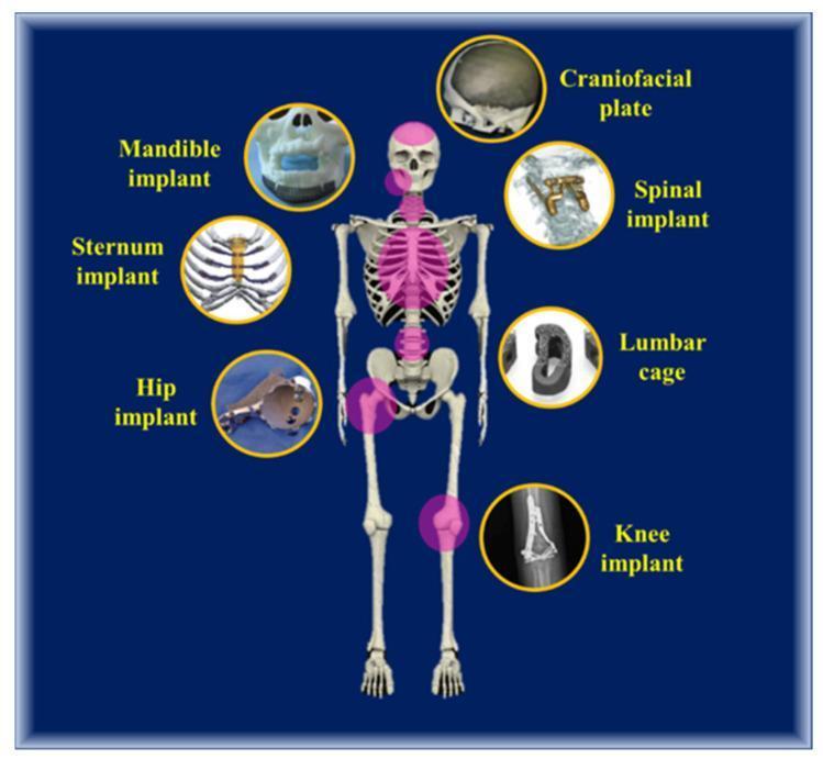
An educational infographic showcasing various orthopedic and maxillofacial metallic implants used in human surgery. A central anterior-view human skeleton acts as a reference, with pink highlights indicating the anatomical regions for implantation. Circular insets surrounding the skeleton provide close-up views of specific devices: 1) Craniofacial plate for skull reconstruction; 2) Mandible implant for jaw stabilization; 3) Sternum implant with a latticework structure for chest wall reconstruction; 4) Hip implant featuring a femoral stem and acetabular cup; 5) Spinal implant showing stabilization hardware in a cervical model; 6) Lumbar cage for intervertebral fusion with visible porous/hollow architecture; 7) Knee implant shown via a lateral x-ray radiography view. These implants are primarily manufactured from biocompatible titanium alloys (Ti-6Al-4V) using additive manufacturing (3D printing) techniques like Direct Metal Laser Sintering (DMLS). The infographic illustrates how patient-specific, bio-mimetic designs facilitate load-bearing support and tissue integration across the axial and appendicular skeleton.
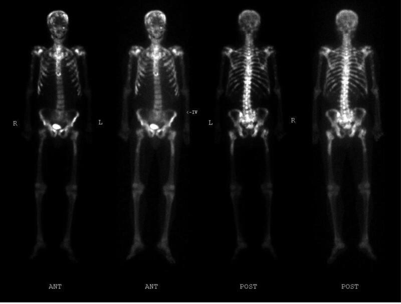
This diagnostic image is a whole-body bone scintigraphy (technetium-99m MDP bone scan) featuring both anterior (ANT) and posterior (POST) planar views of a human skeleton. The scan demonstrates a widespread, multifocal pattern of heterogeneously increased radiotracer uptake, characteristic of extensive osteoblastic activity. Abnormal hot spots are visible throughout the axial and appendicular skeleton, including the calvarium, multiple levels of the spinal column (cervical, thoracic, and lumbar vertebrae), numerous ribs bilaterally, the sternum, the pelvis (sacrum and iliac wings), and the proximal long bones (humeri and femurs). In the anterior view, physiological tracer excretion is noted in the urinary bladder. The diffuse distribution and intensity of these findings are highly suggestive of metastatic bone disease, consistent with advanced malignancies such as castrate-resistant prostate cancer (CRPC). This image serves as a classic educational example of a 'superscan' appearance or extensive skeletal metastases for oncology and nuclear medicine training.
The image presents a series of six preoperative whole-body bone scans (labeled a through f) obtained via electrical capacitance tomography (ECT). Each scan provides an anterior view of the full human skeletal system, including the skull, axial skeleton (spine and rib cage), pelvis, and appendicular skeleton (long bones of the upper and lower extremities). The scans illustrate preoperative staging for patients with pelvic tumors. Visual findings highlight varying degrees of focal radiopharmaceutical uptake in the pelvic region, indicating tumor involvement. Specifically, images (a) and (b) demonstrate significant signal intensity in pelvic regions II and III, while images (c) and (d) show involvement in regions I and II. Images (e) and (f) also depict concentrated signal in the pelvis. These diagnostic images are used to assess the extent of primary low-grade malignant tumors, such as chondrosarcoma, and to rule out distant skeletal metastases before 3D-printed hemipelvic replacement surgery. The presentation facilitates comparison of tumor localization and signal distribution across different clinical cases.
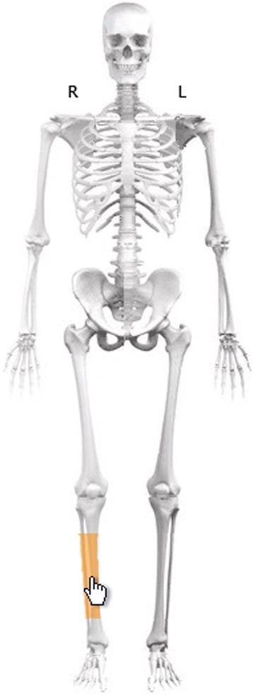
This anatomical diagram displays a complete human skeletal system in an anterior view, utilized as a digital interface for medical data entry and fracture classification. The skeleton is marked with 'R' and 'L' indicators above the shoulders for orientation. The primary focus of the visual is the right lower leg, where the diaphysis of the tibia is highlighted in orange. A hand-shaped cursor is positioned over this highlighted segment, demonstrating the interactive selection process within a clinical software environment. The illustration includes all major bony landmarks of the axial and appendicular skeletons, including the cranium, rib cage, vertebral column, pelvis, and long bones of the limbs. This tool is designed to facilitate the systematic recording of orthopedic injuries, specifically allowing physicians to select a bone segment to initiate AO/OTA fracture classification protocols.
Part 1: Anatomical basics and the skeletal system
A. The basics
Two ways to study anatomy
- Regional approach: you study one region at a time (thorax, abdomen, limbs) and everything in it. This works best with dissection.
- Systemic approach: you follow one system (for example the cardiovascular system) through the whole body. This helps you see how a system is continuous.
Anatomical position
Planes
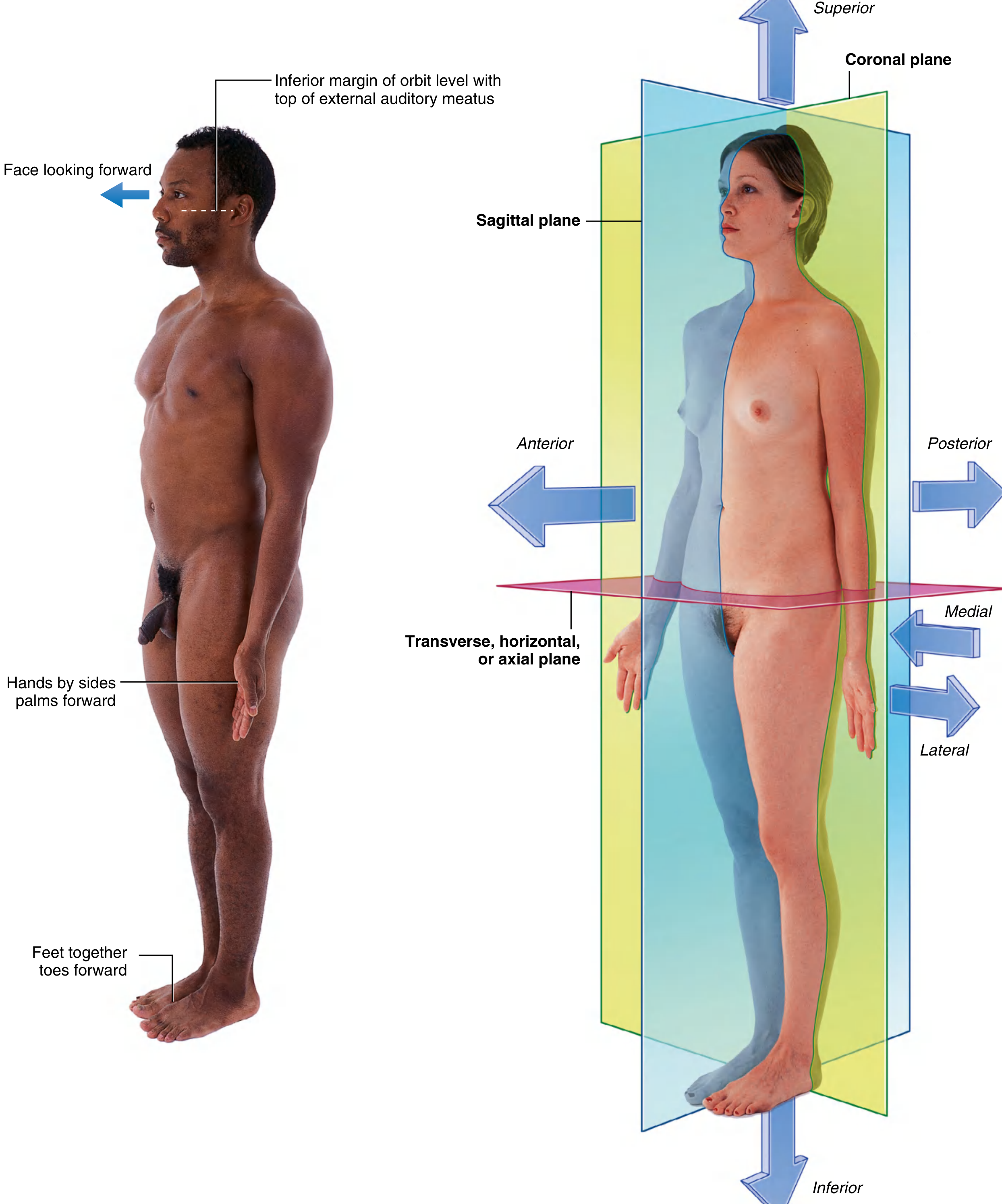
| Plane | Orientation | Divides body into |
|---|---|---|
| Coronal | vertical | anterior and posterior |
| Sagittal | vertical, at right angles to coronal | right and left (the median sagittal plane gives equal halves) |
| Transverse (horizontal, axial) | horizontal | superior and inferior |
Directional terms
- Anterior (ventral) / posterior (dorsal): front and back. The nose is anterior to the ears.
- Medial / lateral: nearer to or farther from the midline. The thumb is lateral to the little finger.
- Superior / inferior: above and below. The head is superior to the shoulders. Cranial and caudal mean toward the head and toward the tail.
- Proximal / distal: nearer to or farther from the origin, mainly in limbs. The hand is distal to the elbow. These terms also apply to branches of vessels and nerves.
- Superficial / deep: nearer to or farther from the body surface. The sternum is superficial to the heart.
- Rostral: toward the nose, mainly used in the head and brain.
B. The skeletal system
1. Divisions
- Axial skeleton: skull, vertebral column, ribs, sternum. It forms the central axis.
- Appendicular skeleton: bones of the upper and lower limbs, including the shoulder and pelvic girdles that attach them to the axis.
2. Cartilage (a basic you must know)
| Type | Feature | Example |
|---|---|---|
| Hyaline | most common, moderate collagen | articular surfaces, costal cartilage |
| Elastic | many elastic fibers | external ear |
| Fibrocartilage | dense collagen, strongest | intervertebral discs |
3. Bone
- Support
- Protection of vital organs
- Storage of calcium and phosphorus
- Levers for muscles to move
- A home for blood-forming marrow
- Compact (cortical) bone: dense outer shell.
- Spongy (trabecular or cancellous) bone: a lattice of spicules enclosing marrow spaces.
| Shape | Description | Examples |
|---|---|---|
| Long | tubular | humerus, femur |
| Short | cuboidal | carpals, tarsals |
| Flat | two compact plates with spongy bone between | skull bones |
| Irregular | various shapes | facial bones, vertebrae |
| Sesamoid | oval bones that develop inside tendons | patella (the largest) |
4. Blood supply and nerves of bone
- A nutrient artery, usually one per bone, enters the bone and supplies the marrow, spongy bone and inner compact bone.
- The periosteum is the fibrous membrane covering the outside of bone, except where articular cartilage is present. It supplies the outer compact bone and can form new bone. A bone stripped of its periosteum will not survive.
- Bone itself has few sensory fibers, but the periosteum has many. That is why fractures and periosteal injury are so painful.
5. How bones form
- Intramembranous ossification: bone forms directly in a mesenchymal membrane. Flat skull bones form this way.
- Endochondral ossification: a cartilage model forms first and is then replaced by bone. Long bones form this way, and the growth plate (physis) drives their lengthening.
6. Bone marrow
- Red marrow makes red cells, platelets and most white cells. It is dominant at birth.
- Yellow marrow is mostly fat. With age, red marrow in the long bones is replaced by yellow marrow.
- Marrow has two stem cell types. Hemopoietic stem cells form blood cells. Mesenchymal stem cells form bone, cartilage and muscle.
- In adults, red marrow persists mainly in the vertebrae, pelvis, sternum, ribs and skull.
7. Parts of a typical long bone
- Diaphysis: the shaft.
- Epiphysis: the end. It lies beyond the growth plate and is covered by articular cartilage.
- Metaphysis: the flared region between them, next to the growth plate.
- Medullary cavity: the marrow-filled space in the shaft.
8. Joints (the "pro" link between bones and muscles)
| Class | Connection | Movement | Example |
|---|---|---|---|
| Fibrous | fibrous tissue | little or none | skull sutures, tibiofibular syndesmosis |
| Cartilaginous | cartilage | slight | pubic symphysis, intervertebral discs |
| Synovial | joint cavity with synovial fluid | free | shoulder, hip, knee |
- Hinge: elbow
- Ball and socket: hip, shoulder
- Pivot: atlantoaxial joint
- Plane: intercarpal joints
- Saddle: first carpometacarpal joint
- Condyloid: wrist
9. Clinical links
- Fractures:
- A fracture happens when bone gives way to abnormal load. It can also happen with normal load if the bone is poor quality, as in osteoporosis.
- Healing runs from blood clot to soft callus to osteoblast-made bone matrix to hard callus.
- Treatment is reduction and then immobilization, or internal or external fixation.
- Avascular necrosis: bone dies when its blood supply is lost. It most often affects the femoral head.
- Bone marrow transplant: healthy hemopoietic stem cells replace diseased marrow, as in leukemia.
- Accessory and sesamoid bones: normal variants, mainly in the hands and feet. Do not mistake them for fractures on X-ray.
C. Pro-level skeleton: next layer
- Skull: neurocranium and viscerocranium, sutures, foramina and the structures passing through them.
- Vertebral column: 7 cervical, 12 thoracic, 5 lumbar, 5 fused sacral and 3 to 4 coccygeal vertebrae. Know the curves and the features of a typical vertebra.
- Thorax: 12 pairs of ribs (7 true, 3 false, 2 floating) and the sternum.
- Upper limb: clavicle, scapula, humerus, radius, ulna, carpals, metacarpals, phalanges.
- Lower limb: hip bone (ilium, ischium, pubis), femur, patella, tibia, fibula, tarsals, metatarsals, phalanges.
Quick self-test
- Which plane separates the body into front and back?
- Name the three cartilage types and one example of each.
- Why can a fracture be very painful when bone itself has few sensory nerves?
- How does endochondral ossification differ from intramembranous ossification?
- Is the clavicle part of the axial or appendicular skeleton?