
Show a image of fatty liver
Reading File
Now let me search the medical library for fatty liver images.
Finding Sources
Finding Sources
Good - found relevant sections. Let me read the Robbins Pathology block for images on fatty liver/steatosis.
Reading File
Reading File
Found two great images. Let me fetch them both in parallel.
Loading Image
Reading File
Here is the histological image of fatty liver (hepatic steatosis) from Robbins & Kumar Basic Pathology:
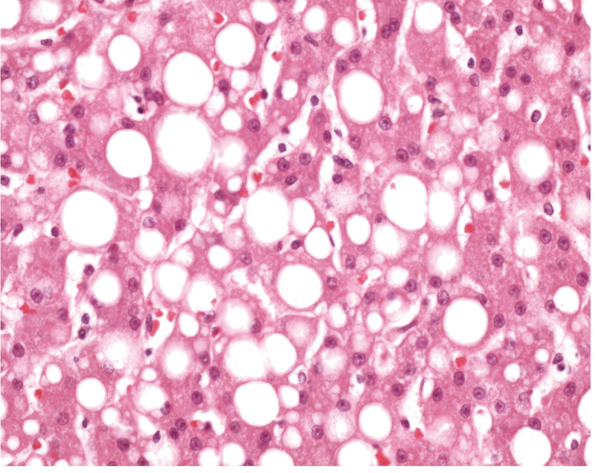
Fig. 2.30 - Fatty Liver (Steatosis), High-Power H&E stain
(Courtesy Dr. James Crawford, Hofstra Northwell School of Medicine)
What you are seeing:
- The large, round, clear (white) vacuoles filling the hepatocytes are fat droplets (triglycerides) - this is called macrovesicular steatosis
- The smaller clear spaces are microvesicular fat droplets
- In many cells, the nucleus is displaced and squeezed to the periphery by the large fat vacuole
- The pink background is the normal liver parenchyma (hepatocytes)
Key pathological points:
- Fatty change (steatosis) represents abnormal accumulation of triglycerides within hepatocytes
- Most common causes: excessive alcohol use, obesity, type 2 diabetes, and nonalcoholic fatty liver disease (NAFLD)
- Fat accumulation typically begins in centrilobular hepatocytes and spreads outward
- Macroscopically, a fatty liver appears large (4-6 kg), soft, yellow, and greasy
- Early fatty change is completely reversible if the underlying cause is removed
Source: Robbins & Kumar Basic Pathology, Chapter 14 (Liver)
This is a shared conversation. Sign in to Orris to start your own chat.