
Give a detailed description of adrenergic drugs which should include its introduction, classification... Mode of actions, pharmacological action, therapeutic uses. Side effects and contraindications.
adrenergic receptor classification sympathomimetic drugs diagram
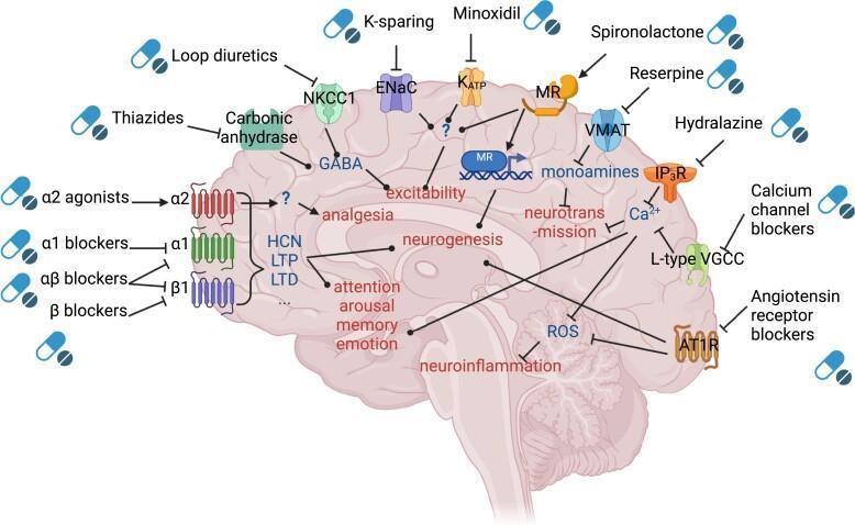
This pathophysiology diagram illustrates the main mechanisms of action for anti-hypertensive drugs within the brain. Set against a sagittal cross-section of the human brain, the illustration maps various pharmacological classes to their molecular targets and downstream physiological effects. Major drug classes identified include alpha-2 agonists, alpha-1 blockers, alpha-beta blockers, and beta blockers acting on adrenergic receptors; diuretics such as thiazides and loop diuretics targeting carbonic anhydrase and NKCC1; and calcium channel blockers (CCBs) acting on L-type voltage-gated calcium channels (VGCC). The diagram also features K-sparing diuretics, spironolactone, minoxidil, and angiotensin receptor blockers (ARBs) targeting AT1R. Key downstream mechanisms include modulation of neurotransmission, neurogenesis, neuroinflammation, and excitability. Specific pathways shown include the inhibition of Reactive Oxygen Species (ROS) by AT1R blockers and CCBs, and the influence of various agents on cognitive domains like memory, emotion, arousal, and attention. The schematic uses standard BioRender iconography to distinguish between stimulatory (sharp arrows) and inhibitory (flat-headed arrows) interactions, highlighting the complex neurobiological impact of systemic anti-hypertensives on central nervous system function.
![This pathophysiology diagram illustrates the role of the Beta-2 Adrenergic Receptor (ADRB2) in modulating inflammatory responses during Rhinovirus (RV) infection. The visual is divided into three primary sections: [1] depicts RV particles binding to Intercellular Adhesion Molecule 1 (ICAM-1) on the surface of airway epithelial cells (ECs). [2] demonstrates that ADRB2 activation inhibits the expression of ICAM-1 and the subsequent release of cytokines and chemokines from these epithelial cells. [3] focuses on systemic immune cell modulation, showing that ADRB2 signaling exerts inhibitory effects (indicated by red T-bars) on multiple cell types: in macrophages (Mφ), it suppresses TNF-α, IL-6, IL-1β, and various CCL chemokines; in mast cells (MC), it inhibits TNF-α, histamine, and leukotriene release; in CD4+ T cells, it suppresses IFN-γ production; and in CD8+ T cells, it blocks TNF-α, IFN-γ, and cytolytic activity. The diagram serves as an educational resource for understanding neuro-immune interactions and the therapeutic mechanism of beta-agonists in dampening virus-induced airway inflammation.](https://cdn.orris.care/cdss_images/pmc_clinical_VQA_bdc00632428101dee8d73e300a2b0ae2a72521d5b25266eddbef38d212ed1f84.jpg)
This pathophysiology diagram illustrates the role of the Beta-2 Adrenergic Receptor (ADRB2) in modulating inflammatory responses during Rhinovirus (RV) infection. The visual is divided into three primary sections: [1] depicts RV particles binding to Intercellular Adhesion Molecule 1 (ICAM-1) on the surface of airway epithelial cells (ECs). [2] demonstrates that ADRB2 activation inhibits the expression of ICAM-1 and the subsequent release of cytokines and chemokines from these epithelial cells. [3] focuses on systemic immune cell modulation, showing that ADRB2 signaling exerts inhibitory effects (indicated by red T-bars) on multiple cell types: in macrophages (Mφ), it suppresses TNF-α, IL-6, IL-1β, and various CCL chemokines; in mast cells (MC), it inhibits TNF-α, histamine, and leukotriene release; in CD4+ T cells, it suppresses IFN-γ production; and in CD8+ T cells, it blocks TNF-α, IFN-γ, and cytolytic activity. The diagram serves as an educational resource for understanding neuro-immune interactions and the therapeutic mechanism of beta-agonists in dampening virus-induced airway inflammation.
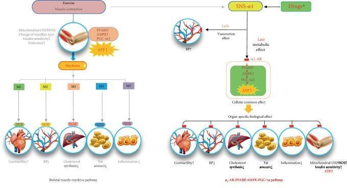
A pathophysiology diagram illustrating the comparative metabolic pathways of exercise-induced skeletal muscle contraction versus α1-adrenergic receptor (α1-AR) activation. The left side details the 'Skeletal muscle-myokine pathway,' where exercise triggers muscle contraction, leading to increased mitochondrial oxidative phosphorylation (OXPHOS), PGC-1α signaling, and ATP production. This results in the secretion of myokines (M1–M5) that interact with specific receptors (M1R–M5R) to influence multiple organs: increasing cardiac contractility, reducing blood pressure (BP), decreasing liver cholesterol synthesis, reducing adipose tissue fat, and lowering systemic inflammation. The right side illustrates the 'α1-AR-PPARδ-AMPK-PGC-1α pathway,' initiated by the sympathetic nervous system (SNS-α1) or drugs like midodrine. This pathway features an early vasomotion effect (BP elevation) and a late metabolic effect. The metabolic cascade involves PPARδ, AMPK, and PGC-1α, leading to increased ATP and mirrored organ-specific benefits, including improved skeletal muscle insulin sensitivity and mitochondrial OXPHOS. The diagram serves as an educational tool to explain how pharmacological α1-AR agonists can mimic several systemic health benefits typically associated with physical exercise.
![This physiological diagram displays nine time-series graphs (A–I) illustrating intracellular calcium ([Ca2+]i) dynamics in adipocytes, measured via Fura-2 fluorescence ratios. The graphs demonstrate the synergistic and potentiation effects of various agonists on calcium signaling. Panels A and B show periodic, high-amplitude [Ca2+]i oscillations induced by the combination of Acetylcholine (ACh) and Norepinephrine (NE). Panel C depicts the inhibition of these oscillations using the M3-cholinergic receptor antagonist p-F-HHSID, resulting in only a single transient spike. Panels D–H illustrate diverse dynamic regimes triggered by ACh and the α1-adrenergic agonist Phenylephrine, including fast oscillations (D), impulse-shaped oscillations (E), relaxation-type oscillations (F), and transitions to new steady states (G, H). Panel I shows impulse-shaped oscillations elicited by the α2-adrenergic agonist UK-14,304 in the presence of ACh. The data illustrate complex nonlinear signaling pathways and the convergence of M3-cholinergic and α-adrenergic receptor activation in adipocyte calcium homeostasis.](https://cdn.orris.care/cdss_images/pmc_clinical_VQA_89f4396e44260123ca58985d1422823adf03181a9bbd5bbbe3f5c85a21f8880e.jpg)
This physiological diagram displays nine time-series graphs (A–I) illustrating intracellular calcium ([Ca2+]i) dynamics in adipocytes, measured via Fura-2 fluorescence ratios. The graphs demonstrate the synergistic and potentiation effects of various agonists on calcium signaling. Panels A and B show periodic, high-amplitude [Ca2+]i oscillations induced by the combination of Acetylcholine (ACh) and Norepinephrine (NE). Panel C depicts the inhibition of these oscillations using the M3-cholinergic receptor antagonist p-F-HHSID, resulting in only a single transient spike. Panels D–H illustrate diverse dynamic regimes triggered by ACh and the α1-adrenergic agonist Phenylephrine, including fast oscillations (D), impulse-shaped oscillations (E), relaxation-type oscillations (F), and transitions to new steady states (G, H). Panel I shows impulse-shaped oscillations elicited by the α2-adrenergic agonist UK-14,304 in the presence of ACh. The data illustrate complex nonlinear signaling pathways and the convergence of M3-cholinergic and α-adrenergic receptor activation in adipocyte calcium homeostasis.
adrenergic receptor alpha beta subtypes classification table pharmacology
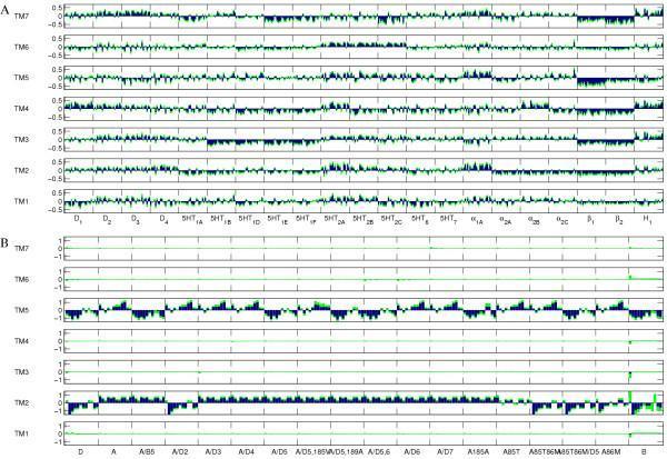
Two-panel bar chart (A and B) representing proteochemometric modeling data of G protein-coupled receptor (GPCR) ligand affinity. Both panels decompose ligand affinity contributions across the seven transmembrane (TM) regions (TM1 to TM7), plotted for various receptor subtypes. Panel A shows data for amine GPCRs, including dopamine (D1-D4), serotonin (5HT1A-5HT7), alpha-adrenergic (α1A-α2C), beta-adrenergic (β1-β2), and histamine (H1) receptors. Blue bars indicate the average contribution of each TM region to ligand binding affinity, while green bars represent one standard deviation, illustrating model variability. Panel B focuses on alpha-1 adrenoreceptors, showing relative contributions for specific ligands. In panel B, TM2 and TM5 show substantial positive or negative contributions to affinity, while regions TM1, TM3, TM4, TM6, and TM7 demonstrate minimal or negligible roles. The charts illustrate structural-functional relationships in pharmacodynamics, specifically identifying which helical domains are primary determinants for ligand binding across different GPCR families and receptor-ligand combinations.
![<table><thead><tr><th colspan="3">2014 Location</th><th>2014 Recommendation Text</th><th>2014 Strength of Recommendation</th><th>Recommendation Category</th><th>2020 Recommendation</th></tr><tr><th>Section</th><th>Number</th><th>Page</th><th></th><th></th><th></th><th></th></tr></thead><tbody><tr><td>4.6</td><td>44</td><td>p. 52</td><td>We recommend against the use of more than one of the following three drug classes together in the same patient: angiotensin-converting-enzyme inhibitors, angiotensin II receptor blockers, or direct renin inhibitors.</td><td>Strong against</td><td>Not reviewed, Not changed</td><td>Recommendation 26</td></tr><tr><td>4.6</td><td>45</td><td>p. 52</td><td>We recommend additional therapy in refractory hypertension (for those who do not tolerate or are not adequately controlled with triple therapy [i.e., thiazidetype diuretics, ACEI or ARB, and CCBs] described in Recommendation 43) or as supplementary therapy in some clinical indications. Drug classes for consideration can include (not in priority order):<br>a. Aldosterone/mineralocorticoid receptor antagonists (e.g., spironolactone, eplerenone)<br>b. Other potassium-sparing diuretic (i.e., amiloride)<br>c. Alpha adrenergic blockers<br>d. Beta adrenergic blockers<br>e. Non-dihydropyridine calcium channel blockers<br>f. Combined alpha-beta adrenergic blockers<br>g. Peripherally acting antiadrenergic agents (reserpine, pending availability)<br>h. Direct acting vasodilators (e.g., hydralazine, minoxidil)<br>i. Centrally acting antiadrenergic drugs (e.g., clonidine, methyldopa)</td><td>Strong for</td><td>Reviewed, New-replaced</td><td>Recommendation 28</td></tr><tr><td>4.6</td><td>46</td><td>p. 54</td><td>We recommend against the use of alpha-adrenergic blockers as monotherapy, but this class of agents may be used as supplemental therapy or if warranted by comorbid conditions (e.g., symptomatic prostatic hypertrophy). (Modified from 2004 VA/DoD HTN CPG)</td><td>Strong against</td><td>Not reviewed, Deleted</td><td>--</td></tr><tr><td>4.7</td><td>47</td><td>p. 54</td><td>In patients with hypertension and chronic kidney disease (reduced kidney function with albuminuria), we recommend treatment with an angiotensin-converting enzyme inhibitor, or angiotensin II receptor blocker for improving kidney outcomes. (Modified from 2004 VA/DoD HTN CPG)</td><td>Strong for</td><td>Reviewed, Deleted</td><td>--</td></tr><tr><td>4.7</td><td>48</td><td>p.56</td><td>In African American patients with hypertension, we recommend against using an angiotensin-converting-enzyme inhibitor or angiotensin II receptor blocker as monotherapy.</td><td>Strong against</td><td>Reviewed, Not changed</td><td>Recommendation 24</td></tr></tbody></table>](https://cdn.orris.care/cdss_images/GLGCA_41043_1763756169158_6114997f-b826-4a5a-9e6b-8bd00369eda2.png)
<table><thead><tr><th colspan="3">2014 Location</th><th>2014 Recommendation Text</th><th>2014 Strength of Recommendation</th><th>Recommendation Category</th><th>2020 Recommendation</th></tr><tr><th>Section</th><th>Number</th><th>Page</th><th></th><th></th><th></th><th></th></tr></thead><tbody><tr><td>4.6</td><td>44</td><td>p. 52</td><td>We recommend against the use of more than one of the following three drug classes together in the same patient: angiotensin-converting-enzyme inhibitors, angiotensin II receptor blockers, or direct renin inhibitors.</td><td>Strong against</td><td>Not reviewed, Not changed</td><td>Recommendation 26</td></tr><tr><td>4.6</td><td>45</td><td>p. 52</td><td>We recommend additional therapy in refractory hypertension (for those who do not tolerate or are not adequately controlled with triple therapy [i.e., thiazidetype diuretics, ACEI or ARB, and CCBs] described in Recommendation 43) or as supplementary therapy in some clinical indications. Drug classes for consideration can include (not in priority order):<br>a. Aldosterone/mineralocorticoid receptor antagonists (e.g., spironolactone, eplerenone)<br>b. Other potassium-sparing diuretic (i.e., amiloride)<br>c. Alpha adrenergic blockers<br>d. Beta adrenergic blockers<br>e. Non-dihydropyridine calcium channel blockers<br>f. Combined alpha-beta adrenergic blockers<br>g. Peripherally acting antiadrenergic agents (reserpine, pending availability)<br>h. Direct acting vasodilators (e.g., hydralazine, minoxidil)<br>i. Centrally acting antiadrenergic drugs (e.g., clonidine, methyldopa)</td><td>Strong for</td><td>Reviewed, New-replaced</td><td>Recommendation 28</td></tr><tr><td>4.6</td><td>46</td><td>p. 54</td><td>We recommend against the use of alpha-adrenergic blockers as monotherapy, but this class of agents may be used as supplemental therapy or if warranted by comorbid conditions (e.g., symptomatic prostatic hypertrophy). (Modified from 2004 VA/DoD HTN CPG)</td><td>Strong against</td><td>Not reviewed, Deleted</td><td>--</td></tr><tr><td>4.7</td><td>47</td><td>p. 54</td><td>In patients with hypertension and chronic kidney disease (reduced kidney function with albuminuria), we recommend treatment with an angiotensin-converting enzyme inhibitor, or angiotensin II receptor blocker for improving kidney outcomes. (Modified from 2004 VA/DoD HTN CPG)</td><td>Strong for</td><td>Reviewed, Deleted</td><td>--</td></tr><tr><td>4.7</td><td>48</td><td>p.56</td><td>In African American patients with hypertension, we recommend against using an angiotensin-converting-enzyme inhibitor or angiotensin II receptor blocker as monotherapy.</td><td>Strong against</td><td>Reviewed, Not changed</td><td>Recommendation 24</td></tr></tbody></table>
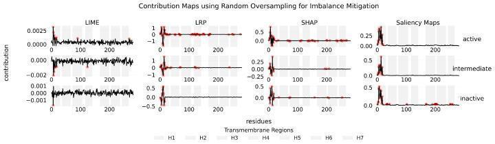
This Comparison Chart displays contribution maps for a 1D-CNN model used in protein conformational state classification, specifically focusing on the beta-2 adrenergic receptor (β2AR). The visualization illustrates how four different explainability methods—LIME, LRP, SHAP, and Saliency Maps—assign importance to amino acid residues across three states: active, intermediate, and inactive. The data presented utilized Random Oversampling for class-imbalance mitigation. Each graph plots contribution (y-axis) against residue position (x-axis), with shaded vertical bars indicating transmembrane regions H1 through H7. Red points denote relevant residues that significantly influence the model's predictions. LIME, LRP, and SHAP demonstrate both positive and negative contributions (bidirectional fluctuations), while Saliency Maps show only positive magnitudes. The charts highlight distinct residue importance patterns within the transmembrane helices, particularly around residues 0-50, aiding in the identification of key structural features that define the transitions between receptor conformations.
Adrenergic Drugs: A Comprehensive Overview
1. Introduction
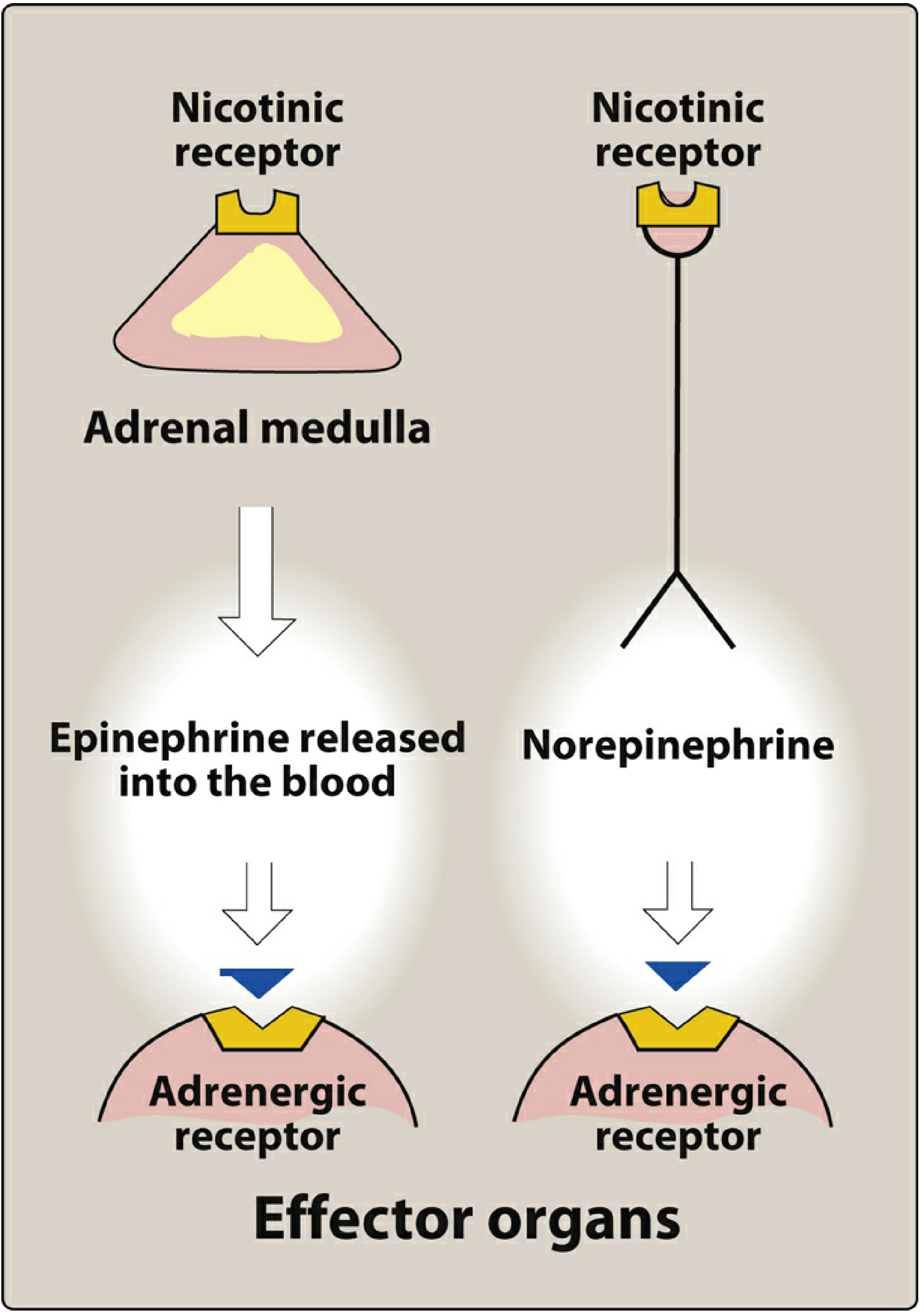
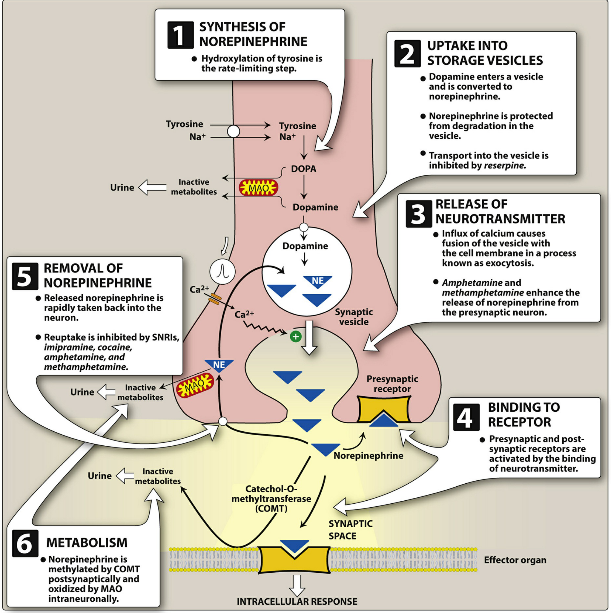
Neurotransmission at Adrenergic Neurons
- Synthesis: Tyrosine → DOPA (rate-limiting step, by tyrosine hydroxylase) → Dopamine → Norepinephrine (in vesicles, by dopamine-β-hydroxylase)
- Storage: Norepinephrine is stored in synaptic vesicles (reserpine blocks this transport)
- Release: An action potential triggers Ca²⁺ influx → vesicle exocytosis
- Receptor binding: Norepinephrine binds presynaptic (α₂ autoreceptors) and postsynaptic adrenergic receptors
- Removal: Primary route is neuronal reuptake (Uptake 1, blocked by cocaine, amphetamines, SNRIs); secondary metabolism by COMT (postsynaptic) and MAO (intraneuronal)
2. Classification
A. By Mechanism of Action
| Class | Mechanism | Examples |
|---|---|---|
| Direct-acting | Bind and activate adrenergic receptors directly | Epinephrine, norepinephrine, dopamine, phenylephrine, clonidine, albuterol, dobutamine |
| Indirect-acting | Enhance NE release or inhibit its reuptake/metabolism | Amphetamine, cocaine, tyramine |
| Mixed-acting | Both direct receptor activation + NE release | Ephedrine, pseudoephedrine |
B. By Chemical Structure
- Epinephrine, norepinephrine, dopamine, isoproterenol, dobutamine
- Features: high potency, rapid inactivation (by COMT and MAO), poor CNS penetration (polar), only parenteral/inhaled use
- Phenylephrine, ephedrine, amphetamine, albuterol, terbutaline
- Features: longer half-life (not metabolized by COMT), oral bioavailability, better CNS penetration
C. By Receptor Selectivity (Most Clinically Useful Classification)
| Subgroup | Drugs | Primary Receptor |
|---|---|---|
| α₁ agonists | Phenylephrine, methoxamine, midodrine | α₁ |
| α₂ agonists | Clonidine, methyldopa, dexmedetomidine, apraclonidine | α₂ |
| β₁ agonists (selective) | Dobutamine | β₁ |
| β₂ agonists (selective) | Albuterol (salbutamol), terbutaline, salmeterol, formoterol | β₂ |
| β₃ agonists | Mirabegron | β₃ |
| Non-selective α+β | Epinephrine | α₁, α₂, β₁, β₂ |
| Non-selective β | Isoproterenol | β₁, β₂ |
| Non-selective α+β (primarily α) | Norepinephrine | α₁, α₂, β₁ |
| Dopamine + adrenergic | Dopamine (dose-dependent) | D₁, β₁, α₁ |
| Mixed acting | Ephedrine, pseudoephedrine | α + β (indirect + direct) |
3. Adrenergic Receptor Types and Subtypes
- α receptors: Epi ≥ NE >> Isoproterenol
- β receptors: Isoproterenol > Epi ≥ NE
| Receptor | Subtype | G Protein | Second Messenger | Key Locations |
|---|---|---|---|---|
| α₁ | α₁A, α₁B, α₁D | Gq | ↑ IP₃, DAG → ↑ Ca²⁺ | Vascular smooth muscle, iris dilator, bladder sphincter, prostate |
| α₂ | α₂A, α₂B, α₂C | Gi | ↓ cAMP | Presynaptic terminals, platelets, pancreatic β cells, fat cells |
| β₁ | - | Gs | ↑ cAMP | Heart (SA node, AV node, ventricle), kidney (JGA) |
| β₂ | - | Gs | ↑ cAMP | Bronchial/vascular smooth muscle, liver, skeletal muscle, uterus |
| β₃ | - | Gs | ↑ cAMP | Adipose tissue, urinary bladder detrusor |
| D₁ | - | Gs | ↑ cAMP | Renal/mesenteric/coronary vasculature |
| D₂ | - | Gi | ↓ cAMP | Presynaptic terminals, pituitary |
4. Modes of Action
α₁ Receptor Signaling
α₂ Receptor Signaling
β Receptor Signaling (β₁ and β₂)
Indirect-Acting Mechanism
5. Pharmacological Actions (by System)
Cardiovascular System
- Heart (β₁): ↑ heart rate (positive chronotropy), ↑ contractility (positive inotropy), ↑ conduction velocity, ↑ automaticity → increased cardiac output; increases myocardial O₂ demand
- Blood vessels (α₁): Vasoconstriction in skin, mucous membranes, viscera → ↑ peripheral resistance
- Blood vessels (β₂): Vasodilation in skeletal muscle, liver, coronary vasculature
- Net BP effect of epinephrine: ↑ systolic BP (β₁ cardiac stimulation) + slight ↓ diastolic BP (β₂ vasodilation in muscle); mean arterial pressure may be unchanged or slightly elevated
- Net BP effect of norepinephrine: ↑ systolic and diastolic BP (predominantly α); reflex bradycardia
- Net BP effect of phenylephrine (pure α₁): ↑ BP with reflex bradycardia; no cardiac stimulation
Respiratory System (β₂)
- Bronchodilation via ↑ cAMP → smooth muscle relaxation
- Inhibit release of bronchoconstricting mediators from mast cells
- May reduce microvascular leakage and increase mucociliary transport
- Adverse: tachycardia (β₁ spillover), skeletal muscle tremor, ↓ serum potassium
Metabolic Effects
- Liver (β₂, α₁): Glycogenolysis → ↑ blood glucose
- Adipose tissue (β₃): Lipolysis → ↑ free fatty acids and glycerol
- Pancreas (β₂): ↑ insulin secretion; (α₂): ↓ insulin secretion
- Potassium (β₂): ↑ cellular K⁺ uptake → ↓ plasma K⁺ (hypokalemia)
Eyes
- α₁ (iris dilator muscle): Mydriasis (pupil dilation)
- β₂ (ciliary muscle): Reduced aqueous humor production
- α₂ (ciliary body): Reduce aqueous secretion - used in glaucoma (brimonidine)
GI Tract
- α₁ and β₂: Decreased intestinal motility and tone (sphincter contraction by α₁)
- Reduced peristalsis
Urinary Bladder
- α₁ (trigone, sphincter): Contraction → urinary retention
- β₃ (detrusor): Relaxation → increases bladder capacity (mirabegron for overactive bladder)
CNS (lipid-soluble noncatecholamines)
- Low doses: increased alertness, reduced fatigue, euphoria, anorexia
- High doses: anxiety, restlessness, tremors, insomnia
- Centrally acting α₂ agonists (clonidine): sedation, reduced sympathetic tone
Uterus
- β₂: Uterine relaxation (tocolysis - used to delay preterm labor)
- α: Uterine contraction
6. Individual Drug Profiles
Epinephrine (Adrenaline)
- Receptor profile: α₁, α₂, β₁, β₂ (all four major subtypes)
- Pharmacological action: At low doses β effects predominate (vasodilation); at high doses α effects predominate (vasoconstriction). Produces positive inotropy and chronotropy, bronchodilation, vasoconstriction of skin/mucosa, glycogenolysis
- Therapeutic uses: Anaphylaxis (first-line), cardiac arrest (CPR), acute severe bronchospasm, addition to local anesthetics (to prolong action and reduce systemic absorption), open-angle glaucoma (topical), hemostasis in surgery
- Route: IV, IM (autoinjector in anaphylaxis), SC, inhaled, topical
Norepinephrine (Noradrenaline)
- Receptor profile: α₁ (dominant), α₂, β₁ (minimal β₂)
- Pharmacological action: Potent vasoconstriction of all vascular beds, increases systolic and diastolic BP, triggers reflex bradycardia (via baroreceptor activation offsetting direct β₁ effect), minimal bronchodilation
- Therapeutic uses: Vasopressor of first choice in septic shock and other distributive shock states; maintains mean arterial pressure. Does not cause arrhythmias as readily as dopamine
- Route: IV infusion only
Dopamine
- Receptor profile (dose-dependent):
- Low dose (1-2 mcg/kg/min): D₁ receptors → renal/mesenteric vasodilation
- Moderate dose (2-10 mcg/kg/min): β₁ → ↑ cardiac output
- High dose (>10 mcg/kg/min): α₁ → vasoconstriction
- Therapeutic uses: Cardiogenic shock, heart failure with hypotension, hemodynamic instability. Note: Evidence now favors norepinephrine over dopamine in shock due to higher arrhythmia risk with dopamine
- Route: IV infusion
Dobutamine
- Receptor profile: Primarily β₁; minor β₂ and α₁ effects
- Pharmacological action: Increases heart rate, stroke volume, and cardiac output without major vascular effects. Does not elevate myocardial O₂ demand as much as other sympathomimetics
- Therapeutic uses: Acute decompensated heart failure, inotropic support post-cardiac surgery. Used in stress echocardiography
- Route: IV infusion; tolerance may develop with prolonged use
Isoproterenol
- Receptor profile: β₁ + β₂ (no α activity)
- Pharmacological action: Intense cardiac stimulation (↑ HR, ↑ contractility), peripheral vasodilation (↓ diastolic BP), bronchodilation
- Therapeutic uses: Rarely used now. Historic use in bradyarrhythmias, heart block. Largely replaced by more selective agents
- Route: IV
Phenylephrine
- Receptor profile: Selective α₁
- Pharmacological action: Pure vasoconstriction → ↑ BP with reflex bradycardia; no direct cardiac stimulation
- Therapeutic uses: Nasal decongestant, hypotension during anesthesia/spinal anesthesia, ophthalmic mydriasis (pupil dilation for examination). Topical nasal decongestant
- Route: IV, topical, intranasal, ophthalmic drops
Albuterol (Salbutamol) and β₂ Agonists
- Receptor profile: Selective β₂ (some β₁ at high doses)
- Pharmacological action: Bronchodilation via ↑ cAMP → smooth muscle relaxation; inhibit mast cell degranulation; increase mucociliary clearance
- SABAs (short-acting: albuterol, terbutaline): onset 5 min, duration 4-6 hrs; rescue use
- LABAs (long-acting: salmeterol, formoterol): duration 12+ hrs; maintenance therapy in asthma/COPD
- Therapeutic uses: Asthma (acute and chronic), COPD, exercise-induced bronchospasm; terbutaline: tocolysis (preterm labor)
Clonidine
- Receptor profile: Selective α₂ (centrally and peripherally)
- Pharmacological action: Acts centrally on brainstem α₂ receptors → ↓ sympathetic outflow → ↓ BP, ↓ HR
- Therapeutic uses: Hypertension (second-line), ADHD (with guanfacine), withdrawal from opiates/alcohol/tobacco, analgesia (epidural)
- Side effects: Sedation, dry mouth, constipation, bradycardia. Critical: abrupt discontinuation causes dangerous rebound hypertension
Midodrine
- Receptor profile: Prodrug converted to active desglymidodrine; selective α₁ agonist (peripheral)
- Therapeutic uses: Orthostatic hypotension (first-line oral agent). Avoid doses within 4 hours of bedtime to prevent supine hypertension
Ephedrine and Pseudoephedrine
- Mechanism: Mixed-acting (direct α+β agonism + indirect NE release)
- Therapeutic uses: Nasal decongestant, bronchodilator (older use), hypotension during spinal anesthesia (ephedrine)
- CNS penetration: Good (lipophilic, noncatecholamine) - stimulant effects, abuse potential
7. Therapeutic Uses (Summary by Indication)
| Clinical Condition | Drug of Choice | Rationale |
|---|---|---|
| Anaphylaxis | Epinephrine IM | α₁ (↑ BP, ↓ edema) + β₁ (cardiac) + β₂ (bronchodilation) |
| Septic shock | Norepinephrine (IV) | Vasopressor of first choice; balanced α with some β |
| Cardiogenic shock | Dobutamine ± norepinephrine | β₁ inotrope; NE for BP support |
| Cardiac arrest | Epinephrine (IV) | Improves coronary perfusion pressure during CPR |
| Acute asthma | Albuterol (inhaled) | Selective β₂ → bronchodilation with fewer cardiac effects |
| Chronic asthma/COPD | Salmeterol/Formoterol (LABA) | Long-acting β₂ bronchodilation |
| Orthostatic hypotension | Midodrine (oral) | Peripheral α₁ → increases vascular tone |
| Hypertension | Clonidine (α₂ centrally) | ↓ sympathetic outflow |
| Nasal congestion | Phenylephrine, oxymetazoline | α₁ → mucosal vasoconstriction |
| Glaucoma | Brimonidine (α₂ topical) | ↓ aqueous humor production |
| Overactive bladder | Mirabegron (β₃) | Bladder detrusor relaxation |
| Preterm labor | Terbutaline (β₂) | Uterine relaxation (tocolysis) |
| ADHD | Clonidine, guanfacine (α₂) | Prefrontal cortex modulation |
| With local anesthetics | Epinephrine | Vasoconstriction prolongs local anesthetic action |
| Bradyarrhythmias | Isoproterenol (β₁β₂) | Increases heart rate |
8. Side Effects
Cardiovascular
- Tachycardia, palpitations: From β₁ stimulation (epinephrine, isoproterenol, albuterol at high doses)
- Hypertension: From α₁-mediated vasoconstriction (phenylephrine, high-dose dopamine)
- Arrhythmias: All catecholamines increase cardiac automaticity and risk of dysrhythmias; dopamine has the highest risk
- Angina / myocardial ischemia: Due to increased O₂ demand with β₁ stimulation
- Rebound hypertension: Abrupt clonidine withdrawal
Respiratory
- Tachycardia with β₂ agonists: β₁ spillover at high doses
- Paradoxical bronchospasm: Can occur with inhaled sympathomimetics (preservative-related)
Metabolic
- Hypokalemia: β₂ agonists drive K⁺ into cells
- Hyperglycemia: Glycogenolysis (α₁, β₂ in liver); inhibition of insulin release (α₂ on pancreas)
- Lipolysis: ↑ free fatty acids (β agonism)
CNS (mainly noncatecholamines/indirect-acting)
- Anxiety, restlessness, tremor, insomnia
- Headache
- Seizures at toxic doses (amphetamines)
- Cerebral hemorrhage (severe hypertension from α agonists)
GI
- Nausea, vomiting
- Decreased GI motility, constipation (especially with α₂ agonists like clonidine)
- Dry mouth (α₂ - clonidine)
Urogenital
- Urinary retention: α₁ stimulation (bladder sphincter contraction)
- Reduced renal blood flow at high doses of vasopressors
Local
- Tissue necrosis/ischemia: Extravasation of vasopressors (norepinephrine, dopamine) causes local vasoconstriction and tissue necrosis; treat with phentolamine (α blocker) injection locally
- Tachyphylaxis with indirect-acting agents (store depletion)
Ocular
- Mydriasis (angle-closure glaucoma can be precipitated in susceptible individuals)
9. Contraindications
| Drug/Class | Contraindicated In | Reason |
|---|---|---|
| Epinephrine | Closed-angle glaucoma | Pupillary dilation worsens angle closure |
| Epinephrine | Hyperthyroidism | Exaggerated cardiovascular response |
| Epinephrine | Hypertension/cardiac arrhythmias (relative) | Further ↑ BP and arrhythmias |
| All vasopressors (α agonists) | Hypovolemia (unless volume-replaced) | Vasoconstriction without volume = tissue ischemia |
| β₂ agonists | Tachyarrhythmias | Further increase heart rate |
| Clonidine | History of rebound hypertension; severe bradycardia | Worsens bradycardia; abrupt withdrawal dangerous |
| Non-selective β agonists (isoproterenol) | Coronary artery disease | Increased O₂ demand → ischemia |
| Dopamine/catecholamines | Pheochromocytoma | Hypertensive crisis |
| Indirect-acting (amphetamines, ephedrine) | Patients on MAO inhibitors | MAO inhibitors block NE breakdown → life-threatening hypertensive crisis |
| Sympathomimetics (vasoconstrictors) | Digital/extremity anesthesia (fingers, toes, nose, ears, penis) | Ischemic necrosis from local vasospasm |
| Phenylephrine | Severe hypertension | Worsens hypertension |
| All sympathomimetics | Halothane anesthesia | Sensitizes myocardium to catecholamine-induced arrhythmias |
| β₂ agonists (LABAs) | Asthma as monotherapy (without ICS) | Associated with increased asthma-related mortality when used alone |
Special Population Cautions
- Pregnancy: Vasopressors can reduce uterine blood flow; ephedrine and phenylephrine are used for anesthesia-induced hypotension in obstetrics (phenylephrine preferred for spinal hypotension)
- Elderly: Increased risk of hypertensive events, arrhythmias
- Diabetes: Sympathomimetics can mask hypoglycemia symptoms and worsen hyperglycemia
10. Key Pharmacokinetics Summary
| Drug | Route | Onset | Duration | Metabolism |
|---|---|---|---|---|
| Epinephrine | IM, IV, SC, inhaled | Minutes | 15-30 min | COMT, MAO |
| Norepinephrine | IV infusion | Rapid | 1-2 min after stopping | COMT, MAO |
| Dopamine | IV infusion | Rapid | 10 min after stopping | COMT, MAO |
| Dobutamine | IV infusion | 2-5 min | 10 min after stopping | COMT, conjugation |
| Albuterol | Inhaled | 5 min | 4-6 hrs | Hepatic |
| Salmeterol | Inhaled | 10-20 min | 12 hrs | Hepatic |
| Phenylephrine | IV, intranasal, topical | Minutes | 20-40 min (IV) | MAO |
| Clonidine | Oral, transdermal | 30-60 min (oral) | 8-12 hrs | Renal (50%), hepatic |
| Midodrine | Oral | 30 min | 2-3 hrs | Hydrolysis to desglymidodrine |
| Ephedrine | Oral, IV | 15-30 min (oral) | 4-6 hrs | MAO (slow), renal |
- Lippincott Illustrated Reviews: Pharmacology, Chapter 6 - Adrenergic Drugs (pp. 208-240)
- Katzung's Basic and Clinical Pharmacology, 16th Edition, Chapter 9 - Adrenoceptor-Activating Drugs (pp. 218-240)
- Goodman & Gilman's The Pharmacological Basis of Therapeutics - Sympathomimetic Amines