
White matter
White matter is the part of the brain and spinal cord made up primarily of myelinated axons. It gets its pale color from myelin, the fatty sheath that insulates nerve fibers and speeds up signal transmission. Unlike gray matter (which contains neuron cell bodies), white matter functions mainly as the brain's "wiring" - connecting different regions and allowing them to communicate. It makes up about 50% of the adult brain volume.
Searching sources...
Reading File
Finding Sources
Reading File
Reading File
Reading File
Here is a thorough overview of white matter, drawing from Kaplan & Sadock's Comprehensive Textbook of Psychiatry and Grainger & Allison's Diagnostic Radiology.
White Matter
What Is White Matter?
White matter is the part of the central nervous system made up primarily of myelinated axons (nerve fibers) along with supporting glial cells (mainly oligodendrocytes) and penetrating blood vessels. Its characteristic white/pale appearance comes from the lipid-rich myelin sheaths produced by oligodendrocytes (in contrast to the peripheral nervous system, where Schwann cells form myelin). White matter occupies roughly 42% of the volume of the cerebral hemispheres in humans - and this proportion has grown disproportionately over mammalian evolution compared to the neocortex itself. - Kaplan & Sadock's Comprehensive Textbook of Psychiatry
On imaging, the lipid content gives white matter:
- Low density on CT
- High signal on T1 and low signal on T2 MRI (relative to gray matter)
Three Major Categories of White Matter Tracts
White matter fibers are classically organized into three functional types:
1. Association Fibers (intrahemispheric)
Connect cortical regions within the same hemisphere.
- U-fibers (subcortical) - short, juxtacortical fibers linking adjacent gyri; notably spared in Binswanger disease and CADASIL
- Arcuate fasciculus - connects superior/middle frontal gyri to the temporal lobe; also links parietal and occipital lobes via the superior longitudinal fasciculus (SLF)
- Uncinate fasciculus - orbitofrontal cortex to temporal cortex
- Cingulum bundle - runs under the cingulate gyrus, connecting frontal and parietal lobes to the parahippocampal gyrus
- Inferior longitudinal fasciculus / inferior fronto-occipital fasciculus
2. Commissural Fibers (interhemispheric)
Connect corresponding or different regions across the two hemispheres.
- Corpus callosum - the largest fiber bundle in the brain, containing ~190-300 million axons. It interconnects homotopic (and some heterotopic) cortical regions. Described anatomically from front to back as: rostrum, genu, body, isthmus, and splenium. Its fibers fan out as the forceps minor (anteriorly) and forceps major (posteriorly). Absent in marsupials, birds, reptiles - an evolutionary innovation in placental mammals.
- Anterior commissure - interconnects temporal lobes
- Posterior commissure
3. Projection Fibers (cortico-subcortical)
Link the cortex with lower centers (thalamus, brainstem, spinal cord).
- Corona radiata - fan-shaped structure above the internal capsule; contains ascending/descending fibers
- Internal capsule - compact bundle divided into 5 regions:
- Anterior limb - frontopontine fibers + frontal-thalamic connections
- Genu - corticobulbar fibers
- Posterior limb (largest) - corticospinal fibers
- Retrolenticular limb - optic radiation (lateral geniculate to primary visual cortex)
- Sublenticular limb - auditory radiation (medial geniculate to primary auditory cortex)
- Corticospinal, corticopontine, corticobulbar tracts descend through the internal capsule and cerebral peduncle
- Medial forebrain bundle - connects limbic structures with brainstem monoaminergic systems
Anatomical Landmarks on Imaging
White matter lesions are described by location:
| Zone | Description |
|---|---|
| Subcortical | U-fibers immediately below the cortex |
| Deep | White matter core (centrum semiovale / corona radiata) |
| Periventricular | Thin band adjacent to the ventricular ependyma |
The centrum semiovale refers to the oval white matter core above the lateral ventricles; the corona radiata refers to the same fibers below this level.
Function
White matter primarily serves connectivity. Importantly, the vast majority (~95%) of input into the primary sensory cortex comes from other cortical areas via white matter tracts, not from the thalamus. This means white matter is essentially the brain's long-distance communication network - integrating separated cortical regions into coherent function.
Clinical Relevance
| Condition | White Matter Involvement |
|---|---|
| Multiple sclerosis | Demyelinating plaques, often periventricular |
| Binswanger disease / hypertensive SVD | Lacunar infarcts + white matter degeneration from small vessel disease; U-fibers spared |
| CADASIL | Hereditary small vessel disease; U-fibers also spared |
| Subcortical dementia | Result of progressive loss of cortico-cortical and cortico-subcortical connectivity |
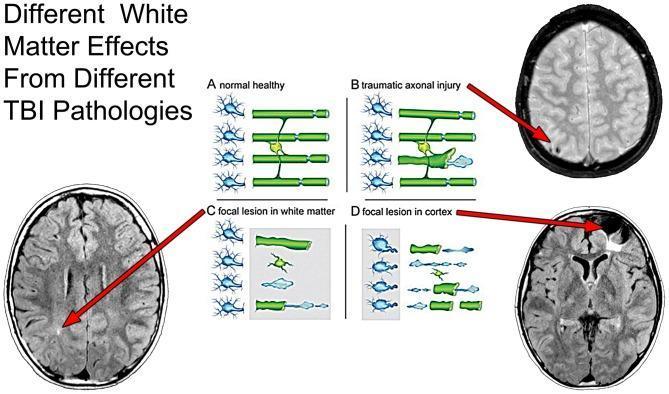
| Traumatic axonal injury (TAI) | Diffuse white matter shear injury from TBI |
| Vanishing white matter disease | Genetic leukodystrophy with progressive white matter loss |
| Periventricular leukomalacia | White matter injury in premature infants |
| Corpus callosum lesions | Disconnection syndromes (alien hand, interhemispheric transfer deficits) |
| Fornix / mammillothalamic tract injury | Amnesia |
Damage to frontal white matter tracts - particularly in the anterior limb of the internal capsule - causes predominantly behavioral and executive dysfunction with relatively few sensorimotor signs.
Imaging: DTI and Tractography
Modern diffusion tensor imaging (DTI) and tractography have revolutionized the study of white matter by revealing the intricate architecture of fiber bundles in the living brain. Fractional anisotropy (FA), radial diffusivity (RD), and axial diffusivity (AD) are key metrics used to assess white matter integrity.
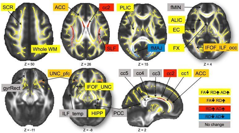
DTI tractography showing major white matter pathways including corpus callosum, corona radiata, superior longitudinal fasciculus, and internal capsule.
Sources: Kaplan & Sadock's Comprehensive Textbook of Psychiatry (White Matter Tracts; White Matter and Cerebral Connectivity) | Grainger & Allison's Diagnostic Radiology (White Matter, Ch. 53)
This is a shared conversation. Sign in to Orris to start your own chat.