
Course and branches of vagus nerve
Reading File
The vagus nerve (CN X) is the longest cranial nerve, with a complex course from the brainstem through the neck, thorax, and abdomen. It carries motor, sensory, and parasympathetic fibers. Let me pull detailed content from the medical library.
Searching sources...
Finding Sources
Finding Sources
Good hits. Let me read the most comprehensive anatomy sources - Gray's Anatomy for Students and Scott-Brown's.
Reading File
Reading File
Finding Sources
Excellent content. Let me read more from Scott-Brown's (continuing the recurrent laryngeal nerve) and also get Gray's overview of vagus in the neck.
Reading File
Finding Sources
Reading File
Reading File
Now let me fetch the anatomical diagrams for visual support.
Loading Image
Loading Image
Loading Image
I now have comprehensive information from multiple authoritative sources. Let me compile the full answer.
Vagus Nerve (Cranial Nerve X) - Course and Branches
Overview
The vagus nerve takes its name from the Latin vagus ("wandering"), reflecting its remarkably extensive course from the brainstem to the abdomen. It is the Xth cranial nerve and the nerve of the 4th branchial arch. It carries four functional components:
- GSE (General somatic efferent) - via nucleus ambiguus: striated muscle of pharynx, larynx, and upper esophagus
- GVE (General visceral efferent) - via dorsal motor nucleus: preganglionic parasympathetic to thoracic and abdominal viscera
- GSA (General somatic afferent) - via jugular (superior) ganglion: skin of posterior ear/external auditory meatus
- GVA (General visceral afferent) - via nodose (inferior) ganglion: visceral sensory from larynx down to the transverse colon
Origin and Ganglia
- 6-8 rootlets emerge from the posterior sulcus (postolivary sulcus) of the lateral medulla oblongata, dorsal to the inferior olive, in close association with CN IX (glossopharyngeal).
- These rootlets unite into a single trunk that exits the skull via the jugular foramen (middle compartment), sharing a dural sheath with CN XI (spinal accessory).
- Two sensory ganglia are found at or just below the jugular foramen:
- Jugular (superior) ganglion - within the foramen; contains cell bodies for general somatic afferents (auricular and meningeal branches)
- Nodose (inferior) ganglion - just below the foramen; contains cell bodies for visceral afferents of all other branches
The accessory nerve (CN XI) contributes a branch to the vagus immediately after exit, providing motor innervation to all visceral striated muscle supplied by the vagus.
Course and Branches - Region by Region
1. Intracranial Portion
Branches given off here:
- Meningeal branch - arises between the two ganglia; supplies dura mater of the posterior cranial fossa
2. Cervical Portion
After exiting the jugular foramen, the vagus descends in the carotid sheath, running posteriorly between the internal jugular vein (IJV) and the common carotid artery (or internal carotid artery in the upper neck).
Branches given off in the neck:
| Branch | Origin | Distribution |
|---|---|---|
| Auricular branch (Arnold's nerve) | Between jugular and nodose ganglia | Passes through mastoid canaliculus; sensory to posterior pinna, posterior EAC, and tympanic membrane. Also mediates referred otalgia |
| Pharyngeal branch | Below nodose ganglion | Crosses the internal carotid artery to join the pharyngeal plexus (with CN IX and sympathetics); motor to pharyngeal constrictors and soft palate (except tensor veli palatini) |
| Superior laryngeal nerve (SLN) | Near nodose ganglion | Passes inferior to ICA, runs anteroinferiorly toward larynx. Divides at the level of the hyoid into: (i) Internal laryngeal nerve - pierces thyrohyoid membrane above superior laryngeal artery; sensory to laryngeal mucosa above vocal folds + piriform fossa; (ii) External laryngeal nerve - runs with superior thyroid artery; motor to cricothyroid muscle (the only intrinsic laryngeal muscle not supplied by RLN) |
| Cervical cardiac branches | Lower neck | Descend to form the cardiac plexus; superior and inferior cervical cardiac branches |
| Carotid body branches | Cervical portion | Supplement CN IX branches to the carotid body chemoreceptors |
3. Thoracic Portion
The two vagus nerves enter the thorax via different routes:
Right vagus nerve:
- Enters the superior mediastinum lying between the right brachiocephalic vein and the brachiocephalic trunk
- Passes posterolaterally to the trachea, is crossed by the arch of the azygos vein, then passes posterior to the root of the right lung
- Gives off the right recurrent laryngeal nerve (RLN) at the root of the neck, which loops under the right subclavian artery and ascends in the tracheoesophageal groove
Left vagus nerve:
- Enters the superior mediastinum posterior to the left brachiocephalic vein, between the left common carotid and left subclavian arteries
- Passes over the arch of the aorta (deep to mediastinal parietal pleura), then posterior to the root of the left lung
- Gives off the left recurrent laryngeal nerve (RLN), which loops under the arch of the aorta at the inferior margin of the arch, just lateral to the ligamentum arteriosum, then ascends in the tracheoesophageal groove
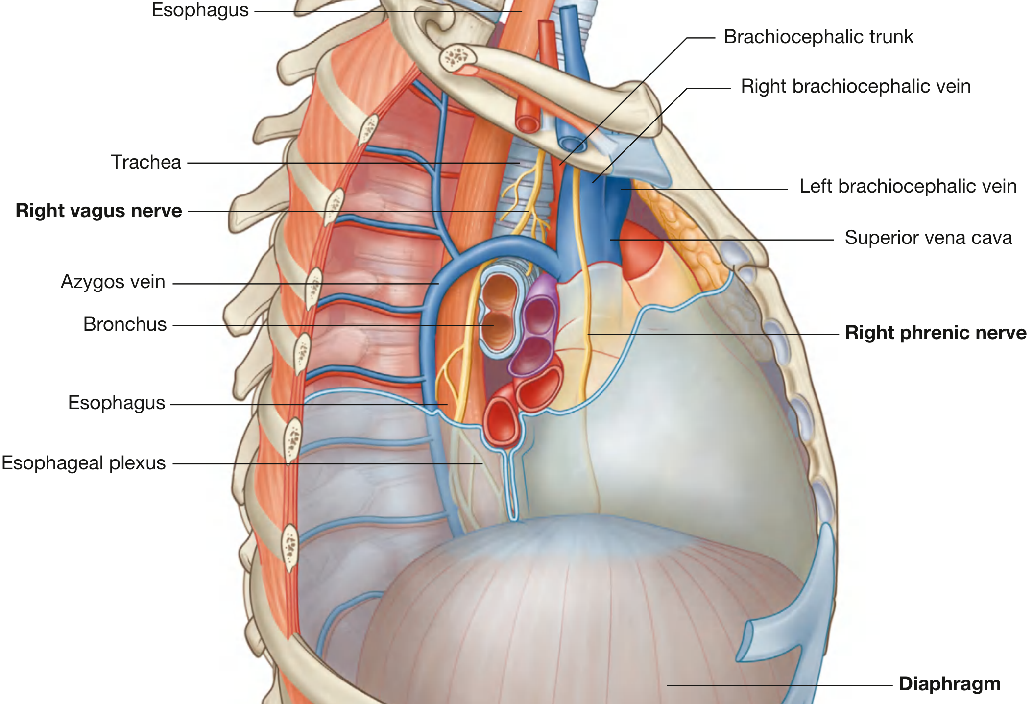
Right vagus nerve in the superior mediastinum - Gray's Anatomy for Students
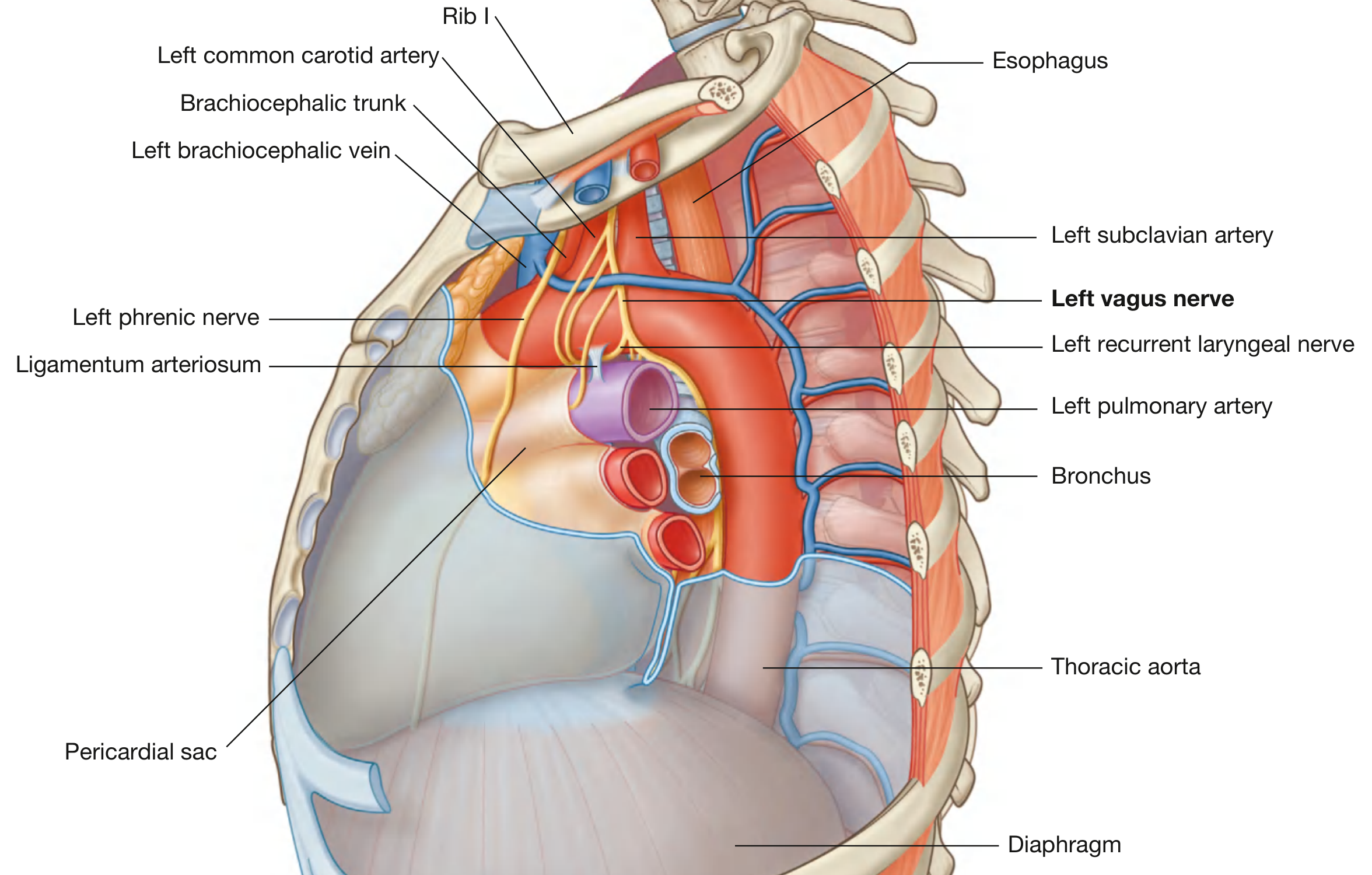
Left vagus nerve with the left recurrent laryngeal nerve looping around the ligamentum arteriosum - Gray's Anatomy for Students
Thoracic branches:
| Branch | Distribution |
|---|---|
| Recurrent laryngeal nerve (RLN) | Motor to ALL intrinsic laryngeal muscles except cricothyroid; sensory to laryngeal mucosa below vocal folds; also supplies trachea and upper esophagus |
| Thoracic cardiac branches | Contribute to cardiac plexus |
| Pulmonary branches | Join the pulmonary plexus anterior and posterior to the lung roots; parasympathetic to bronchi and lungs |
| Esophageal branches | Form the esophageal plexus around the esophagus in the posterior mediastinum |
Both vagi form a plexus around the esophagus, then reconstitute as:
- Anterior vagal trunk (predominantly left vagus) - lies on the anterior surface of the esophagus
- Posterior vagal trunk (predominantly right vagus) - lies on the posterior surface
4. Abdominal Portion
Both trunks enter the abdomen through the esophageal opening (T10 level) of the diaphragm.
Anterior vagal trunk branches:
- Gastric branches - to anterior surface of the stomach
- Hepatic branches - pass in the lesser omentum to the liver and gallbladder
- Pyloric branches - to the pylorus
Posterior vagal trunk branches:
- Gastric branches - to posterior surface of stomach
- Celiac branches - largest branch; passes to the celiac plexus and distributes with its branches to supply the small intestine, ascending colon, and transverse colon
Parasympathetic supply reaches as far as the left (splenic) flexure of the colon (the "Cannon-Böhm point"), beyond which supply comes from the pelvic splanchnic nerves.
Recurrent Laryngeal Nerve - Key Points
The asymmetry of the two RLNs is due to embryology (nerve of the 6th arch following its artery):
- Right RLN: loops under the right subclavian artery (right 4th arch artery); more variable - non-recurrent course in ~1% of individuals (when there is an aberrant subclavian artery)
- Left RLN: loops under the aortic arch at the ligamentum arteriosum (remnant of ductus arteriosus); longer intrathoracic course
Both nerves ascend in the tracheoesophageal groove and enter the larynx by passing deep to the lower border of the inferior constrictor.
A key surgical landmark for identifying the RLN is Beahr's triangle, formed by the common carotid artery laterally, the inferior thyroid vessels superiorly, and the RLN medially.
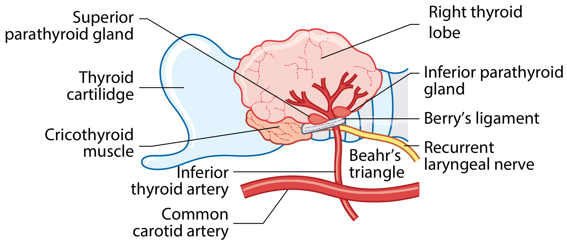
Beahr's triangle for identifying the RLN during thyroid surgery - Scott-Brown's Otorhinolaryngology
Summary Table of All Branches
| Region | Branch | Function |
|---|---|---|
| Intracranial | Meningeal branch | Sensory - posterior fossa dura |
| Jugular foramen | Auricular branch (Arnold's nerve) | Sensory - posterior ear, EAC, tympanic membrane |
| Cervical | Pharyngeal branch | Motor - pharyngeal constrictors, soft palate |
| Cervical | Superior laryngeal nerve (internal) | Sensory - larynx above vocal folds |
| Cervical | Superior laryngeal nerve (external) | Motor - cricothyroid muscle |
| Cervical | Cervical cardiac branches | Parasympathetic/sensory - cardiac plexus |
| Cervical | Carotid body branches | Chemoreceptor supplementation |
| Thoracic | Recurrent laryngeal nerve | Motor - all intrinsic laryngeal muscles except cricothyroid; sensory - larynx below folds |
| Thoracic | Thoracic cardiac branches | Cardiac plexus |
| Thoracic | Pulmonary branches | Pulmonary plexus |
| Thoracic | Esophageal branches | Esophageal plexus |
| Abdominal | Hepatic branches | Liver, gallbladder |
| Abdominal | Gastric branches | Stomach (anterior + posterior) |
| Abdominal | Celiac branches | Small bowel, ascending + transverse colon |
Sources:
- Localization in Clinical Neurology, 8th ed., p. 866-867
- Scott-Brown's Otorhinolaryngology Head & Neck Surgery, p. 601-602
- Bradley and Daroff's Neurology in Clinical Practice
- Gray's Anatomy for Students, p. 261-262
This is a shared conversation. Sign in to Orris to start your own chat.