
Juvenile nasopharyngeal angiofibroma
I'll read the medical library skill first, then research this topic in parallel.
Reading File
Now I'll search the library and PubMed in parallel.
Finding Sources
Finding Sources
Searching PubMed
juvenile nasopharyngeal angiofibroma
The Scott-Brown's ENT textbook has a dedicated section. Let me read it along with the Cummings section in parallel.
Reading File
Finding Sources
Excellent. Now let me read the comprehensive Cummings section and Bailey & Love's section in parallel.
Reading File
Reading File
Reading File
Excellent - I have very rich content. Let me now read more of the Cummings section (which was truncated) and fetch the key images in parallel.
Reading File
Loading Image
Loading Image
The Cummings section continued to osteoma content - the JNA section was truncated. Let me go back and read the full JNA content from Cummings which starts at line 769, reading around lines 787-840.
Reading File
I now have comprehensive content from multiple authoritative sources. Let me compile the full answer.
Juvenile Nasopharyngeal Angiofibroma (JNA)
Overview
Juvenile angiofibroma (JA) is a benign but locally aggressive fibrovascular tumor that occurs almost exclusively in adolescent males, typically between ages 8 and 20. Despite its benign histology, it demonstrates invasive behavior, bone erosion, and a recurrence rate approaching 20%. It is more common in northern India, though the reason is unknown. - Bailey & Love's Short Practice of Surgery, 28th ed.
Pathogenesis and Origin
Histology
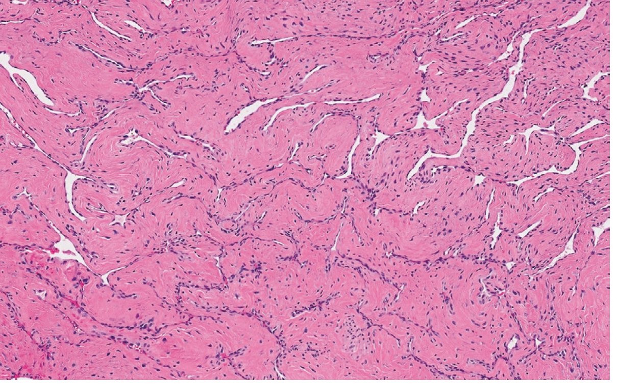
On microscopy, JNA consists of numerous thin-walled vascular channels (lacking a proper smooth muscle coat) embedded in a dense fibrous stroma with stellate fibroblasts. The vessels have an endothelial layer but no muscle layer, making them unable to contract and explaining the profuse bleeding tendency.
Nature: Hamartoma or Neoplasm?
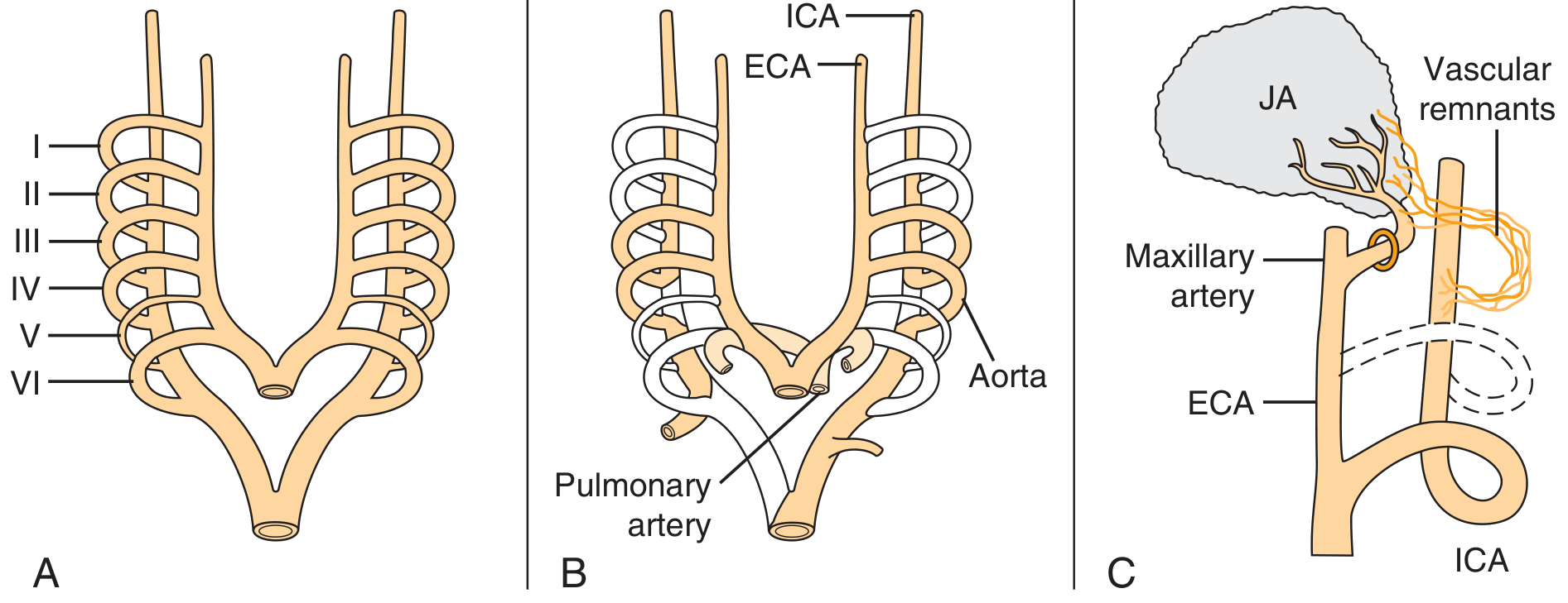
Immunohistochemical and electron microscopy studies suggest JNA is better classified as a vascular malformation (hamartoma) rather than a true neoplasm. Schick et al. proposed that JA develops from incomplete regression of the first branchial arch artery, which forms between days 22-24 of embryogenesis. Incomplete regression leaves vascular remnants that are then stimulated at puberty. This theory is supported by the finding that JA vessels express laminin α2, a marker of early angiogenesis. - Cummings Otolaryngology Head & Neck Surgery
Hormonal Role
The target cells of nasal mucosa are thought to develop into muscularized vascular channels secondary to testosterone stimulation and estrogen hypersensitivity at puberty, explaining why JNA is virtually exclusive to adolescent males. - Scott-Brown's ORL
Molecular Biology
Mutations in CTNNB1 (encoding β-catenin) are present in the majority of sporadic cases. JNA can also develop in association with familial adenomatous polyposis (FAP) - in these syndromic cases it is driven by germline APC gene mutations, which also lead to β-catenin pathway activation. Molecular studies have shown upregulation of VEGFA and activation of VEGF signaling, as well as evidence implicating the FGFR pathway in angiogenesis. - Robbins Pathologic Basis of Disease; Cummings
Site of Origin and Spread
Pathognomonic epicenter: The pterygopalatine fossa - specifically the area of the sphenopalatine foramen or the base of the pterygoid process / superior choana.
Patterns of Spread (follow foramina and fissures of the skull base):
| Direction | Route | Structure Involved |
|---|---|---|
| Medial | Sphenopalatine foramen | Nasopharynx, nasal cavity |
| Posterior | Along vidian nerve | Floor of sphenoid sinus |
| Lateral | Pterygomaxillary fissure | Infratemporal fossa |
| Anterior | Direct extension | Posterior wall of maxillary sinus pushed forward |
| Superior | Inferior/superior orbital fissure, maxillary nerve | Orbit, parasellar region |
| Anterior skull base | Via ethmoid (less common) | Anterior cranial fossa |
Bone involvement occurs via two mechanisms: pressure resorption (subperiosteal growth) or invasion of cancellous bone at the root of the pterygoid process. Transdural growth is very rare. - Cummings
Blood Supply
- Primary: Branches of the external carotid artery - internal maxillary artery and ascending pharyngeal artery
- Advanced lesions: Frequently receive afferents from the internal carotid artery and bilateral carotid systems
- A systematic analysis of 828 cases found ICA supply in 35.6% of lesions, with 30.8% receiving bilateral supply - Cummings
Clinical Features
Classic Presentation (adolescent male)
- Unilateral nasal obstruction (most common with smaller lesions)
- Recurrent severe epistaxis - can lead to anaemia; a hallmark feature
- Purulent rhinorrhoea
- Advanced disease: cheek swelling (infratemporal fossa), proptosis and diplopia (orbital), headache (cranial fossa), vision loss (optic nerve compression)
Endoscopic Finding
A smooth, hypervascularized mass originating behind the middle turbinate in a teenage boy is pathognomonic - the middle turbinate is typically laterally displaced. This clinical picture in the right demographic rarely requires biopsy. - Cummings
Diagnosis
Imaging
CT scan: Best demonstrates bony extent and erosion. Diffuse/patchy enhancement with bone expansion suggests invasion. The key feature is the pathognomonic pterygopalatine fossa epicenter with erosion of the base of the medial pterygoid plate.
MRI: Defines soft-tissue extent. Key MRI features:
- Intense heterogeneous enhancement on T1 post-contrast
- Multiple signal voids on T1 and T2 (= flow voids from major intralesional vessels) - corroborates the diagnosis
- Dural enhancement may suggest intracranial extension
DSA (Digital Subtraction Angiography): Typically shows enlarged intratumoral vessels mixed with more regular arterioles. The venous phase shows an intense inhomogeneous blush showing full tumor extent. Also provides a detailed vascular map prior to embolization.
Biopsy
Generally avoided - high risk of hemorrhage. Clinical and radiological diagnosis in the right patient profile is usually definitive. Biopsy is only justified if CT/MRI are non-diagnostic. - Bailey & Love's
Differential Diagnosis
Other lesions with similar enhancement patterns to consider: lobular capillary hemangioma, hemangiopericytoma, schwannoma - but these do not involve the pterygopalatine fossa and occur in different age groups.
Staging
Several staging systems exist. The Chandler classification is among the earliest, and Andrews/Fisch staging is widely used in surgical planning, broadly dividing tumors into those confined to the nasopharynx/nasal cavity vs. those with pterygopalatine/infratemporal fossa involvement vs. intracranial extension.
Management
Preoperative Embolization
Introduced in the early 1970s, preoperative embolization - performed 24-48 hours before surgery - has transformed JNA management by:
- Dramatically reducing intraoperative blood loss
- Improving surgical access and tumor shrinkage
- Making tumor border identification at dissection more accurate
Transarterial embolization with particles (polyvinyl alcohol, PVA) or liquid agents (NBCA) is standard for ECA-supplied tumors. When intracranial spread involves the ICA, transarterial embolization is risky. In such cases, direct tumor puncture with injection of liquid embolic agents (Onyx or NBCA) was introduced to achieve better devascularization. - Scott-Brown's; Cummings
A 2023 systematic review (PMID 36808756) examined preoperative embolization techniques in JNA.
Surgical Resection
Complete surgical removal is the primary treatment. Approaches:
- Endoscopic resection: Now preferred for smaller, confined lesions (Stages I-II without infratemporal fossa involvement). Some surgeons now omit preoperative embolization for these smaller tumors
- Midfacial degloving: Allows wide exposure and ligation of the feeding maxillary artery
- Lateral rhinotomy: For larger lesions
- Open approaches allow ligation of the maxillary artery and better access to the infratemporal fossa in advanced cases
Radiation Therapy
An alternative for unresectable, recurrent, or intracranial tumors. Primary radiation therapy and stereotactic radiosurgery (CyberKnife) have been used. Radiation carries a small risk of malignant transformation.
Hormonal / Medical Therapy
Historically, flutamide (anti-androgen) was used to reduce tumor size preoperatively, exploiting the androgen receptor expression. Not in routine use.
Prognosis
- Recurrence rates can approach 20%
- 9% of cases can be fatal (from uncontrolled hemorrhage or intracranial extension) - Robbins
- Spontaneous involution has been described in untreated cases, consistent with the hamartoma theory, but is not predictable enough to be managed expectantly
- Residual disease post-surgery: serial MRI monitoring shows that small residual lesions may remain stable or involute without re-intervention
Recent Evidence
- Early-onset JNA systematic review (PMID 38115030) - J Otolaryngol Head Neck Surg, Dec 2023 - reviews cases presenting before the classic adolescent age range
- Preoperative embolization techniques systematic review (PMID 36808756) - Otolaryngol Head Neck Surg, Sep 2023 - comparative review of embolization approaches
- MRI features of JNA systematic review (PMID 37164909) - J Neuroimaging, Sep 2023 - characterizes MRI findings across case series
Sources: Cummings Otolaryngology Head & Neck Surgery | Scott-Brown's Otorhinolaryngology Head & Neck Surgery Vol. 1 | Bailey & Love's Short Practice of Surgery, 28th ed. | Robbins, Cotran & Kumar Pathologic Basis of Disease
This is a shared conversation. Sign in to Orris to start your own chat.