
Teach me the attachment, blood and nerve supply of maxilla
"maxilla"[MeSH Terms] AND "blood supply"
maxilla bone lateral view anatomy
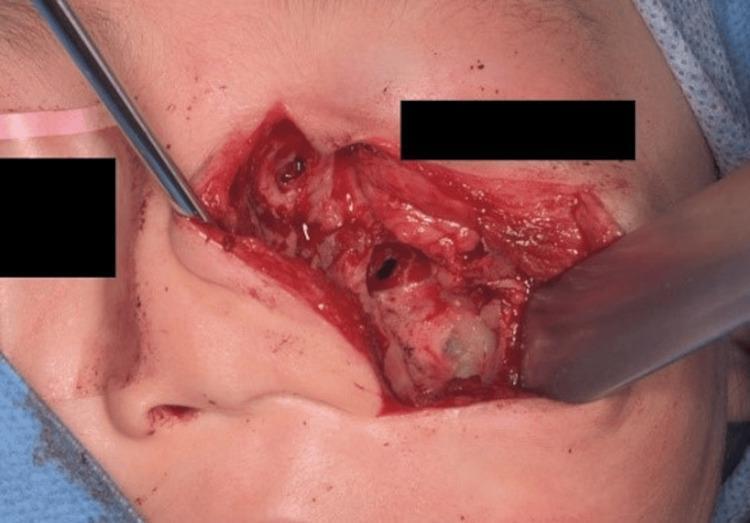
This clinical photograph captures an intraoperative view of a mid-face surgical procedure involving the resection of a tumor from the left nasal dorsum and maxilla. The surgical field demonstrates a deep incision with skin and subcutaneous tissue retracted to expose the underlying anatomy of the mid-face. A significant bone resection defect is visible at the inferior orbital rim and the anterior wall of the maxilla, where the tumor had previously invaded. Visible surgical instrumentation includes a metal probe or retractor on the medial (nasal) side and a flat-bladed surgical elevator on the lateral side, used to retract tissue and provide access to the deep structures. Anatomical landmarks include the lateral wall of the nasal cavity and the exposed bone of the infraorbital region. The image illustrates the clinical application of radical surgical excision for solitary nucleus-associated myofibroma (SNM) or similar invasive soft-tissue tumors, emphasizing the need for partial bone resection when periosteal or osseous invasion is present.
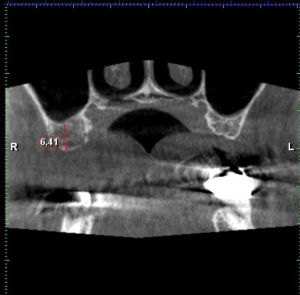
A Cone Beam Computed Tomography (CBCT) scan showing a coronal cross-sectional view of the maxilla and surrounding structures. The imaging highlights the anatomy of the maxillary sinuses and the nasal cavity. In the right posterior maxilla, specifically the first molar region, a vertical red measurement line indicates a residual bone height of 6.41 mm between the alveolar ridge and the floor of the maxillary sinus. On the left side of the scan, a high-density radiopaque structure consistent with a dental implant or surgical material is visible, accompanied by some beam-hardening artifacts. Notably, the bony canal for the Alveolar Antral Artery (AAA) is not visualized within the lateral wall of the maxillary sinus on this slice. This imaging is clinically relevant for pre-operative planning in oral surgery, such as sinus lift procedures and dental implant placement, to assess available bone volume and identify potential vascular risks.
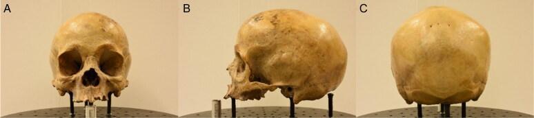
Three clinical photographs showing a human skull specimen from different orientations: (A) anterior, (B) left lateral, and (C) posterior views. The anterior view (A) highlights the mid-face anatomy, including the piriform (nasal) aperture, the anterior nasal spine, and the orbital rims. The lateral view (B) demonstrates the cranial vault's curvature, the temporal fossa, the zygomatic arch, and the external auditory meatus. The posterior view (C) shows the occipital bone and the lambdoid suture. This visual material is used for osteological analysis and morphometric studies, focusing on characterizing variation in anatomical landmarks such as the rhinion and dacryon. Key educational concepts illustrated include cranial topography, forensic anthropology, and the structural relationships between the maxilla, frontal, and parietal bones.
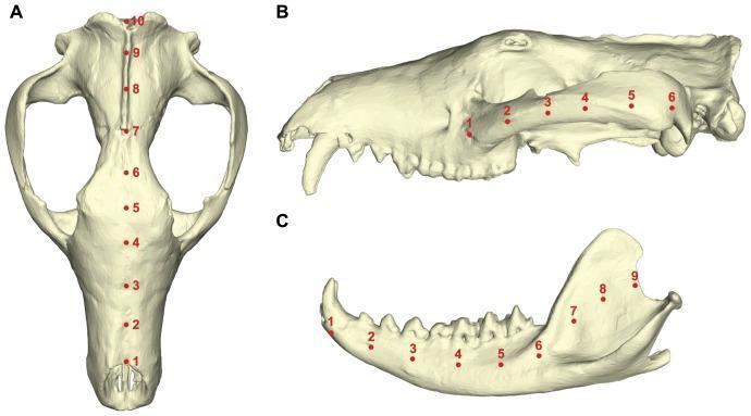
Anatomical diagram and diagnostic model of a reconstructed cranium and mandible, demonstrating anatomical landmarks used for biomechanical stress analysis. Figure A shows a superior (dorsal) view of the skull with 10 red nodes positioned along the mid-sagittal plane, spanning from the anterior nasal bone, through the frontal bone, to the posterior sagittal crest. Figure B displays a lateral view of the cranium, featuring 6 nodes aligned along the zygomatic arch, extending from the zygomatic process of the maxilla toward the squamosal bone. Figure C illustrates a lateral view of the mandible, with 9 nodes distributed along the dentary; nodes 1-6 are spaced equidistantly along the horizontal mandibular body, while nodes 7-9 ascend the ramus toward the coronoid and condylar processes. This visual serves as an educational model for studying the distribution of von Mises stress across different anatomical structures of the skull, aiding in the understanding of biomechanical loads and structural integrity in maxillofacial anatomy.
Maxilla: attachments, blood supply, nerve supply
1. Quick anatomy
2. Attachments
A. Muscles and ligaments
| Muscle | Attachment on the maxilla |
|---|---|
| Levator labii superioris | Just above the infraorbital foramen |
| Levator labii superioris alaeque nasi | Frontal process |
| Levator anguli oris | Canine fossa, below the infraorbital foramen |
| Nasalis | Transverse part: above the incisive fossa. Alar part: above the lateral incisor |
| Depressor septi nasi | Incisive fossa, above the central incisor |
| Orbicularis oculi (orbital and lacrimal parts) | Medial palpebral ligament, frontal process, anterior lacrimal crest |
| Inferior oblique | Orbital surface, near the nasolacrimal groove |
| Buccinator | Outer surface of the alveolar process opposite the molars |
| Masseter (superficial head) | Zygomatic process |
| Medial pterygoid (small superficial head) | Maxillary tuberosity |
- The medial palpebral ligament attaches to the frontal process and the anterior lacrimal crest.
- The buccinator attachment is clinically important. Infection from maxillary molar roots that breaks out above it enters the buccal space. Infection that breaks out below it enters the vestibule (Cummings Otolaryngology).
- Bony articulations: nasal, frontal, lacrimal, ethmoid, zygomatic, palatine, inferior concha, vomer, the opposite maxilla, and the sphenoid pterygoid process (at the tuberosity).
- The posterior surface forms the anterior wall of the infratemporal fossa. It carries the foramen for the posterosuperior alveolar nerve and vessels. Its superior margin forms the inferior border of the inferior orbital fissure (Gray's Anatomy for Students).
B. Memory aid
- Upper lip and nose elevators attach around the infraorbital foramen and the frontal process.
- The cheek (buccinator) attaches to the alveolar process.
- Mastication (masseter and medial pterygoid) attaches to the zygomatic process and the tuberosity.
3. Blood supply
| Artery | Origin | Supplies |
|---|---|---|
| Posterior superior alveolar (PSAA) | 3rd part of the maxillary artery | Molars and premolars, adjacent bone, the maxillary sinus, buccal gingiva |
| Infraorbital artery | Maxillary artery, entering via the inferior orbital fissure | Orbital floor, anterior maxilla, cheek |
| Anterior superior alveolar (ASAA) | Infraorbital artery | Upper incisors, canine and sinus mucosa, via the canalis sinuosus |
| Middle superior alveolar (often present) | Infraorbital artery | Premolars |
| Greater (descending) palatine | Maxillary artery | Hard palate, palatal gingiva |
| Sphenopalatine | Maxillary artery | Nasal septum, lateral wall and nasopalatine region (via the incisive canal) |
- The alveolar antral artery runs in the lateral sinus wall. It can bleed during a sinus lift or Caldwell-Luc procedure.
- Most of the maxilla's blood supply is rich and overlapping. This is why it heals well and why the maxilla has a lower risk of osteonecrosis than the mandible.
4. Nerve supply
- Posterior superior alveolar nerve (PSAN): leaves V2 in the pterygopalatine fossa and pierces the infratemporal surface of the maxilla. It supplies the molars (except the mesiobuccal root of the first molar in many people), the sinus lining, and the buccal gingiva and cheek.
- Middle superior alveolar nerve (MSAN): arises from the infraorbital nerve in the infraorbital groove. It supplies the premolars. It is variable.
- Anterior superior alveolar nerve (ASAN): arises from the infraorbital nerve near the midpoint of its canal and runs through the canalis sinuosus. It supplies the incisors, canine and anterior sinus wall.
- Greater palatine nerve: palatal mucosa and gingiva from the molars to the premolars.
- Nasopalatine nerve: anterior palate and palatal gingiva behind the incisors and canine, through the incisive canal.
- Lesser palatine nerve: soft palate.
- Buccal gingiva: PSAN in the molar region, and the infraorbital nerve anteriorly. The long buccal nerve (V3) supplies the buccal gingiva of the molars. Palatal gingiva is supplied by the palatine nerves.
- Maxillary teeth are usually anaesthetised by local infiltration, because the buccal cortical bone is thin. Infraorbital and PSAN blocks are used for larger areas.
- Infraorbital numbness after midface fractures or tumour invasion points to injury to the infraorbital nerve.