
Human skeleton anatomy
Reading File
Now let me search the library for skeleton anatomy content and images in parallel.
Finding Sources
Finding Sources
Searching the Web
human skeleton anatomy overview bones labeled diagram
Finding Sources
Loading Image
Loading Image
Now let me read the Gray's Anatomy and Developing Human textbook sections on the skeletal system:
Reading File
Reading File
Let me also fetch the Gray's Anatomy diagram and read more from the textbook:
Loading Image
Reading File
I now have excellent material from Gray's Anatomy for Students plus two detailed labeled diagrams. Here is a thorough overview:
Human Skeleton Anatomy
Labeled Diagrams
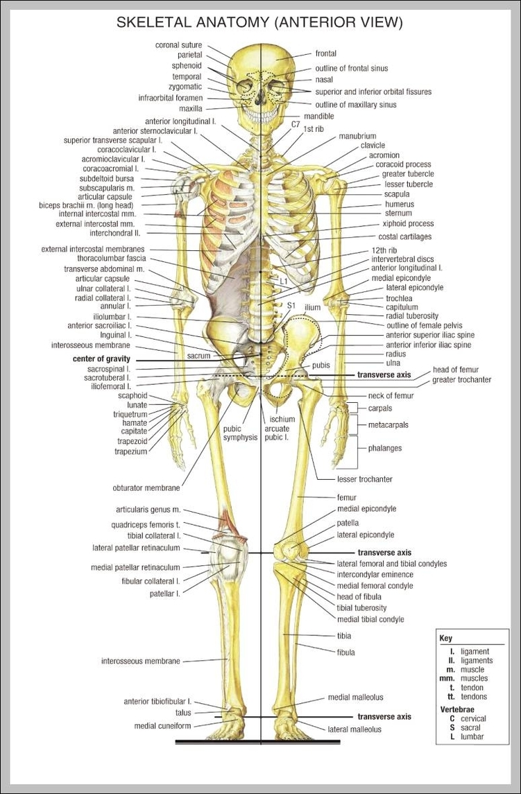

Overview
The adult human skeleton contains 206 bones. These are organized into two major divisions:
| Division | Bones | Count |
|---|---|---|
| Axial skeleton | Skull, vertebral column, ribs, sternum | 80 |
| Appendicular skeleton | Upper limbs, lower limbs, shoulder girdles, pelvic girdle | 126 |
- Gray's Anatomy for Students - "The skeleton can be divided into two subgroups, the axial skeleton and the appendicular skeleton."
1. Axial Skeleton (80 bones)
Skull
- Neurocranium (braincase): frontal, 2 parietal, 2 temporal, occipital, sphenoid, ethmoid
- Viscerocranium (face): nasal, maxilla, lacrimal, zygomatic, palatine, inferior nasal conchae, vomer, mandible
- Hyoid bone (throat, supports tongue)
- Ear ossicles (3 per side): malleus, incus, stapes
Vertebral Column (33 vertebrae total)
| Region | Number | Key Features |
|---|---|---|
| Cervical | 7 (C1-C7) | C1 = Atlas, C2 = Axis; support head, allow rotation |
| Thoracic | 12 (T1-T12) | Articulate with ribs |
| Lumbar | 5 (L1-L5) | Largest vertebrae; bear body weight |
| Sacrum | 5 fused | Forms posterior wall of pelvis |
| Coccyx | 4 fused | Vestigial tailbone |
Thorax
- Sternum: manubrium + body + xiphoid process
- 12 pairs of ribs: True ribs (1-7, attach directly to sternum), False ribs (8-10, attach via costal cartilage), Floating ribs (11-12, no anterior attachment)
2. Appendicular Skeleton (126 bones)
Shoulder Girdle (4 bones)
- Clavicle (collarbone) x2
- Scapula (shoulder blade) x2
Upper Limb (each side = 30 bones)
| Segment | Bones |
|---|---|
| Arm | Humerus |
| Forearm | Radius + Ulna |
| Wrist (carpals) | Scaphoid, lunate, triquetrum, pisiform, trapezium, trapezoid, capitate, hamate (8 per wrist) |
| Palm (metacarpals) | 5 metacarpals |
| Fingers (phalanges) | 14 phalanges (2 in thumb, 3 in each finger) |
Pelvic Girdle (2 hip bones)
- Each hip bone = ilium + ischium + pubis (fused in adults)
- Together with sacrum they form the bony pelvis
- Key landmarks: anterior superior iliac spine (ASIS), pubic symphysis, obturator foramen, acetabulum
Lower Limb (each side = 30 bones)
| Segment | Bones |
|---|---|
| Thigh | Femur (longest bone in body) |
| Kneecap | Patella (largest sesamoid bone) |
| Leg | Tibia + Fibula |
| Ankle (tarsals) | Talus, calcaneus, navicular, cuboid, 3 cuneiforms (7 per ankle) |
| Foot (metatarsals) | 5 metatarsals |
| Toes (phalanges) | 14 phalanges |
Bone Structure & Types
Bone Types by Shape
| Type | Description | Example |
|---|---|---|
| Long bones | Tubular; shaft + two epiphyses | Humerus, femur, tibia |
| Short bones | Cuboidal | Carpals, tarsals |
| Flat bones | Two compact plates + spongy bone between | Skull, sternum, scapula |
| Irregular bones | Varied complex shapes | Vertebrae, facial bones |
| Sesamoid bones | Develop within tendons | Patella, sesamoids of thumb |
Bone Tissue
- Compact bone: Dense outer shell of all bones
- Spongy (trabecular/cancellous) bone: Internal lattice containing marrow cavities; site of blood cell production
Blood Supply & Innervation
- A nutrient artery (one per bone) enters the internal cavity and supplies marrow, spongy bone, and inner compact bone layers
- The periosteum (fibrous outer membrane) covers all bone surfaces except at joint cartilage; it is richly supplied with sensory nerves and is very pain-sensitive
- Bone itself has few sensory nerve fibers; most internal nerve fibers are vasomotor
Cartilage
Three types of cartilage are found in the skeleton:
| Type | Features | Location |
|---|---|---|
| Hyaline | Most common; moderate collagen | Articular surfaces, costal cartilages, growth plates |
| Elastic | Collagen + elastic fibers | External ear, epiglottis |
| Fibrocartilage | Dense collagen, few cells | Intervertebral discs, pubic symphysis, knee menisci |
Cartilage is avascular - nourished entirely by diffusion. It has no blood vessels, lymphatics, or nerves.
Functions of the Skeleton
The skeleton serves five main functions:
- Support - structural framework for the body
- Protection - shields vital organs (skull protects brain; rib cage protects heart and lungs)
- Mineral reservoir - stores calcium and phosphorus
- Movement - levers for muscular action
- Haematopoiesis - spongy bone cavities house bone marrow for blood cell production
Bone Development
- Intramembranous ossification: Mesenchyme ossifies directly without a cartilage model (flat bones of the cranium)
- Endochondral ossification: Cartilaginous model forms first, then ossifies; used for most bones including long bones and the appendicular skeleton
- Ossification centers appear by the end of the embryonic period (about 56 days)
- Skeletal maturity is typically reached between ages 20-25
Skeletal Age Assessment
Skeletal maturity can be assessed by radiographing the non-dominant (left) hand and comparing to standard reference radiographs. This is clinically useful in evaluating growth disorders, malnutrition, and hypothyroidism.
For a great visual walkthrough, this video covers the bones of the skeleton clearly:
Sources: Gray's Anatomy for Students; The Developing Human: Clinically Oriented Embryology; Wikipedia - List of bones
This is a shared conversation. Sign in to Orris to start your own chat.