Tell me all piles like disease of anal region

Reading File
Finding Sources
Finding Sources
Finding Sources
Finding Sources
Finding Sources
Finding Sources
Reading File
Finding Sources
Finding Sources
Reading File
Reading File
Searching Images

hemorrhoids piles classification internal external anal canal
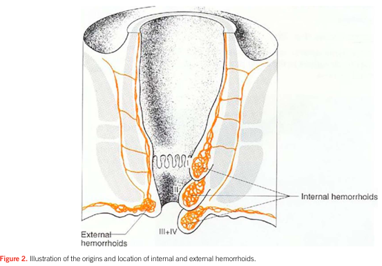
Summary : This illustration shows the anatomical origins and locations of internal and external hemorrhoids in relation to the anal canal and rectum.

illustration:
# Scene Overview :
  • Main subject is a cross-sectional anatomical diagram of the lower rectum and anal canal.
  • The image highlights the positions of internal and external hemorrhoids using orange outlines.
  • The anal canal is centrally depicted, with the rectum above and the perianal skin below.

# Technical Details :
  • Internal hemorrhoids are shown as bulging structures within the anal canal, above the dentate line.
  • External hemorrhoids are depicted as bulges below the dentate line, near the anal verge.
  • Labels indicate "Internal hemorrhoids" and "External hemorrhoids" with arrows pointing to their respective locations.
  • The illustration includes reference to hemorrhoid grades (III, III+IV) near the anal opening.

# Spatial Relationships :
  • Internal hemorrhoids are located higher up within the anal canal, above the dentate line.
  • External hemorrhoids are situated outside or below the anal canal, near the skin surface.
  • The diagram provides a clear distinction between the two types based on their anatomical position.

# Analysis :
  • The figure visually clarifies the difference in origin and location between internal and external hemorrhoids, emphasizing their anatomical separation by the dentate line. Internal hemorrhoids are positioned within the anal canal, while external hemorrhoids are found at or below the anal verge.
Summary : This illustration shows the anatomical origins and locations of internal and external hemorrhoids in relation to the anal canal and rectum. illustration: # Scene Overview : • Main subject is a cross-sectional anatomical diagram of the lower rectum and anal canal. • The image highlights the positions of internal and external hemorrhoids using orange outlines. • The anal canal is centrally depicted, with the rectum above and the perianal skin below. # Technical Details : • Internal hemorrhoids are shown as bulging structures within the anal canal, above the dentate line. • External hemorrhoids are depicted as bulges below the dentate line, near the anal verge. • Labels indicate "Internal hemorrhoids" and "External hemorrhoids" with arrows pointing to their respective locations. • The illustration includes reference to hemorrhoid grades (III, III+IV) near the anal opening. # Spatial Relationships : • Internal hemorrhoids are located higher up within the anal canal, above the dentate line. • External hemorrhoids are situated outside or below the anal canal, near the skin surface. • The diagram provides a clear distinction between the two types based on their anatomical position. # Analysis : • The figure visually clarifies the difference in origin and location between internal and external hemorrhoids, emphasizing their anatomical separation by the dentate line. Internal hemorrhoids are positioned within the anal canal, while external hemorrhoids are found at or below the anal verge.

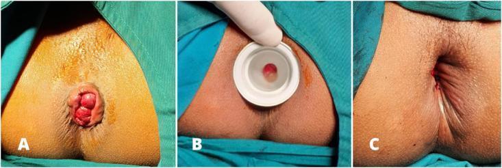
This clinical photograph sequence depicts three stages of a surgical hemorrhoidectomy for prolapsed internal hemorrhoids (Grade III/IV). Panel A shows the preoperative state with large, circumferential, erythematous hemorrhoidal piles protruding through the anal verge. The mucosa appears congested and edematous. Panel B demonstrates the intraoperative phase of anal dilatation. A white, circular, rigid plastic dilator is inserted into the anal canal to provide surgical exposure and reduce internal anal sphincter hypertonicity. A portion of the hemorrhoidal tissue is visible through the central aperture of the dilator. Panel C shows the immediate postoperative result following excision and radial wound closure. The protruding masses have been successfully resected. The anal canal is restored to its anatomical position with visible simple interrupted sutures (multifilament absorbable type) placed radially. Minor punctate bleeding is noted at the suture lines, which is typical for this stage of the procedure. The surrounding perianal skin remains intact and draped in a sterile surgical field.
This clinical photograph sequence depicts three stages of a surgical hemorrhoidectomy for prolapsed internal hemorrhoids (Grade III/IV). Panel A shows the preoperative state with large, circumferential, erythematous hemorrhoidal piles protruding through the anal verge. The mucosa appears congested and edematous. Panel B demonstrates the intraoperative phase of anal dilatation. A white, circular, rigid plastic dilator is inserted into the anal canal to provide surgical exposure and reduce internal anal sphincter hypertonicity. A portion of the hemorrhoidal tissue is visible through the central aperture of the dilator. Panel C shows the immediate postoperative result following excision and radial wound closure. The protruding masses have been successfully resected. The anal canal is restored to its anatomical position with visible simple interrupted sutures (multifilament absorbable type) placed radially. Minor punctate bleeding is noted at the suture lines, which is typical for this stage of the procedure. The surrounding perianal skin remains intact and draped in a sterile surgical field.

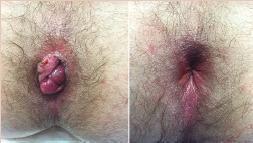
This comparison clinical photograph illustrates a pre-operative and post-operative view of the anal region in a patient with symptomatic internal hemorrhoids. The left panel shows significant internal hemorrhoidal prolapse, characterized by several engorged, reddish, and lobulated mucosal masses protruding through the anal sphincter. The tissue appears swollen with visible folds and irregular surface texture. The right panel displays the clinical outcome following Transanal Hemorrhoidal Dearterialization (THD) and mucopexy. In this view, the prolapsed hemorrhoidal tissue has been successfully reduced and lifted back into the distal rectum. The anal opening appears anatomically normal with no visible protrusions, though there is mild residual erythema of the surrounding perianal skin. This side-by-side comparison demonstrates the effectiveness of the mucopexy technique in restoring the normal anatomy of the anal canal by lifting and repositioning pathological hemorrhoidal piles.
This comparison clinical photograph illustrates a pre-operative and post-operative view of the anal region in a patient with symptomatic internal hemorrhoids. The left panel shows significant internal hemorrhoidal prolapse, characterized by several engorged, reddish, and lobulated mucosal masses protruding through the anal sphincter. The tissue appears swollen with visible folds and irregular surface texture. The right panel displays the clinical outcome following Transanal Hemorrhoidal Dearterialization (THD) and mucopexy. In this view, the prolapsed hemorrhoidal tissue has been successfully reduced and lifted back into the distal rectum. The anal opening appears anatomically normal with no visible protrusions, though there is mild residual erythema of the surrounding perianal skin. This side-by-side comparison demonstrates the effectiveness of the mucopexy technique in restoring the normal anatomy of the anal canal by lifting and repositioning pathological hemorrhoidal piles.

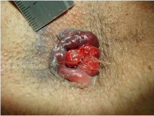
This clinical photograph displays a detailed view of the perianal region demonstrating circular perianal thrombosis and prolapsed hemorrhoids. The central focus shows bright red, lobulated, prolapsing internal hemorrhoids protruding through the anal canal, characterized by a glossy, friable appearance indicative of active or recent bleeding. Surrounding these is a prominent, circular area of dark purple to bluish discoloration consistent with thrombosed external hemorrhoids. Significant edema and swelling are visible in the associated perianal tissue, particularly involving concomitant anal tags on the left-lateral aspect. The surrounding skin is hirsute and shows localized bruising. A medical ruler is partially visible at the top left of the frame for size reference. This image serves as an educational example of advanced hemorrhoidal disease, illustrating the coexistence of grade IV internal prolapse with acute external venous thrombosis and tissue edema.

This clinical photograph displays a detailed view of the perianal region demonstrating circular perianal thrombosis and prolapsed hemorrhoids. The central focus shows bright red, lobulated, prolapsing internal hemorrhoids protruding through the anal canal, characterized by a glossy, friable appearance indicative of active or recent bleeding. Surrounding these is a prominent, circular area of dark purple to bluish discoloration consistent with thrombosed external hemorrhoids. Significant edema and swelling are visible in the associated perianal tissue, particularly involving concomitant anal tags on the left-lateral aspect. The surrounding skin is hirsute and shows localized bruising. A medical ruler is partially visible at the top left of the frame for size reference. This image serves as an educational example of advanced hemorrhoidal disease, illustrating the coexistence of grade IV internal prolapse with acute external venous thrombosis and tissue edema.

Searching Images

anal fissure chronic fissure-in-ano

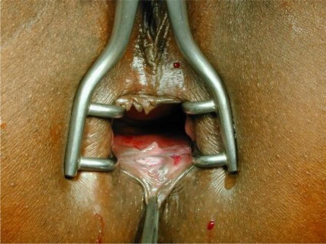
This clinical photograph shows a chronic anal fissure located at the 6 o'clock position in a patient in the supine (lithotomy) position. A metallic Sims (Schulze-Bergmann) rectal speculum is inserted to provide surgical exposure of the anal canal. The fissure is visible as a distinct, longitudinal tear or linear ulcer in the anoderm, surrounded by thickened, inflamed margins and characteristic secondary changes such as a hypertrophied anal papilla or skin tag. The surgical field demonstrates the relationship between the perianal skin, the anal verge, and the internal sphincter fibers. Small amounts of serosanguinous fluid are present on the mucosa. This image serves as a teaching tool for proctology and colorectal surgery, specifically illustrating the preoperative state before a fissurectomy or anocutaneous advancement flap procedure.
This clinical photograph shows a chronic anal fissure located at the 6 o'clock position in a patient in the supine (lithotomy) position. A metallic Sims (Schulze-Bergmann) rectal speculum is inserted to provide surgical exposure of the anal canal. The fissure is visible as a distinct, longitudinal tear or linear ulcer in the anoderm, surrounded by thickened, inflamed margins and characteristic secondary changes such as a hypertrophied anal papilla or skin tag. The surgical field demonstrates the relationship between the perianal skin, the anal verge, and the internal sphincter fibers. Small amounts of serosanguinous fluid are present on the mucosa. This image serves as a teaching tool for proctology and colorectal surgery, specifically illustrating the preoperative state before a fissurectomy or anocutaneous advancement flap procedure.

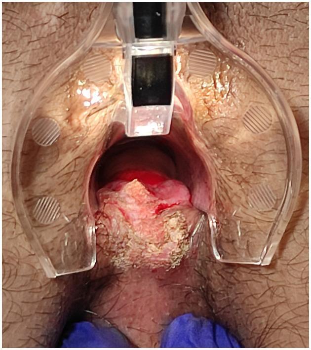
This clinical photograph displays a surgical intervention for a chronic anal fissure located in the posterior midline. An anoscope/retractor is utilized to provide clear visualization of the anal canal. The central pathology features a posterior fissure associated with a prominent sentinel skin tag at the anal verge. The image illustrates the intraoperative state following CO2 laser scanner vaporization and debridement. The treated tissue exhibits a heterogeneous, rough, and irregular texture with areas of whitish-yellowish discoloration, characteristic of superficial thermal debridement and laser-induced vaporization of the sentinel tag. Reddish, vascularized regions are visible deeper within the canal, indicating inflamed mucosa or areas of recent surgical manipulation. The surrounding perianal skin is normal in appearance, with visible hair follicles, providing a clear contrast to the textured, treated surgical site. This image serves as a clinical reference for proctological surgical procedures using laser technology to manage chronic anal fissures and associated hypertrophic skin tags.

This clinical photograph displays a surgical intervention for a chronic anal fissure located in the posterior midline. An anoscope/retractor is utilized to provide clear visualization of the anal canal. The central pathology features a posterior fissure associated with a prominent sentinel skin tag at the anal verge. The image illustrates the intraoperative state following CO2 laser scanner vaporization and debridement. The treated tissue exhibits a heterogeneous, rough, and irregular texture with areas of whitish-yellowish discoloration, characteristic of superficial thermal debridement and laser-induced vaporization of the sentinel tag. Reddish, vascularized regions are visible deeper within the canal, indicating inflamed mucosa or areas of recent surgical manipulation. The surrounding perianal skin is normal in appearance, with visible hair follicles, providing a clear contrast to the textured, treated surgical site. This image serves as a clinical reference for proctological surgical procedures using laser technology to manage chronic anal fissures and associated hypertrophic skin tags.

Searching Images

fistula in ano Parks classification perianal abscess
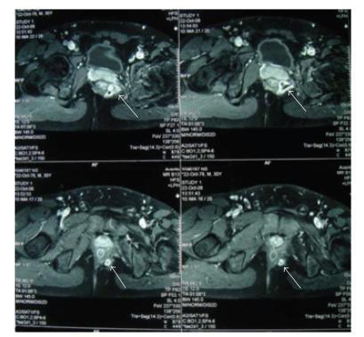
Axial pelvic Magnetic Resonance Imaging (MRI) scans demonstrate a complex suprasphincteric fistula in ano with associated abscess formation. The series of T2-weighted or contrast-enhanced images shows a high-signal-intensity fluid collection representing an abscess cavity, approximately 2 cm in diameter, located superior to the levator ani muscle. White arrows indicate the fistula tract, which presents as a distinct linear pathway of altered signal intensity extending from an internal opening in the rectum through the pelvic musculature toward the perianal region. The images highlight the relationship between the inflammatory process and key anatomical landmarks, including the rectum, levator ani, and perianal soft tissues. This diagnostic imaging is characteristic of complex perianal disease, showing both the localized suppuration (abscess) and the extensive 10 cm fistulous tract. Such findings are critical for surgical planning in cases of deep pelvic infections or complex fistula management.
Axial pelvic Magnetic Resonance Imaging (MRI) scans demonstrate a complex suprasphincteric fistula in ano with associated abscess formation. The series of T2-weighted or contrast-enhanced images shows a high-signal-intensity fluid collection representing an abscess cavity, approximately 2 cm in diameter, located superior to the levator ani muscle. White arrows indicate the fistula tract, which presents as a distinct linear pathway of altered signal intensity extending from an internal opening in the rectum through the pelvic musculature toward the perianal region. The images highlight the relationship between the inflammatory process and key anatomical landmarks, including the rectum, levator ani, and perianal soft tissues. This diagnostic imaging is characteristic of complex perianal disease, showing both the localized suppuration (abscess) and the extensive 10 cm fistulous tract. Such findings are critical for surgical planning in cases of deep pelvic infections or complex fistula management.

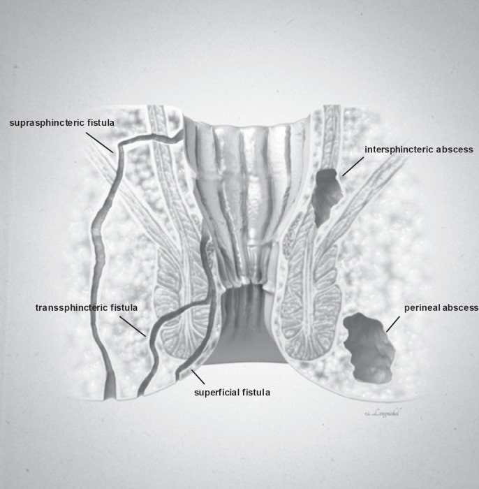
**Imaging Modality:** Medical illustration/Anatomical diagram.

**Anatomical Region:** Coronal cross-section of the anorectal region, including the anal canal, rectum, internal and external anal sphincters, and surrounding perianal soft tissues.

**Observed Pathology:** The illustration classifies various types of anorectal fistulas and abscesses based on their anatomical trajectory and location.

**Characteristic Visual Features:**
*   **Fistula Tracts:** Three distinct epithelial-lined tracks are labeled. A **superficial fistula** is shown confined to the perianal skin and distal anal canal. A **transsphincteric fistula** is depicted crossing both the internal and external sphincter muscles into the ischioanal fossa. A **suprasphincteric fistula** is illustrated looping above the puborectalis muscle before descending to the skin surface.
*   **Abscess Formations:** Two localized collections of fluid/pus are identified. An **intersphincteric abscess** is positioned within the space between the internal and external sphincter muscles. A **perineal (perianal) abscess** is shown as a larger pocket located in the subcutaneous tissue adjacent to the anal verge.

**Key Diagnostic Features:** The diagram emphasizes the relationship between inflammatory tracts and the sphincter complex, following the Parks classification system for fistula-in-ano. Suitable for clinical education on perianal sepsis and surgical planning.

**Imaging Modality:** Medical illustration/Anatomical diagram. **Anatomical Region:** Coronal cross-section of the anorectal region, including the anal canal, rectum, internal and external anal sphincters, and surrounding perianal soft tissues. **Observed Pathology:** The illustration classifies various types of anorectal fistulas and abscesses based on their anatomical trajectory and location. **Characteristic Visual Features:** * **Fistula Tracts:** Three distinct epithelial-lined tracks are labeled. A **superficial fistula** is shown confined to the perianal skin and distal anal canal. A **transsphincteric fistula** is depicted crossing both the internal and external sphincter muscles into the ischioanal fossa. A **suprasphincteric fistula** is illustrated looping above the puborectalis muscle before descending to the skin surface. * **Abscess Formations:** Two localized collections of fluid/pus are identified. An **intersphincteric abscess** is positioned within the space between the internal and external sphincter muscles. A **perineal (perianal) abscess** is shown as a larger pocket located in the subcutaneous tissue adjacent to the anal verge. **Key Diagnostic Features:** The diagram emphasizes the relationship between inflammatory tracts and the sphincter complex, following the Parks classification system for fistula-in-ano. Suitable for clinical education on perianal sepsis and surgical planning.

Searching Images

rectal prolapse perianal skin tag pilonidal sinus
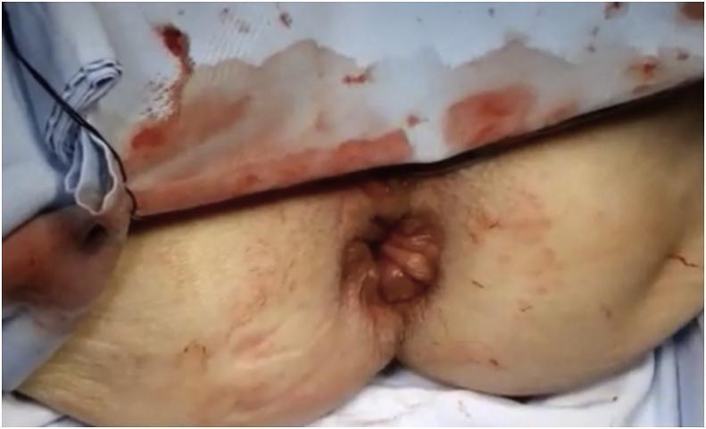
Clinical photograph of the perianal region demonstrating a full-thickness rectal prolapse. The image shows a reddish-brown, rounded mass of rectal mucosa and wall protruding through the anal sphincter. The prolapsed tissue exhibits a characteristic glistening, moist surface with concentric mucosal folds. The surrounding perianal skin shows signs of irritation and erythema. A surgical context is evident, with a blood-stained medical drape positioned above the site and minor blood markings on the skin of the buttocks, suggesting a perioperative or immediate postoperative state. This visual is typical for educational materials regarding colorectal pathology and surgical interventions like Perineal Stapled Prolapsectomy (PSP). The image highlights the morphological characteristics of external rectal prolapse and the clinical appearance of the anal region during or following corrective surgery.
Clinical photograph of the perianal region demonstrating a full-thickness rectal prolapse. The image shows a reddish-brown, rounded mass of rectal mucosa and wall protruding through the anal sphincter. The prolapsed tissue exhibits a characteristic glistening, moist surface with concentric mucosal folds. The surrounding perianal skin shows signs of irritation and erythema. A surgical context is evident, with a blood-stained medical drape positioned above the site and minor blood markings on the skin of the buttocks, suggesting a perioperative or immediate postoperative state. This visual is typical for educational materials regarding colorectal pathology and surgical interventions like Perineal Stapled Prolapsectomy (PSP). The image highlights the morphological characteristics of external rectal prolapse and the clinical appearance of the anal region during or following corrective surgery.

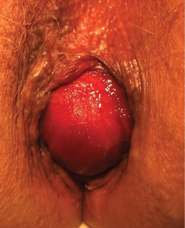
A clinical photograph of the perianal region showing an isolated anterior rectal prolapse in an 84-year-old female. The most prominent feature is a large, globular, erythematous mass protruding through the anal orifice. The prolapsed tissue exhibits a smooth, glistening mucosal surface and is notably lacking the concentric circular folds typically characteristic of full-thickness rectal intussusception. This absence of concentric rings suggests a different underlying mechanism, such as a herniation of an enterocele. The surrounding perianal skin shows signs of atrophy and fine wrinkling consistent with aging. The mass is significant in size, occupying the majority of the visible anal aperture. This image is an educational example for gastroenterology and urogynecology, highlighting the clinical distinction between various types of rectal bulges and the physical signs of anterior rectal wall prolapse secondary to pelvic floor descent or enterocele.
A clinical photograph of the perianal region showing an isolated anterior rectal prolapse in an 84-year-old female. The most prominent feature is a large, globular, erythematous mass protruding through the anal orifice. The prolapsed tissue exhibits a smooth, glistening mucosal surface and is notably lacking the concentric circular folds typically characteristic of full-thickness rectal intussusception. This absence of concentric rings suggests a different underlying mechanism, such as a herniation of an enterocele. The surrounding perianal skin shows signs of atrophy and fine wrinkling consistent with aging. The mass is significant in size, occupying the majority of the visible anal aperture. This image is an educational example for gastroenterology and urogynecology, highlighting the clinical distinction between various types of rectal bulges and the physical signs of anterior rectal wall prolapse secondary to pelvic floor descent or enterocele.

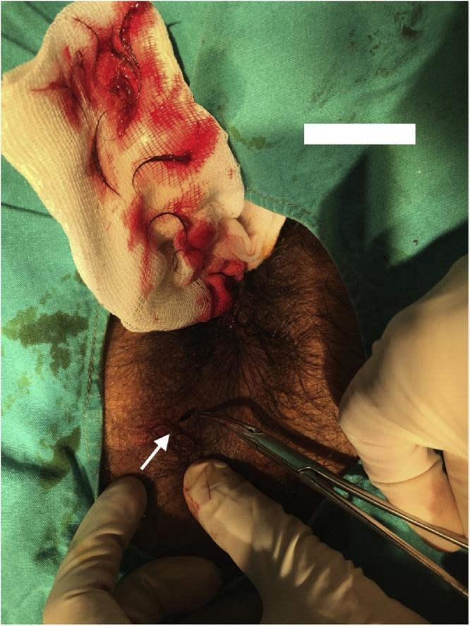
Clinical photograph of a surgical procedure addressing a perianal pilonidal sinus in a 24-year-old male. The patient is positioned in the prone position, and the perianal skin is visible with significant terminal hair growth. A gloved surgeon is using curved surgical forceps to explore a small punctum (sinus opening) located at the 7 o’clock position relative to the anus, highlighted by a white arrow. Adjacent to the operative site is a surgical gauze pad containing blood and several extracted hairs, which were removed from the subcutaneous cavity. The image demonstrates the hallmark clinical finding of pilonidal disease: a sinus tract containing a nest of hair. The surrounding field is draped with sterile green surgical drapes. This visual highlights the presentation and initial surgical management (debridement and hair removal) of a rare perianal manifestation of pilonidal sinus disease, which is distinct from the more common sacrococcygeal location.

Clinical photograph of a surgical procedure addressing a perianal pilonidal sinus in a 24-year-old male. The patient is positioned in the prone position, and the perianal skin is visible with significant terminal hair growth. A gloved surgeon is using curved surgical forceps to explore a small punctum (sinus opening) located at the 7 o’clock position relative to the anus, highlighted by a white arrow. Adjacent to the operative site is a surgical gauze pad containing blood and several extracted hairs, which were removed from the subcutaneous cavity. The image demonstrates the hallmark clinical finding of pilonidal disease: a sinus tract containing a nest of hair. The surrounding field is draped with sterile green surgical drapes. This visual highlights the presentation and initial surgical management (debridement and hair removal) of a rare perianal manifestation of pilonidal sinus disease, which is distinct from the more common sacrococcygeal location.

Here is a thorough, textbook-grounded overview of all piles-like (and related) diseases of the anal region, organized by category.

Diseases of the Anal Region (Piles and Piles-like Conditions)


1. Haemorrhoids (Piles)

Haemorrhoids are the classic "piles." They arise from congestion and sliding downward of the vascular anal cushions - normally present in the left lateral, right anterior, and right posterior positions of the anal canal. Bleeding from haemorrhoids is arterial (not venous) due to direct arteriovenous shunts in the cushions.

Internal Haemorrhoids

Located above the dentate line, covered by transitional/columnar epithelium. Because of autonomic innervation above the dentate line, they are painless unless complicated.
GradeFeatures
Grade IBleed but do not prolapse
Grade IIProlapse on straining, reduce spontaneously
Grade IIIProlapse on straining, require manual reduction
Grade IVPermanently prolapsed, irreducible

External Haemorrhoids

Located below the dentate line, covered by anoderm (richly innervated somatic). Thrombosis causes significant pain.

Thrombosed / Incarcerated Haemorrhoids

Prolapsed Grade IV piles can become incarcerated and strangulate - a surgical emergency causing severe pain, oedema, and necrosis.
Internal and external hemorrhoids anatomy
Anatomy of internal vs external hemorrhoids relative to the dentate line - Sabiston Textbook of Surgery

2. Anal Fissure (Fissure-in-Ano)

A painful linear ulcer (tear) of the distal anal canal, extending from the anal verge up toward the dentate line. Almost always in the posterior midline (anterior midline in women during childbirth).
  • Acute fissure: fresh tear, heals with conservative treatment
  • Chronic fissure: triad of fissure + sentinel skin tag at the distal end + hypertrophied anal papilla at the proximal end
Key feature: Pain starts with defecation and persists for a period after - classically described as "passing broken glass." Associated with raised internal anal sphincter tone.
Causes: Hard stools, diarrhoea, anal trauma, anal receptive intercourse.
Chronic anal fissure with sentinel skin tag

3. Anorectal Abscess

A collection of pus in the perirectal/perianal spaces. Most arise from infection of the anal glands (cryptoglandular theory) - glands in the intersphincteric plane become blocked, then infected, and spread along tissue planes.
Types (by anatomical space):
  • Perianal abscess - most common, superficial, below the dentate line
  • Ischiorectal (ischioanal) abscess - in the ischiorectal fossa, large and deep
  • Intersphincteric abscess - between internal and external sphincters
  • Supralevator abscess - above the levator ani, rare and serious
  • Horseshoe abscess - tracks circumferentially behind the rectum
Presentation: Throbbing pain (characteristic), swelling, fever, fluctuance. An untreated abscess either bursts spontaneously or is drained - either can result in a fistula-in-ano.

4. Fistula-in-Ano

A granulation-tissue lined track with an internal opening in the anal canal (usually at a crypt) and an external opening on the perianal skin. The most common cause of a perineal fistula.
Parks Classification:
TypeDescription
IntersphinctericTrack stays in intersphincteric plane (most common)
TranssphinctericCrosses both internal and external sphincters into ischiorectal fossa
SuprasphinctericGoes above puborectalis before descending
ExtrasphinctericOutside the sphincter complex entirely (rare)
Goodsall's Rule: External openings anterior to a transverse line through the anus track directly to the dentate line; those posterior track to the posterior midline.
Key feature: Intermittent pain - increases as discharge accumulates when the tract closes, then disappears when it discharges.
Fistula-in-ano Parks classification diagram

5. Rectal Prolapse (Procidentia)

Protrusion of rectal wall tissue through the anal canal.
TypeDescription
Mucosal prolapseOnly mucosa and submucosa prolapse (partial)
Full-thickness (Procidentia)All layers of the rectum prolapse; >2 inches in length; has concentric circular folds
Internal intussusceptionRectum telescopes inside but does not protrude
Key feature distinguishing from prolapsed haemorrhoids: Rectal prolapse shows concentric folds; prolapsed haemorrhoids show radial folds/grooves between piles.
Symptoms: Palpable anal mass, bleeding, mucus discharge, poor perianal hygiene, faecal incontinence.
Full-thickness rectal prolapse

6. Perianal Haematoma (Thrombosed External Haemorrhoid)

A sudden, acutely painful bluish lump at the anal verge from rupture of a small external vein with blood clot formation in the subcutaneous perianal tissue. Often precipitated by straining, heavy lifting, or coughing. Not a true haemorrhoid but mimics one closely. Treated by incision and evacuation within 48-72 hours, or conservative management thereafter.

7. Anal Skin Tags

Redundant folds of skin at the anal verge. Usually arise after resolution of a thrombosed external haemorrhoid or as the sentinel tag of a chronic fissure. Asymptomatic but may cause hygiene difficulties or pruritus. Excised if symptomatic.

8. Pilonidal Sinus

A sinus or abscess in the sacrococcygeal region, classically in the natal cleft, containing hair. Rarer perianal location also exists. Caused by penetration of loose hairs into the skin creating a foreign-body reaction and sinus formation. Presents as a discharging sinus, abscess, or pain. Common in young hirsute males.
Perianal pilonidal sinus with sinus opening

9. Hidradenitis Suppurativa

Chronic inflammatory disease of apocrine sweat glands in the perianal and perineal skin (also axillary, inguinal). Presents with recurrent painful nodules, abscesses, and discharging sinuses. Can mimic fistula-in-ano. Characterised by multiple sinuses and scarring, but the internal opening is not in the anal canal (unlike fistula-in-ano).

10. Anal Polyp

A pedunculated or sessile growth in the anal canal. Types include:
  • Hypertrophied anal papilla (fibrous polyp at the dentate line)
  • Adenomatous/villous polyp - pre-malignant, can prolapse
  • Juvenile polyp - in children, causes rectal bleeding
Polyps may prolapse through the anus during defecation, mimicking prolapsed haemorrhoids.

11. Carcinoma of the Anal Canal and Anal Margin

Squamous Cell Carcinoma (SCC) of the Anal Canal

  • Most common primary anal malignancy
  • Strongly associated with HPV (types 16, 18)
  • Risk factors: HIV, MSM, immunosuppression
  • Precursor: Anal Intraepithelial Neoplasia (AIN)
  • Treatment: Nigro protocol (chemoradiation with 5-FU + mitomycin C) - avoids surgery

Adenocarcinoma

  • Arises from proximal (rectal-type) mucosa above dentate line
  • Treated as rectal cancer (neoadjuvant CRT + APR)

Anal Margin Carcinoma

  • SCC of the perianal skin, behaves like skin SCC
  • Better prognosis than canal carcinoma

12. Pruritus Ani

Not a disease itself but a symptom complex of persistent perianal itching. Causes include:
  • Local: haemorrhoids, fissure, fistula, skin tags, poor hygiene
  • Dermatological: contact dermatitis, psoriasis, lichen sclerosus
  • Infective: threadworms (Enterobius - commonest in children), fungal (Candida), STIs
  • Systemic: diabetes, jaundice, lymphoma
  • Dietary: spicy foods, caffeine, citrus

13. Anal Stenosis (Stricture)

Narrowing of the anal canal. Causes:
  • Post-surgical (most common) - after haemorrhoidectomy with excess skin/mucosa removal
  • Inflammatory - tuberculosis, lymphogranuloma venereum, Crohn's disease
  • Congenital - failure of anal membrane to rupture
  • Neoplastic - annular carcinoma
  • Radiation - post-pelvic radiotherapy
  • Senile - chronic internal sphincter contraction in elderly

14. Sexually Transmitted Infections (STIs) of the Anal Region

Several STIs cause anal pathology resembling or coexisting with haemorrhoids:
  • Condyloma acuminata (anal warts): HPV-induced perianal/anal canal warts; cauliflower-like appearance
  • Syphilis: primary chancre at the anal verge (painless ulcer), condyloma lata (secondary)
  • Gonorrhoea / Chlamydia: proctitis with discharge
  • Herpes simplex: painful perianal ulcers
  • Lymphogranuloma venereum (LGV): causes proctitis and eventually rectal stricture

15. Perianal Crohn's Disease

A specific manifestation of Crohn's disease (up to 30-40% of Crohn's patients) with:
  • Multiple, complex, or unusual fistulae-in-ano
  • Deep "knife-cut" anal fissures (often lateral, not midline)
  • Perianal abscesses
  • Oedematous "elephant ear" skin tags
  • Anal stenosis
Distinguished from ordinary fistula/fissure by lateral or multiple fissures, atypical appearance, and systemic Crohn's features (diarrhoea, weight loss, perianal Crohn's often precedes bowel disease).

Quick Diagnostic Summary Table

ConditionMain SymptomKey Feature
Internal haemorrhoidsPainless bleeding, prolapseAbove dentate line
External haemorrhoidsPain on thrombosisBelow dentate line
Anal fissureSevere pain after defecationPosterior midline, sentinel tag
Perianal abscessThrobbing pain, feverFluctuant swelling
Fistula-in-anoIntermittent discharge + painExternal opening on perianal skin
Rectal prolapseMass per anumConcentric folds, full thickness
Perianal haematomaAcute pain, bluish lumpClot in perianal skin
Pilonidal sinusDischarge/abscess, natal cleftHair-containing sinus
Anal carcinomaBleeding, mass, painHard irregular mass
Condyloma acuminataCauliflower wartsHPV-related
Perianal Crohn'sMultiple/atypical fistulaeLateral fissures, oedematous tags

Sources: S Das - A Manual on Clinical Surgery (13th Edition), Sabiston Textbook of Surgery, Maingot's Abdominal Operations, Sleisenger & Fordtran's Gastrointestinal and Liver Disease, Yamada's Textbook of Gastroenterology, Harrison's Principles of Internal Medicine 22E
This is a shared conversation. Sign in to Orris to start your own chat.