
cd /home/daytona/workspace && python3 -c " import pdfplumber pdf_path = '/home/daytona/workspace/attachments/d6a8d7dd-559f-4af4-b47f-00b216cd9de0/SRI SIR SLIDE14-6.pdf' with pdfplumber.open(pdf_path) as pdf: print(f'Total pages: {len(pdf.pages)}') for i, page in enumerate(pdf.pages): print(f'\n========== PAGE {i+1} ==========') text = page.extract_text() if text: print(text) else: print('[No extractable text on this page]') "
cd /home/daytona/workspace && python3 -c " from pdf2image import convert_from_path import os pdf_path = '/home/daytona/workspace/attachments/d6a8d7dd-559f-4af4-b47f-00b216cd9de0/SRI SIR SLIDE14-6.pdf' output_dir = '/home/daytona/workspace/attachments/d6a8d7dd-559f-4af4-b47f-00b216cd9de0/' images = convert_from_path(pdf_path, dpi=200) for i, img in enumerate(images): out_path = os.path.join(output_dir, f'slide_page_{i+1}.png') img.save(out_path, 'PNG') print(f'Saved: {out_path}') print('Done') "
cd /home/daytona/workspace && python3 -c " import base64, json with open('/home/daytona/workspace/attachments/d6a8d7dd-559f-4af4-b47f-00b216cd9de0/slide_page_1.png', 'rb') as f: data = f.read() print(f'File size: {len(data)} bytes') print('File exists and is readable') "
cd /home/daytona/workspace && python3 -c " import pytesseract from PIL import Image img = Image.open('/home/daytona/workspace/attachments/d6a8d7dd-559f-4af4-b47f-00b216cd9de0/slide_page_1.png') text = pytesseract.image_to_string(img, config='--psm 6') print(text) "
cd /home/daytona/workspace && python3 -c " from PIL import Image # Resize and compress for easier viewing img = Image.open('/home/daytona/workspace/attachments/d6a8d7dd-559f-4af4-b47f-00b216cd9de0/slide_page_1.png') print(f'Original size: {img.size}') # Resize to smaller img_small = img.resize((1600, int(img.height * 1600 / img.width)), Image.LANCZOS) img_small.save('/home/daytona/workspace/attachments/d6a8d7dd-559f-4af4-b47f-00b216cd9de0/slide_small.jpg', 'JPEG', quality=85) print(f'Saved compressed version') import os size = os.path.getsize('/home/daytona/workspace/attachments/d6a8d7dd-559f-4af4-b47f-00b216cd9de0/slide_small.jpg') print(f'Compressed size: {size} bytes') "
cd /home/daytona/workspace && python3 -c " from PIL import Image import os # Make it even smaller img = Image.open('/home/daytona/workspace/attachments/d6a8d7dd-559f-4af4-b47f-00b216cd9de0/slide_page_1.png') img_small = img.resize((800, int(img.height * 800 / img.width)), Image.LANCZOS) img_small.save('/home/daytona/workspace/attachments/d6a8d7dd-559f-4af4-b47f-00b216cd9de0/slide_tiny.jpg', 'JPEG', quality=75) size = os.path.getsize('/home/daytona/workspace/attachments/d6a8d7dd-559f-4af4-b47f-00b216cd9de0/slide_tiny.jpg') print(f'Tiny size: {size} bytes') "
- Black eyebrows and eyeliner
- Red lips
- Orange/skin-toned coloring
- Each mask appears slightly different - some showing asymmetry, drooping, or distortion of facial features
- Top left & top right: More symmetrical, intact-looking faces
- Bottom left: Shows a long black line/streak running from the eye down to the lip area (possibly illustrating a scar, nerve line, or "en coup de sabre" morphea)
- Bottom right: Shows more distortion, with the face appearing more damaged/irregular
Face developmental anomalies
Facial Developmental Anomalies
About the Slide (SLIDE14-6)
Normal Face Development (Embryological Basis)
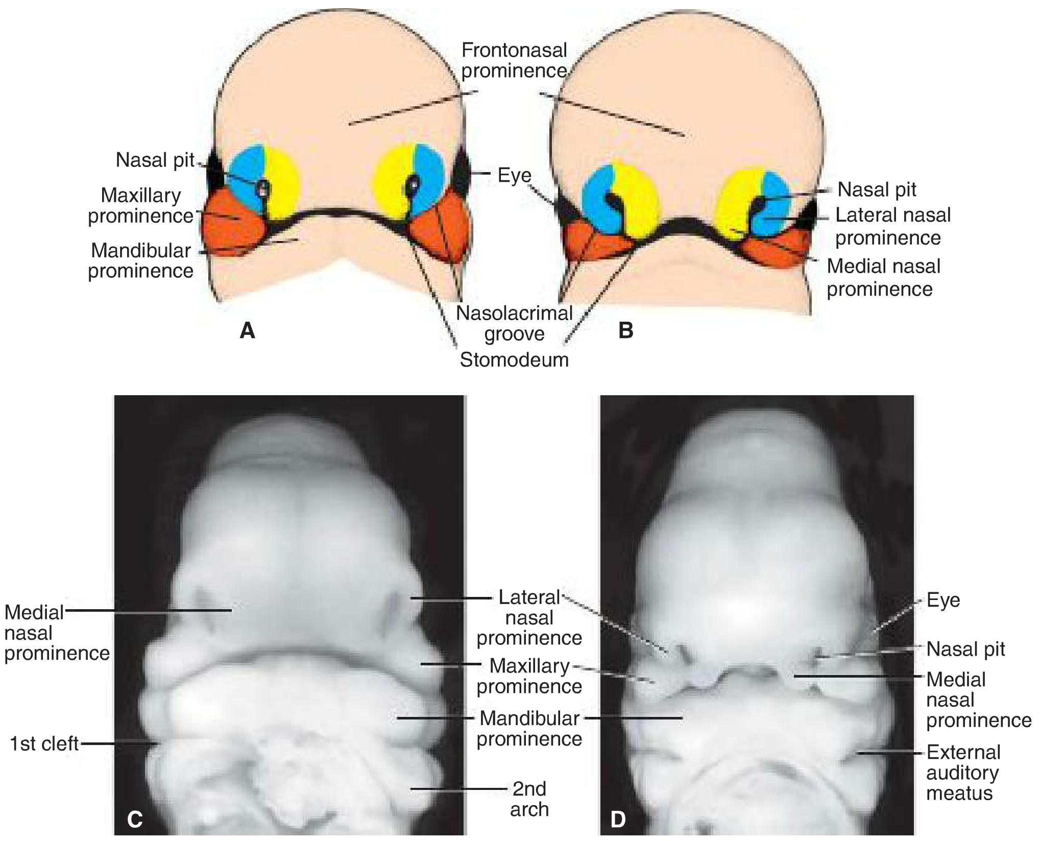
| Prominence | Structures it forms |
|---|---|
| Frontonasal | Forehead, bridge of nose, medial & lateral nasal prominences |
| Medial nasal | Philtrum of upper lip, premaxilla, nasal tip & septum |
| Lateral nasal | Alae of nose |
| Maxillary | Cheeks, lateral upper lip, secondary palate |
| Mandibular | Lower jaw, lower lip |
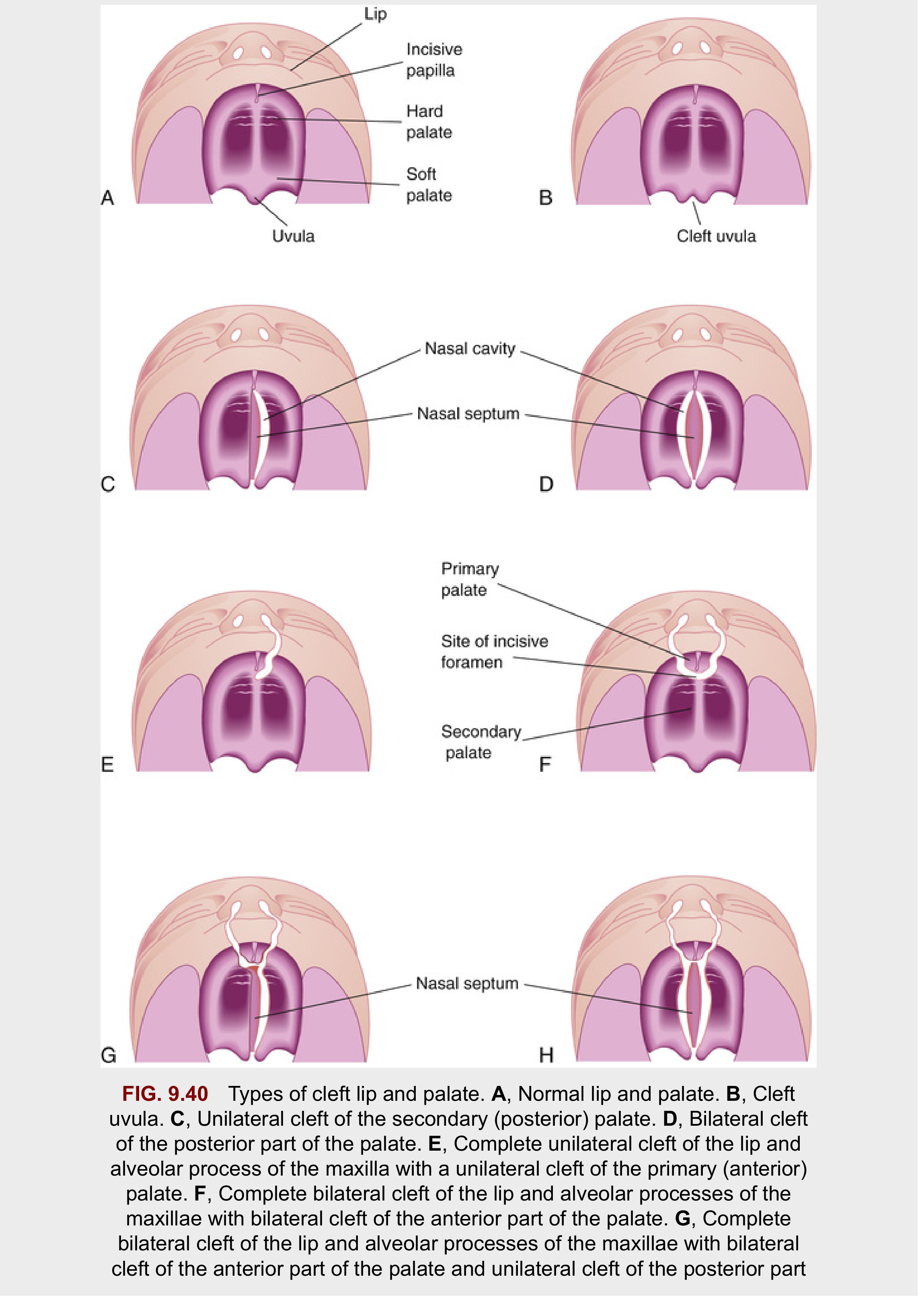
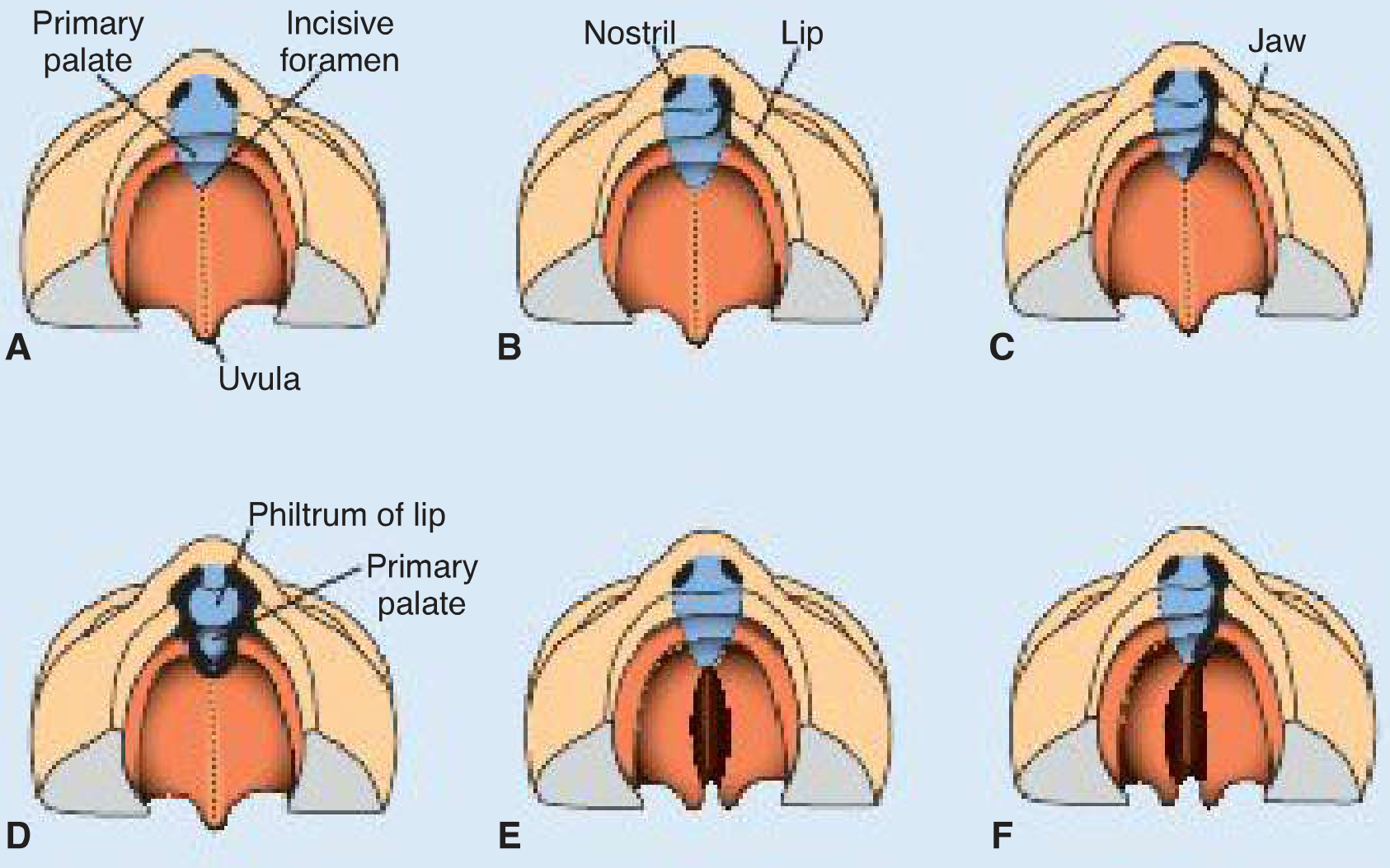
The Incisive Foramen: Key Landmark for Classification
- Anterior clefts (anterior to incisive foramen): cleft lip, cleft upper jaw/alveolus
- Posterior clefts (posterior to incisive foramen): cleft secondary palate, cleft uvula
- Combined: both anterior and posterior components
Types of Facial Clefts
1. Cleft Lip (Cheiloschisis)
- Unilateral cleft lip - one side fails to fuse (more common; ~1 in 1000 births, 60-80% male)
- Bilateral cleft lip - both sides fail
- A partial bridge of tissue called the Simonart band may connect parts in incomplete clefts
- Severity ranges from a notch in the vermilion border to a complete cleft extending into the nostril
2. Cleft Palate (Palatoschisis)
-
Shelves too small
-
Failure to elevate horizontally
-
Inhibition of fusion
-
Tongue fails to drop (micrognathia)
-
Cleft uvula - mildest form (bifid uvula), isolated failure at the posterior tip
-
Partial cleft palate - soft palate only
-
Complete cleft palate - extends through soft and hard palate to incisive foramen
3. Combined Cleft Lip + Cleft Palate
4. Median Cleft Lip (Rare)
- Mutations in SHH (Sonic Hedgehog) gene
- Altered cholesterol biosynthesis
- Teratogens like alcohol in week 3
5. Oblique Facial Cleft
6. Holoprosencephaly with Proboscis + Synophthalmia
- Fusion of both eyes into one (synophthalmia/cyclopia)
- A single tube-like nose (proboscis)
- Single forebrain ventricle
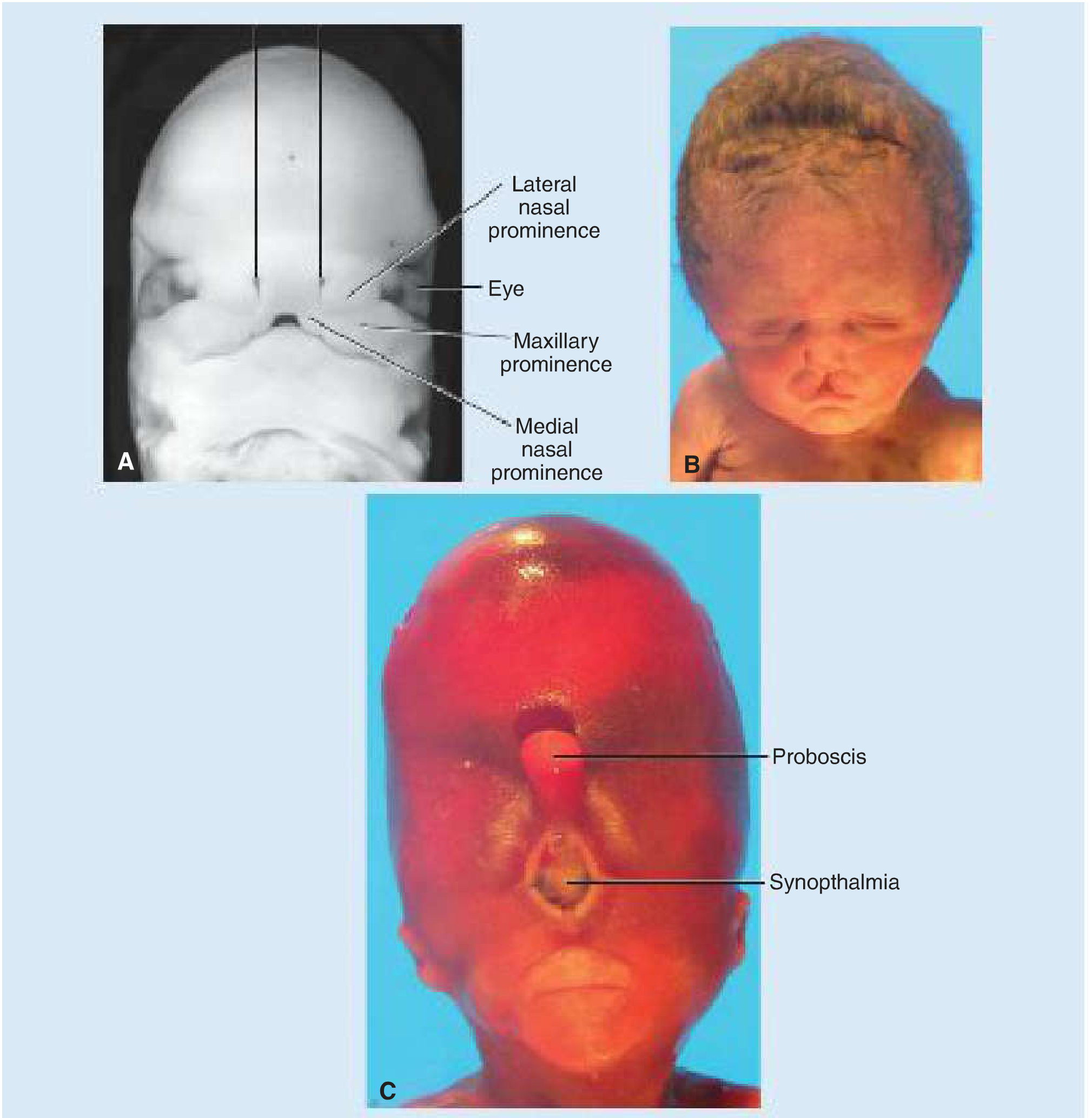
Clinical Photographs
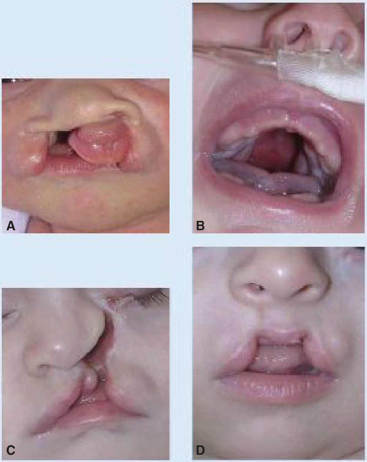
Epidemiology & Etiology
| Feature | Cleft Lip ± Palate | Isolated Cleft Palate |
|---|---|---|
| Incidence | ~1/700 births | ~1/1500 births |
| Sex | More in males (65%) | More in females (55%) |
| Ethnicity | Highest: Asian, Native American (3.5/1000); Lowest: African American (1/1000) | - |
- Genetic: Mutations in IRF6 (Van der Woude syndrome - most common associated syndrome, autosomal dominant), MSX1, SHH
- Teratogens: Valproic acid (anticonvulsant), alcohol, cigarette smoking
- Most cases are multifactorial
Summary: The Four Masks in the Slide
- Top left - Normal face (reference)
- Top right - Unilateral cleft lip (one side)
- Bottom left - Unilateral cleft lip extending to nostril (complete anterior cleft), shown by the long black line running from eye/nostril to lip
- Bottom right - Bilateral or more severe combined cleft (both sides affected, more distorted appearance)