
Lords Plication hydrocele
Lord's plication hydrocele surgery tunica vaginalis
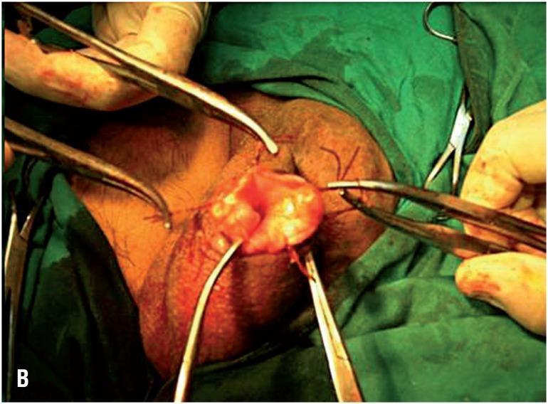
This clinical photograph captures an intraoperative view of a surgical procedure on the male scrotum, specifically a minimally invasive hydrocelectomy using the eversion technique (Lord's or Jaboulay-type modification). The surgical field shows the scrotum stabilized with surgical clamps and forceps. A small incision has been made, through which the parietal tunica vaginalis is everted. The image demonstrates the critical step of suturing the edge of the tunica vaginalis directly to the dartos fascia and scrotal skin. This maneuver aims to keep the visceral surface of the tunica vaginalis exposed to the lymph-rich subcutaneous tissues to facilitate fluid absorption and prevent hydrocele recurrence. Visible elements include surgical instrumentation (hemostats and forceps), interrupted suture lines securing the everted tissue, and the characteristic red, vascularized appearance of the visceral tunica. This content is intended for urological surgical training, illustrating anatomical landmarks and technical execution of scrotal sac eversion.
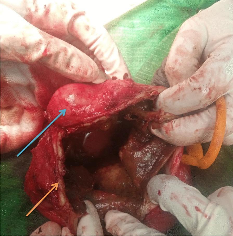
An intraoperative clinical photograph showing a surgical dissection of the scrotum. A blue arrow identifies the left testis, which appears as a smooth, glistening, ovoid structure. An orange arrow points to the thickened and irregular wall of the tunica vaginalis, which demonstrates prominent, whitish, eggshell-like calcific deposits. The surgical field reveals a dark, turbid fluid collection within the scrotal sac, consistent with a chronic hematocele or complex hydrocele. Gloved hands are visible retracting the tissues to expose the internal pathology. The image illustrates a rare complication of long-standing scrotal fluid collections where the parietal layer of the tunica vaginalis undergoes extensive dystrophic calcification, requiring surgical excision (hydrocelectomy). This content is relevant for urological surgery education, specifically regarding the diagnosis and management of chronic scrotal masses and the visual identification of calcified anatomical structures.
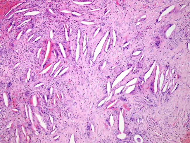
Light microscopy of a thickened hydrocele wall from tunica vaginalis demonstrates cross-sectional histology with dense fibrous stroma, scattered inflammatory cells, and cholesterol clefts. Acute neutrophilic bands and chronic lymphoplasmacytic infiltrates coexist, while multinucleated foreign body giant cells form around cholesterol clefts, reflecting a cholesterol granulomatous reaction. Fibrosis is prominent, with fascicular, collagen-rich architecture and minimal residual lining. The observed histology indicates chronic inflammatory and granulomatous response to leaked or degraded hydrocele contents, superimposed infection, and scarring. Diagnostic significance includes evidence of infectious complication and chronicity; findings support secondary infection in a long-standing hydrocele and help differentiate from neoplastic or purely serous processes. Clinically relevant search terms: hydrocele wall pathology, tunica vaginalis inflammation, cholesterol granuloma, foreign body giant cell reaction, chronic infection, fibrosis, scrotal hydrocele, hydrocelectomy planning, granulomatous scrotal disease.
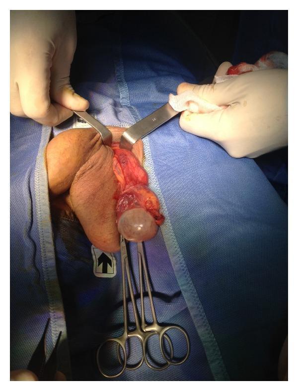
This clinical photograph illustrates an intraoperative view of an open scrotal surgical procedure focusing on the left testis and spermatic cord. The patient is positioned supine, with the surgical field isolated by blue drapes. A pair of Farabeuf retractors is used to widen the scrotal incision, exposing the underlying dartos fascia and spermatic cord. The primary finding is a translucent, fluid-filled cystic structure, identified as a hydrocele of the tunica vaginalis, which is being manipulated with surgical clamps. The testis and associated vascular structures of the cord are visible. The procedure demonstrates the resection and eversion of the tunica vaginalis, a common surgical technique for hydrocele repair (hydrocelectomy). The image serves as an educational resource for urological surgery, specifically highlighting anatomical landmarks such as the external inguinal ring, the spermatic cord, and the relationship between the tunica vaginalis and the testis during surgical intervention.
Lord's Plication for Hydrocele
What is a Hydrocele?
Surgical Options for Hydrocele - Overview
| Technique | Best For | Key Feature |
|---|---|---|
| Lord's Plication | Thin-walled hydrocele | No excision, minimal dissection |
| Jaboulay's (Eversion) | Moderate sac | Sac everted behind testis |
| Simple Excision | Thick-walled, large/chronic | Sac wall removed entirely |
Lord's Plication - The Procedure
Steps
- Scrotal incision - A transverse or midline scrotal incision is made through the skin, dartos, and fascial layers.
- Delivery of the sac - The hydrocele sac (parietal tunica vaginalis) is delivered into the wound.
- Opening the sac - The hydrocele sac is opened along its anterior surface and the fluid is drained.
- Extrusion of testis - The testis is extruded (delivered) through the aperture.
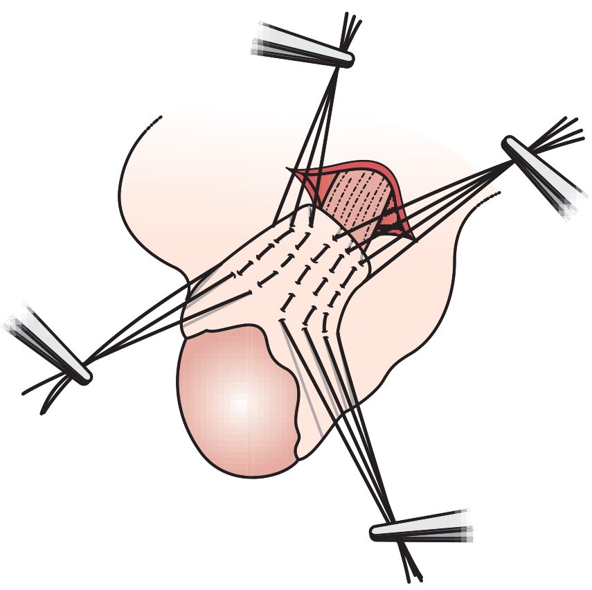
- Plication sutures - A series of interrupted absorbable (chromic) sutures are placed radially in a circumferential fashion around the sac - plicating (bunching/folding) the redundant tunica vaginalis.
- Tying sutures - When these sutures are tied, the tunica bunches up at its attachment to the testis - no tissue is excised.
- Closure - The testis is returned and the scrotum is closed in layers.
Key Advantages
- Minimal dissection - reduces operative trauma
- Lowest risk of haematoma - since no excision is performed, there is no bleeding cut edge
- No drain needed - as there is no raw excised surface
- Faster recovery - less postoperative morbidity
Indications
- Thin-walled hydrocele sac
- Small to moderate volume hydrocele
- Primary (not recurrent) hydrocele
- Patient preference for less invasive approach
Contraindications / When NOT to use Lord's
- Thick-walled sac - cannot be plicated effectively; requires excision
- Large, multiloculated, or chronic hydroceles
- Secondary hydrocele with underlying pathology (e.g., tumour, infection)
Lord's vs. Jaboulay's - Key Comparison
| Feature | Lord's Plication | Jaboulay's Eversion |
|---|---|---|
| Sac wall | Plicated (folded), not excised | Everted and anchored behind testis |
| Dissection | Minimal | More extensive |
| Haematoma risk | Lower | Higher (cut edges can bleed) |
| Suitable sac | Thin-walled | Thin to moderate |
| Drain | Not usually needed | May be placed |
| Recurrence | Slightly higher than excision | Lower than Lord's |
Intraoperative Clinical Image
Bottom Line (Exam Mnemonic)
Lord's = "Plicate the Thin" - No cut, no bleed, minimal dissection, bunching sutures on thin sacs
- Bailey and Love's Short Practice of Surgery, 28th ed., p. 1587
- Campbell-Walsh-Wein Urology, Hydrocelectomy section, p. 2460
Hydrocele
hydrocele types anatomy diagram congenital acquired
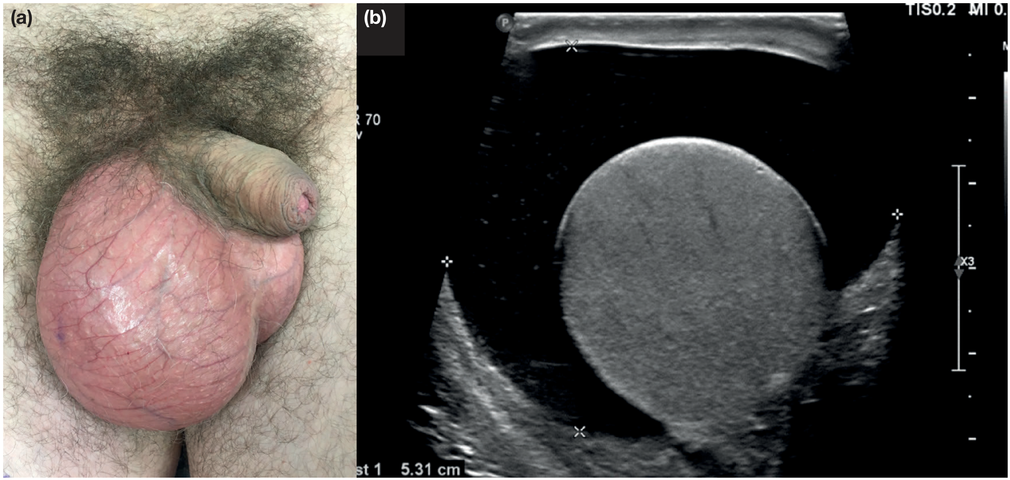
hydrocele scrotal swelling transillumination clinical examination
Hydrocele - Complete Overview
Definition
Incidence
- Affects approximately 1% of adult men
- >80% of newborn boys have a patent processus vaginalis - most close spontaneously within 18 months
Embryology & Pathophysiology
Classification / Types
| Type | Description | Processus Status |
|---|---|---|
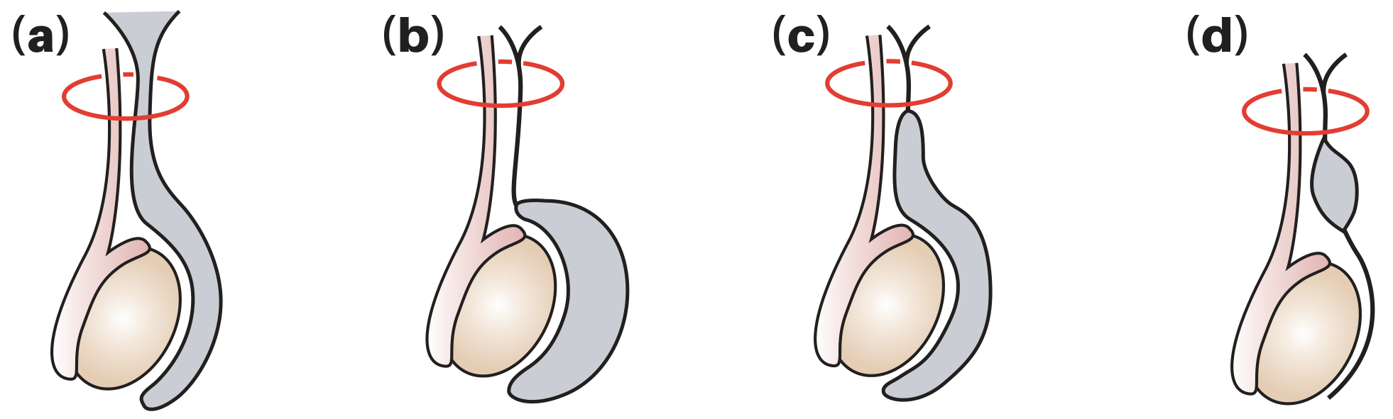
| (a) Communicating (Congenital) | Patent processus vaginalis allows peritoneal fluid to flow into the tunica vaginalis | Patent throughout - may be associated with indirect inguinal hernia |
| (b) Non-communicating (Vaginal/Primary) | Processus closed; fluid accumulates from excessive production or defective lymphatic absorption by tunica vaginalis | Fully closed |
| (c) Infantile Hydrocele | Distal processus closes correctly, mid-portion patent, proximal end open and communicating with tunica | Mid-portion patent, proximal open |
| (d) Hydrocele of the Cord | Distal and proximal processus both closed, mid-portion patent - isolated cyst along cord | Mid-portion patent only |
Non-communicating primary hydrocele is the most common type globally.Secondary hydrocele usually occurs in men >40 years - caused by local injury, infection, neoplasm, or radiotherapy.
Clinical Features
Clinical Examination - 3 Key Questions
- Can you get above the swelling? - You CAN get above a hydrocele (spermatic cord palpable above it). If you cannot, suspect an inguino-scrotal hernia.
- Is the testis/epididymis palpable? - A hydrocele encloses both testis and epididymis, making them impalpable.
- Does it transilluminate? - Hydroceles are brilliantly transilluminant (fluid-filled sac transmits light).
Additional Clinical Points
- Usually painless - may reach large size before presentation
- Beware acute hydrocele in a young man - suspect testicular tumour
- Congenital hydrocele may be intermittent (fluid drains back to peritoneal cavity when lying down)
- Ascites should be considered if bilateral
- Hydrocele of cord - smooth oval swelling above testis, moves down when testis is pulled downward; can mimic inguinal hernia
Hydrocele of the Canal of Nuck (females)
Investigation
- Ultrasound - most valuable investigation; clarifies nature of swelling, assesses whether testis is diseased
- Always exclude testicular malignancy by USS - especially in acute or secondary hydrocele
IMPORTANT: If a tumour is suspected, the hydrocele should NOT be punctured - risk of malignant needle-track implantation.
Treatment
Congenital Hydrocele (Children)
- Most resolve spontaneously by 18 months
- If persistent: ligation of the patent processus vaginalis (herniotomy)
Acquired Hydrocele (Adults)
- Small hydroceles: no treatment needed
- Large, symptomatic: surgical treatment indicated
Surgical Options
| Technique | Indication | Key Feature |
|---|---|---|
| Lord's Plication | Thin-walled sac | No excision - sutures bunch the sac; minimal haematoma risk |
| Jaboulay's Eversion | Moderate sac | Sac everted and anchored behind testis |
| Simple Excision | Thick-walled/large/chronic | Sac wall excised and edges oversewn; lowest recurrence but highest haematoma risk |
| Aspiration ± Sclerosant | Unfit for surgery | Fluid reaccumulates; sclerosant (tetracycline/doxycycline) reduces recurrence; risk of haematocele |
Filarial Hydrocele (Tropical)
- Accounts for up to 80% of hydroceles in tropical countries (endemic Wuchereria bancrofti regions)
- Follows repeated attacks of filarial epididymo-orchitis
- Occasionally fluid is chylous (chylocele)
Summary Box (Bailey & Love)
- A hydrocele is a collection of fluid within the tunica vaginalis
- Hydroceles surround the testis and transilluminate brightly
- Ultrasound is valuable, especially when testis/epididymis are impalpable
- Can be managed conservatively unless large and symptomatic
- Surgery is the mainstay of treatment
- Testicular malignancy is an uncommon but important cause to exclude