
Elaborate anatomy of deep neck spaces and discuss complications due to infection in these spaces in detail for long question answer for ms ent pg exam in simplified manner
deep neck spaces anatomy diagram cross section
Anatomical Diagram: This axial cross-section of a human cadaver illustrates the musculoskeletal anatomy of the ischiofemoral space, a key region in the diagnosis of ischiofemoral impingement syndrome (IIS). The image identifies the medial boundary formed by the ischial tuberosity (IT) and the lateral boundary formed by the lesser trochanter (LT) of the femur. Centrally, the quadratus femoris muscle (QF) is shown spanning the distance between these two bony landmarks, with the obturator externus muscle (OE) situated deep to it. The adductor muscle group—including the adductor longus (ADL), adductor brevis (ADB), and adductor magnus (ADM)—is positioned medially and anteriorly. Posterior to the ischial tuberosity, the tendons of the semitendinosus (ST) and semimembranosus (SM) are visible, emerging from their proximal attachments. The gluteus maximus (GMA) provides the most superficial posterior layer. This clinical imaging resource provides essential context for understanding the spatial relationships and narrowing of the ischiofemoral and quadratus femoris spaces in pelvic pathology.
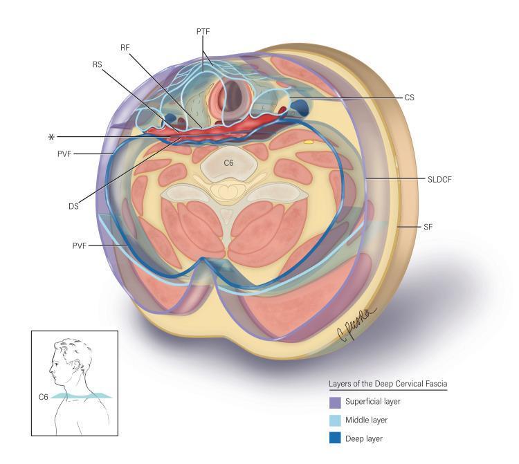
This medical anatomical diagram illustrates a transverse section of the neck at the C6 vertebral level, detailing the layers of the deep cervical fascia and the potential spaces they define. The illustration categorizes the fascia into three layers: superficial (yellow), middle (light blue), and deep (dark blue). Key anatomical structures labeled include the superficial fascia (SF), the superficial layer of deep cervical fascia (SLDCF), and the pretracheal fascia (PTF) enclosing the visceral compartment. Centrally, the diagram highlights the alar fascia (marked with an asterisk), which is situated between the retropharyngeal fascia (RF) anteriorly and the prevertebral fascia (PVF) posteriorly. This arrangement delineates two critical clinical spaces: the retropharyngeal space (RS), located between the RF and the alar fascia, and the 'danger space' (DS), situated between the alar fascia and the PVF. The carotid sheath (CS) is shown laterally. This visualization is essential for understanding the pathways of infection spread between the neck and the mediastinum, as well as providing anatomical guidance for surgical procedures in the prevertebral region.
This composite educational graphic illustrates the cross-sectional anatomy of the anterior neck, focusing on the left thyroid lobe through three modalities: (a) a 3D anatomical diagram, (b) an optoacoustic image, and (c) a correlative ultrasound with Directional Power Doppler. The 3D diagram establishes the spatial orientation of the common carotid artery, jugular vein, trachea, and thyroid lobe. The optoacoustic cross-section (b) displays high-sensitivity vascular mapping, identifying the sternocleidomastoid (s) and infrahyoid (m) muscles, the carotid artery (C), and the trachea (Tr). Intraglandular vascularity (3) and superficial vessels (1, 2) are highlighted in a high-contrast heatmap. The corresponding ultrasound image (c) provides structural grayscale context with superimposed Doppler signals (red and blue) indicating blood flow direction. This multimodal comparison demonstrates how optoacoustic imaging provides superior visualization of microvascular networks within the thyroid gland (T) compared to standard Doppler ultrasound, while maintaining consistent anatomical landmarks such as the trachea's medial position and the lateral placement of the carotid artery.
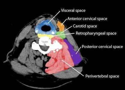
**Modality:** Axial Computed Tomography (CT) scan of the neck. **Anatomical Region:** Mid-cervical level (suprahyoid/infrahyoid junction), displaying the cervical vertebrae, airway, and surrounding soft tissue structures. **Description:** This is an educational anatomical diagram utilizing digital color-coding to delineate the deep cervical fascia compartments on the left side of a normal neck. The image serves as a reference for the topographic anatomy of potential neck spaces. **Key Landmarks and Color-Coded Spaces:** * **Visceral Space (Blue):** Encompasses the midline structures including the trachea, esophagus, and thyroid gland. * **Anterior Cervical Space (Orange):** Located anterior to the carotid space. * **Carotid Space (Green):** Contains the carotid artery, internal jugular vein, and cranial nerves IX–XII. * **Retropharyngeal Space (Yellow):** Positioned between the visceral fascia and the prevertebral fascia. * **Perivertebral Space (Red):** Surrounds the cervical vertebra and paraspinal muscles. * **Posterior Cervical Space (Purple):** Located posterior and lateral to the carotid space, deep to the sternocleidomastoid muscle. **Clinical Significance:** Essential for localizing pathology, such as abscesses or neoplasms, and predicting the spread of infection along fascial planes.
retropharyngeal abscess CT scan neck infection danger space mediastinitis
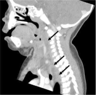
This diagnostic image is a sagittal view of a computerized tomography (CT) scan of the neck and upper thorax. The image demonstrates a large retropharyngeal abscess, characterized by a well-defined hypodense (darker) fluid collection. Black arrows indicate the longitudinal extent of the collection as it occupies the retropharyngeal space, located anterior to the cervical vertebrae and posterior to the pharynx and esophagus. Notably, the abscess tracks caudally from the cervical region, crossing the thoracic inlet and extending into the posterior mediastinum. The surrounding soft tissues show significant edema and displacement. Anatomical landmarks visible include the oral cavity, nasal passages, cervical spine, and superior mediastinal structures. This clinical finding is of high significance due to the risk of descending necrotizing mediastinitis. The image is intended for medical education regarding deep neck space infections and their potential routes of anatomical spread.
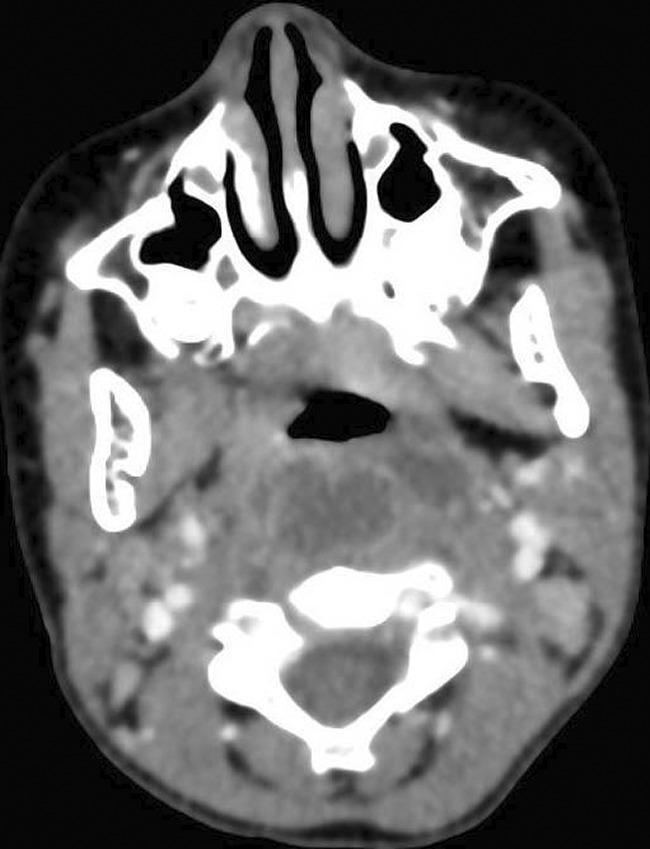
This diagnostic image is an axial CT scan of the head and neck with intravenous contrast. The scan shows a well-defined, low-density fluid collection in the retropharyngeal space, characteristic of a retropharyngeal abscess. The primary lesion is centrally located, measuring approximately 3.7 x 1.5 cm, and exhibits peripheral rim enhancement. Two smaller adjacent satellite collections are visible, suggesting multi-loculated fluid or local spread of infection. Anatomical landmarks include the nasal cavity and maxillary sinuses anteriorly, the oropharynx, and the cervical vertebrae (C1/C2 level) posteriorly. The surrounding soft tissues show evidence of edema and inflammatory changes, manifesting as increased mottled density and obliteration of normal tissue planes. The image is clinically significant for diagnosing deep neck space infections and evaluating potential complications such as airway compromise or posterior extension into the 'danger space' and epidural space. This material is suitable for intermediate to advanced medical learners focusing on radiology, otolaryngology, and emergency medicine.
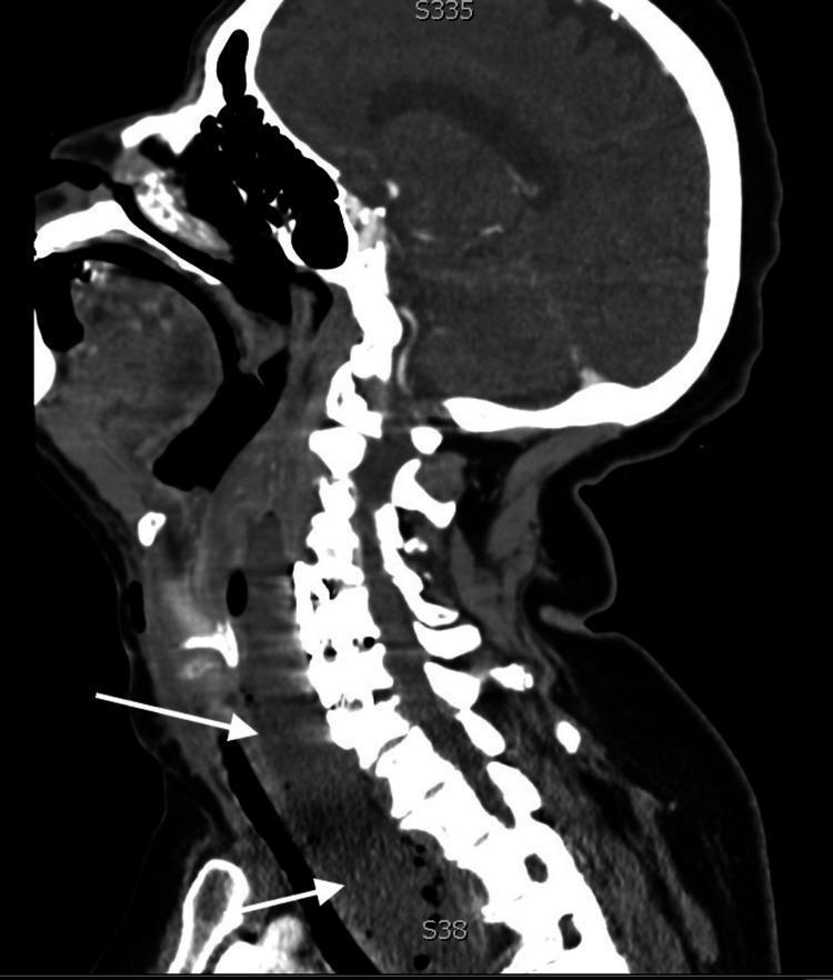
This diagnostic image is a sagittal computed tomography (CT) scan of the head, neck, and upper chest, highlighting a large retropharyngeal abscess. The visual reveals a significant, hypodense, and heterogeneously textured collection in the retropharyngeal space, marked by white arrows. The abscess originates at the level of the cervical spine and extends inferiorly through the deep neck spaces into the superior mediastinum. The mass effect is clinically significant, showing marked anterior displacement and compression of the trachea, along with narrowing of the glottic and subglottic airway regions. The cervical vertebrae are visible posterior to the collection, with some hyperdense artifacts suggesting possible spinal hardware. This image illustrates the critical pathophysiology of descending necrotizing mediastinitis and the acute risk of airway obstruction associated with deep neck space infections. It is a vital educational resource for identifying radiological signs of abscess progression and secondary mass effects on vital aerodigestive structures.
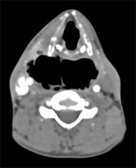
**Modality:** Axial Computed Tomography (CT) of the neck with contrast. **Anatomic Region:** Deep neck spaces at the level of the hypopharynx and cervical vertebrae. **Pathology:** Extensive emphysema/gas collection within the retropharyngeal and "danger" spaces. **Visual Features:** - **Distribution:** Large, confluent pockets of low-attenuation gas (black) occupy the retropharyngeal space. - **Landmarks:** The gas collection is positioned posterior to the pharyngeal constrictor muscles and anterior to the prevertebral fascia. - **Extension:** The gas extends across the midline, indicating potential involvement of the "danger space" (the potential space between the alar and prevertebral fascia). - **Secondary Findings:** There is visible soft tissue swelling and displacement of adjacent vascular and airway structures. Scattered small gas bubbles are also visible in the right lateral soft tissues. **Diagnostic Significance:** This radiologic pattern is characteristic of a deep neck space infection (e.g., retropharyngeal abscess with gas-forming organisms) or traumatic pneumomediastinum/neck emphysema. Cross-midline extension in this anatomical plane is a key feature for identifying potential pathways for descending mediastinitis.
Ludwig angina floor mouth swelling submandibular space
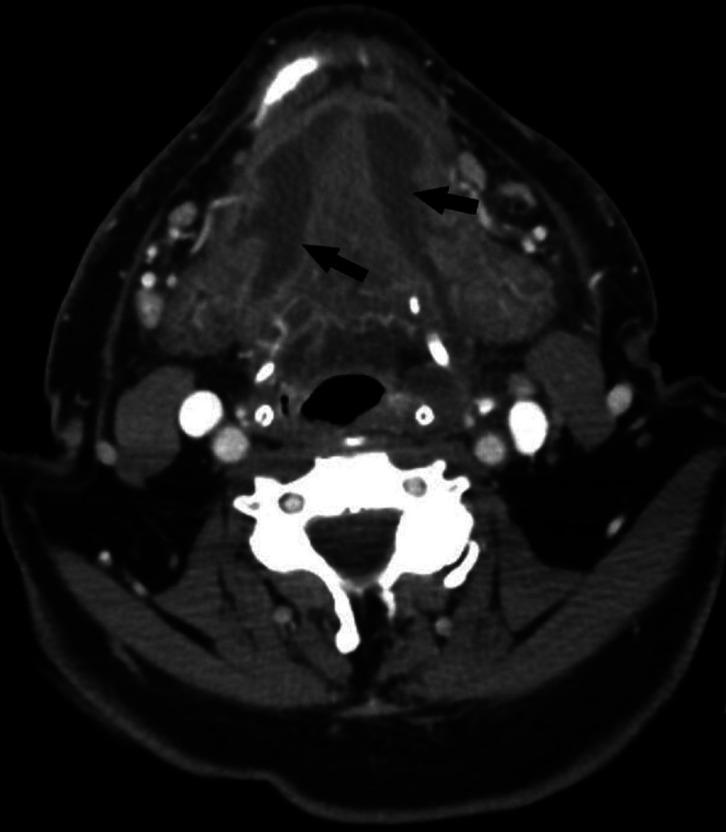
This diagnostic image is an axial contrast-enhanced CT scan of the submandibular and floor-of-mouth region. The image demonstrates a characteristic presentation of Ludwig's angina, an aggressive cellulitis of the submandibular space. Significant pathology includes bilateral hypodense areas within the floor of the mouth (indicated by black arrows), representing the formation of bilateral abscesses. These collections are associated with diffuse soft tissue swelling and a noticeable mass effect on the surrounding structures. Key anatomical landmarks visible include the mandible, the cervical spine, and the oropharyngeal airway, the latter of which shows significant narrowing (stenosis) due to the posterior displacement and swelling of the tongue and sublingual tissues. The pathology originates from the mandibular molar region, highlighting the odontogenic nature of the infection. This image is used in medical education to illustrate life-threatening airway compromise and the diagnostic imaging findings associated with deep neck space infections and maxillofacial emergencies.
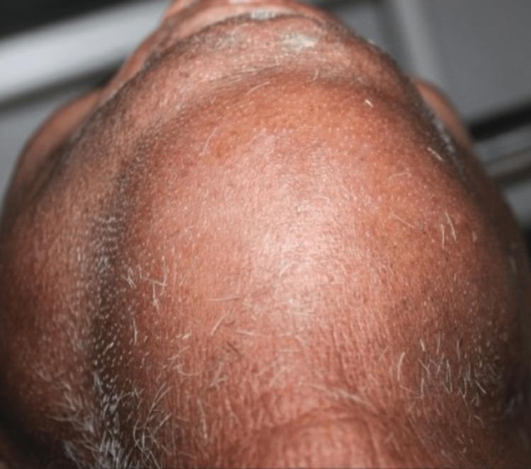
This clinical photograph displays an inferior-to-superior view of a patient's neck and submental region, demonstrating significant, diffuse soft tissue swelling characteristic of Ludwig's angina. The swelling involves the bilateral submandibular and sublingual spaces, resulting in a firm, 'woody' induration that obscures normal anatomical contours. The skin surface appears taut, stretched, and erythematous, indicating underlying acute inflammation. Visible skin wrinkling and grey facial hair suggest an older adult patient. The clinical presentation is highly suggestive of a rapidly spreading cellulitis of the submandibular space, often secondary to odontogenic infections, which poses a severe risk of airway obstruction. This image serves as an educational tool for identifying the external manifestations of floor-of-mouth infections and the necessity for urgent surgical decompression and airway management.
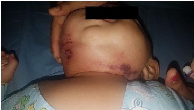
This clinical photograph displays a six-month-old female infant with a severe presentation of Ludwig's angina. The image shows significant, diffuse swelling (cellulitis) involving the submandibular and submental regions, extending toward the right side of the face and neck. Notable skin findings include prominent ecchymosis and dark purple discoloration on the right side of the neck, suggesting vascular congestion or tissue ischemia. Linear abrasions and skin lesions are visible on the left lateral aspect of the face near the ear. The infant's face appears flushed, indicating acute inflammation. This case represents a rare pediatric manifestation of Ludwig's angina, which is typically characterized by rapidly spreading cellulitis of the floor of the mouth and submandibular space. The image highlights the critical need for monitoring airway patency in pediatric patients with progressive submandibular swelling.
ANATOMY OF DEEP NECK SPACES AND COMPLICATIONS OF INFECTION
MS ENT PG Long Answer
INTRODUCTION
FASCIAL LAYERS OF THE NECK (Foundation)
- Invests the platysma muscle
- Skin flaps are raised in the subplatysmal plane (below this) to preserve skin blood supply
| Layer | Also Called | Structures Enclosed |
|---|---|---|
| Superficial/Investing layer | Layer 1 | Envelops entire neck; splits to enclose SCM, trapezius, parotid, submandibular gland |
| Middle/Pretracheal (visceral) layer | Layer 2 | Thyroid, trachea, pharynx, esophagus |
| Deep/Prevertebral layer | Layer 3 | Prevertebral muscles, vertebral column |
- Retropharyngeal space (anterior)
- Danger space (posterior, between alar fascia and prevertebral fascia)
CLASSIFICATION OF DEEP NECK SPACES
- Suprahyoid spaces (above hyoid): Parapharyngeal, peritonsillar, masticator, parotid, submandibular, sublingual
- Infrahyoid spaces: Visceral space
- Entire neck (skull base to mediastinum): Retropharyngeal, danger space, prevertebral, carotid space
ANATOMY OF INDIVIDUAL DEEP NECK SPACES
1. PARAPHARYNGEAL SPACE (PPS) - "The Central Hub"
- Apex: Greater cornu of hyoid bone
- Base: Skull base (petrous apex, sphenoid)
| Wall | Structure |
|---|---|
| Medial | Pharyngeal constrictor muscles, buccopharyngeal fascia |
| Lateral | Medial pterygoid muscle, ramus of mandible, deep lobe of parotid |
| Posterior | Prevertebral fascia |
| Superior | Skull base |
| Inferior | Hyoid bone |
2. RETROPHARYNGEAL SPACE (RPS)
| Direction | Boundary |
|---|---|
| Superior | Skull base (C1 vertebra) |
| Inferior | T1-T2 (fusion of buccopharyngeal + alar fascia) - some sources say as low as carina |
| Anterior | Buccopharyngeal fascia (covering pharyngeal constrictors) |
| Posterior | Alar fascia (anterior leaf of deep cervical fascia) |
| Lateral | Parapharyngeal spaces bilaterally |
- Laterally with both PPS
- Posteriorly with the danger space (via alar fascia)
- Inferiorly with the posterior mediastinum
3. DANGER SPACE (Alar Space)
| Direction | Boundary |
|---|---|
| Superior | Skull base |
| Inferior | Diaphragm |
| Anterior | Alar fascia |
| Posterior | Prevertebral fascia |
4. PREVERTEBRAL SPACE
| Direction | Boundary |
|---|---|
| Superior | Skull base |
| Inferior | Coccyx (entire length of vertebral column) |
| Anterior | Prevertebral fascia |
| Posterior | Vertebral bodies and paraspinal muscles |
| Lateral | Transverse processes of vertebrae |
5. SUBMANDIBULAR SPACE
| Direction | Boundary |
|---|---|
| Superior | Oral mucosa of floor of mouth |
| Inferior | Investing fascia (skin) below mandible |
| Lateral | Inner surface of mandible |
| Medial | Mylohyoid and hyoglossus muscles |
| Posterior | Open - communicates with parapharyngeal space |
- Sublingual space (above mylohyoid): Contains sublingual gland, Wharton's duct, lingual nerve, hypoglossal nerve
- Submandibular space proper/Submylohyoid space (below mylohyoid): Contains submandibular gland
6. MASTICATOR SPACE
7. PERITONSILLAR SPACE
8. PAROTID SPACE
9. VISCERAL SPACE
10. CAROTID SPACE
SUMMARY TABLE OF DEEP NECK SPACES
| Space | Location | Key Contents | Communicates With |
|---|---|---|---|
| Parapharyngeal | Central, inverted pyramid | Fat, maxillary a., carotid sheath (post-styloid) | Nearly ALL spaces - central hub |
| Retropharyngeal | Posterior to pharynx | Loose tissue, lymph nodes | PPS, danger space, posterior mediastinum |
| Danger space | Between alar & prevertebral fascia | Loose areolar tissue | RPS, prevertebral, mediastinum, diaphragm |
| Prevertebral | Anterior to vertebral bodies | Paraspinal muscles, brachial plexus | Danger space |
| Submandibular | Below floor of mouth | Submandibular gland | Sublingual, PPS |
| Sublingual | Floor of mouth, above mylohyoid | Sublingual gland, lingual n. | Submandibular, PPS |
| Masticator | Around mandibular ramus | Muscles of mastication, V3 | PPS |
| Peritonsillar | Around tonsil | Loose areolar tissue | PPS |
| Parotid | Around parotid | Parotid gland, CN VII | PPS |
| Visceral | Infrahyoid midline | Thyroid, trachea, esophagus | Anterior mediastinum |
| Carotid | Carotid sheath | CCA/ICA, IJV, CN X | Skull base to chest |
INFECTIONS OF DEEP NECK SPACES
ETIOLOGY
- Streptococcus viridans (most common)
- Staphylococcus aureus (including MRSA - commonest in children under 2 years)
- Klebsiella pneumoniae (especially in diabetics)
- Bacteroides, Fusobacterium, Peptostreptococcus (anaerobes)
- Beta-lactamase producing organisms in up to 40% of isolates
SPECIFIC SPACE INFECTIONS AND THEIR FEATURES
PERITONSILLAR ABSCESS (Quinsy)
- Most common deep neck infection in adults
- Pus collects between tonsillar capsule and superior constrictor muscle
- Features: Unilateral tonsillar swelling, uvular deviation to opposite side, "hot potato" voice, drooling, trismus (mild), odynophagia
- Treatment: Needle aspiration or incision & drainage + IV antibiotics + tonsillectomy ("interval" or "quinsy tonsillectomy")
PARAPHARYNGEAL ABSCESS
- Often secondary to: peritonsillar abscess, submandibular space infection, or dental infection
- Features:
- Medial displacement of tonsil and lateral pharyngeal wall
- Neck swelling or asymmetry (variable)
- Trismus (pterygoid involvement)
- High fever, dysphagia
- Danger: Airway compromise; spread to RPS and posterior mediastinum
- Treatment: External drainage (submandibular approach or retromandibular approach) + IV antibiotics
RETROPHARYNGEAL ABSCESS
- Febrile, drooling, dysphonia
- Characteristic posture: neck flexed, head extended ("sniffing position")
- Bulging of posterior pharyngeal wall (best seen on lateral view)
- Stridor, respiratory distress
- Neck pain, dysphagia, fever
- Less dramatic presentation
- Often preceded by trauma/instrumentation
LUDWIG'S ANGINA
- Bilateral submandibular space involvement
- Woody, brawny, non-pitting induration (NOT fluctuant - it is cellulitis)
- Elevation of floor of mouth → pushes tongue upward and backward → airway emergency
- Brawny, board-like induration of floor of mouth and submandibular region bilaterally
- Elevation of tongue (cannot protrude)
- Drooling, dysphagia, trismus
- "Woody" feel on palpation
- Stridor, respiratory distress in advanced cases
- High fever, sepsis
- Airway management - paramount (awake fibreoptic intubation preferred; tracheostomy if needed)
- IV antibiotics - high dose penicillin + metronidazole, or ampicillin-sulbactam
- Surgical drainage - multiple incisions in floor of mouth + external approach under chin
COMPLICATIONS OF DEEP NECK SPACE INFECTIONS
CLASSIFICATION:
1. AIRWAY OBSTRUCTION (Most Immediate Threat)
- Mechanisms: Medial displacement of pharyngeal wall, tongue elevation (Ludwig's), tracheal compression (retropharyngeal)
- Management: Awake fibreoptic intubation, or tracheostomy - avoid blind nasal intubation
- Securing the airway must be the first priority
2. DESCENDING NECROTIZING MEDIASTINITIS (DNM)
- Type I: Localized to superior mediastinum (above T4/carina)
- Type IIA: Diffuse below carina (pulmonary hilum)
- Type IIB: Diffuse with tracheobronchial tree involvement
- Symptoms of neck infection PLUS chest pain, dyspnea, worsening sepsis
- Widened mediastinum on CXR
- Subcutaneous emphysema (gas-forming organisms)
- Septic shock - mortality 10-15% even with treatment (was 50% in pre-antibiotic era)
3. JUGULAR VEIN THROMBOSIS (Lemierre's Syndrome)
- Organism: Fusobacterium necrophorum (gram-negative anaerobe)
- Pathophysiology: Tonsillar/peritonsillar infection → IJV thrombophlebitis → Septic emboli to lungs → "Postanginal septicemia"
- Features: Tender, cord-like IJV on palpation, spiking fevers, lung abscesses/cavities on CXR
- Imaging: Doppler US or CT - IJV filling defect
- Treatment: IV antibiotics (metronidazole + beta-lactam), anticoagulation controversial, surgical ligation of IJV in some cases
4. CAROTID ARTERY COMPLICATIONS
- Parapharyngeal or retropharyngeal infection erodes into the carotid sheath
- Presents as: Herald bleed (small warning bleed) → Massive hemorrhage
- Emergency management: Direct pressure, urgent surgery (ligation or repair)
- Less common but equally life-threatening
- Can lead to stroke (cerebral ischemia)
5. CRANIAL NERVE PALSIES
- CN IX palsy: Dysphagia
- CN X palsy: Hoarseness, aspiration
- CN XI palsy: Shoulder drop (trapezius weakness)
- CN XII palsy: Tongue deviation to affected side
- Horner's syndrome: Ptosis, miosis, anhidrosis (sympathetic chain involvement)
6. INTRACRANIAL COMPLICATIONS (via skull base)
- Cavernous sinus thrombosis - most common from dental/periorbital infection spreading via ophthalmic veins
- Proptosis, chemosis, painful ophthalmoplegia (CN III, IV, VI), meningism, fever
- Mortality high; treat with IV antibiotics (coverage for MRSA + anaerobes), anticoagulation
- Intracranial abscess / Epidural abscess
- Meningitis
- Dural sinus thrombosis - sagittal sinus, lateral sinus
7. ORBITAL COMPLICATIONS
- Spread from dental/sinus infection via infraorbital space
- Orbital cellulitis → subperiosteal abscess → orbital abscess
- Vision-threatening if posterior spread to optic nerve
8. NECROTIZING FASCIITIS OF THE NECK
- Rare but rapidly fatal
- Polymicrobial infection with gas-forming organisms (often Group A Streptococcus + anaerobes)
- "Dishwater" pus at surgery, skin necrosis, crepitus
- Treatment: Radical serial surgical debridement, IV antibiotics, hyperbaric oxygen (adjunct)
9. SEPTIC THROMBOPHLEBITIS / SEPTIC EMBOLI
- IJV thrombosis → pulmonary septic emboli → multiple lung abscesses
- Remote abscesses in joints, liver, kidney (hematogenous spread)
10. SPINAL COMPLICATIONS (Prevertebral space involvement)
- Vertebral osteomyelitis
- Epidural abscess → spinal cord compression → paralysis
- Requires urgent MRI spine + neurosurgical consultation
INVESTIGATION OF DEEP NECK INFECTIONS
| Investigation | Purpose |
|---|---|
| CT neck with contrast | Gold standard - delineates space involved, distinguishes cellulitis from abscess (rim enhancement), guides drainage |
| MRI | Better soft tissue differentiation; useful for skull base/intracranial extension |
| Lateral neck X-ray | Quick bedside - shows retropharyngeal widening, air in tissues |
| Chest X-ray | Mediastinal widening, pleural effusion, lung abscess |
| CT chest | Mandatory if DNM suspected |
| Ultrasound | IJV thrombosis (Doppler), guide needle aspiration |
| FBC | Leukocytosis |
| Blood cultures | Bacteremia/septicemia |
| Pus culture & sensitivity | Guide targeted antibiotics |
- Abscess: >2.5 cm diameter hypodense collection with peripheral rim enhancement
- Conservative management more likely to succeed with collections < 2.5 cm in a single space
MANAGEMENT PRINCIPLES
Step 1: AIRWAY
- Always assess and secure airway first
- Awake fibreoptic nasal intubation (preferred in anticipated difficult airway)
- Tracheostomy if intubation not possible
Step 2: IV ANTIBIOTICS
- Penicillin G + Metronidazole
- Clindamycin (penicillin allergy; first-line for MRSA coverage in children <2 years)
- Vancomycin (confirmed MRSA)
- Moxifloxacin (Eikenella corrodens - resistant to clindamycin)
Step 3: SURGICAL DRAINAGE
- Abscess > 2.5 cm or multispace involvement
- No improvement within 24-48 hours of antibiotics
- Airway compromise
- Complications (mediastinitis, carotid involvement)
- Peritonsillar: Needle aspiration or intraoral I&D
- Retropharyngeal: Intraoral (head-down, suction ready) or external
- Parapharyngeal: External (submandibular incision, 2 cm below mandible)
- Ludwig's angina: Floor of mouth + external bilateral submandibular incisions
- DNM: Cervicotomy + mediastinotomy/VATS
CLINICAL SCENARIOS OF SPREAD (for exam - important!)
KEY EXAM POINTS TO REMEMBER
- Parapharyngeal space is the central hub - communicates with almost all deep neck spaces
- Danger space extends from skull base to diaphragm (retropharyngeal space extends only to T1-T2)
- Retropharyngeal lymph nodes disappear after puberty - hence retropharyngeal abscess is predominantly a pediatric disease (typically children 2-4 years)
- Ludwig's angina is a cellulitis (NOT an abscess), is bilateral, involves submandibular + sublingual + submental spaces, and is an airway emergency
- Airway management is always the first priority in deep neck infections
- Descending necrotizing mediastinitis is the most lethal complication, spreading via the danger space
- Lemierre's syndrome = IJV thrombophlebitis due to Fusobacterium necrophorum - "postanginal septicemia"
- MRSA is now the most common organism in community-acquired deep neck infection in children under 2 years
- CT with contrast is the imaging modality of choice for deep neck infection
- Pre-styloid PPS lesions displace carotid posteriorly; post-styloid lesions displace fat pad anteriorly