
Describe the anatomy of the airway and the tracheobronchial tree. Emphasize on the structures relevant to airway management and endotracheal intubation. Anatomy of larynx with nerve supply and explain in detail the different vocal cord palsies and their significance in Anesthesia • • • • • • Long Questions (20 Marks) Describe the anatomy of the airway and the tracheobronchial tree. Emphasize on the structures relevant to airway management and endotracheal intubation. (Jan 2025)
Reading File
Searching sources...
This is a broad, visual anatomy question - launching parallel searches across the medical library.
Finding Sources
Finding Sources
Finding Sources
Finding Sources
Finding Sources
Finding Sources
Excellent - found key sources in Miller's Anesthesia, Morgan & Mikhail, Gray's Anatomy, and Cummings Otolaryngology. Reading these in parallel now.
Reading File
Reading File
Reading File
Good content from Miller's and Gray's. Now reading deeper into Miller's Anesthesia for airway anatomy, nerve supply, and vocal cord palsies, plus searching for more anesthesia-specific content.
Reading File
Finding Sources
Finding Sources
Reading File
Reading File
Searching Images
larynx anatomy cartilages vocal cords diagram
Excellent data from Morgan & Mikhail and Localization in Clinical Neurology. Now fetching the key textbook images from Miller's and Morgan & Mikhail:
Loading Image
Loading Image
Loading Image
I now have comprehensive data. Let me also fetch the Gray's Anatomy larynx diagram and the Morgan & Mikhail vocal cord table image, plus read the Bailey & Love vocal cord palsy section:
Reading File
Reading File
Reading File
Loading Image
Loading Image
I now have all the key content and images. Let me compose the comprehensive answer.
Anatomy of the Airway and Tracheobronchial Tree - With Emphasis on Airway Management and Endotracheal Intubation
PART 1: OVERVIEW OF THE AIRWAY
The airway can be divided into:
- Upper airway - nose, mouth, pharynx, larynx
- Lower airway - trachea, bronchi, bronchioles
PART 2: UPPER AIRWAY ANATOMY
A. Nose and Nasal Cavity
The nasal cavity is the preferred route for nasotracheal intubation. Key features:
- Divided by the nasal septum
- Lined by highly vascular mucosa (Kiesselbach's plexus anteriorly) - vulnerable to epistaxis during nasal intubation
- The turbinates (inferior, middle, superior) project from the lateral wall; the inferior turbinate is the most common obstruction during nasotracheal passage
- The nasopharynx connects posteriorly via the choanae
- Anesthesia relevance: Vasoconstriction (e.g., xylometazoline or cocaine) reduces bleeding risk before nasotracheal intubation. The tube passes below the inferior turbinate along the floor of the nasal cavity.
B. Oral Cavity and Oropharynx
- The oral cavity is the primary route for orotracheal intubation
- The tongue is the most common cause of upper airway obstruction in the unconscious patient
- The tonsillar pillars, uvula, and posterior pharyngeal wall form the oropharynx
- Mallampati classification assesses the visibility of these structures and predicts intubation difficulty
- The vallecula - the space between the base of the tongue and the epiglottis - is the target for the Macintosh laryngoscope blade during intubation
C. Pharynx
Three parts:
- Nasopharynx - behind nose, above soft palate
- Oropharynx - behind oral cavity
- Hypopharynx (laryngopharynx) - behind larynx, extending to C6
The piriform recesses (pyriform sinuses) flank the larynx laterally. Misplaced tubes can pass into these, and local anesthetic injected here (transtracheal block approach) can anesthetize the superior laryngeal nerve. Foreign bodies and guidewires can get lodged here.
PART 3: THE LARYNX
A. Overview and Position
The larynx lies at the level of C3-C6 in adults (higher - C3-C4 - in children and neonates), opposite the 4th-6th cervical vertebrae. It is:
- Continuous below with the trachea
- Opens above into the hypopharynx
- Suspended from the hyoid bone above
- A valve (sphincter) protecting the lower airway AND an instrument for phonation
(Miller's Anesthesia, 10e, p. 5841)
B. Cartilaginous Framework
The larynx has 9 cartilages: 3 unpaired + 3 paired.
Unpaired:
| Cartilage | Description | Anesthesia Relevance |
|---|---|---|
| Thyroid | Largest, 2 laminae fused anteriorly (60° male, 90° female), forms laryngeal prominence ("Adam's apple") | Landmark for nerve blocks; thyroid notch palpable for cricothyrotomy orientation |
| Cricoid | Signet-ring shaped, C6 level | Only complete cartilaginous ring in the airway; landmark for cricothyrotomy; Sellick's maneuver |
| Epiglottis | Leaf-shaped fibroelastic cartilage | Macintosh blade tip placed in vallecula lifts it; Miller blade placed posterior to it |
Paired:
| Cartilage | Function |
|---|---|
| Arytenoid | Posterior attachment of vocal cords; articulate with cricoid; adduction/abduction of cords |
| Corniculate | Atop arytenoids; seen laryngoscopically |
| Cuneiform | In aryepiglottic fold |
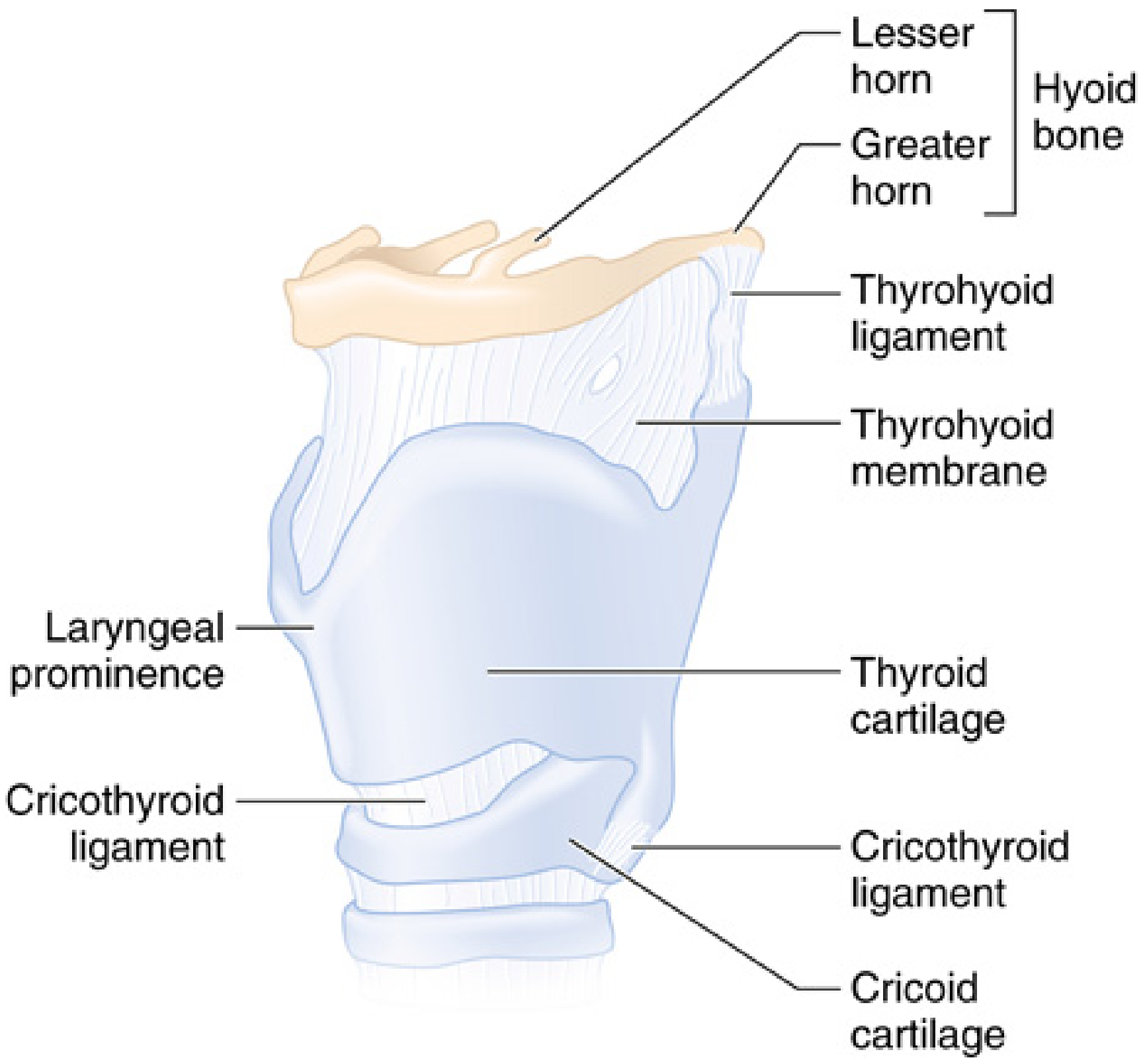
Cartilaginous and membranous components of the larynx (Miller's Anesthesia, 10e)
C. Membranes and Ligaments
- Thyrohyoid membrane - connects hyoid to thyroid cartilage; contains the internal laryngeal nerve and superior laryngeal vessels (a route for superior laryngeal nerve block)
- Cricothyroid membrane (CTM) - connects thyroid to cricoid anteriorly; approximately 9 mm high and 30 mm wide; avascular in its upper two-thirds; site for emergency cricothyrotomy and transtracheal jet ventilation
- Hyoepiglottic ligament - attaches epiglottis to hyoid; renders the vallecula useful as a lever point for Macintosh blade
D. Interior of the Larynx
From the pharynx, as seen during direct laryngoscopy:
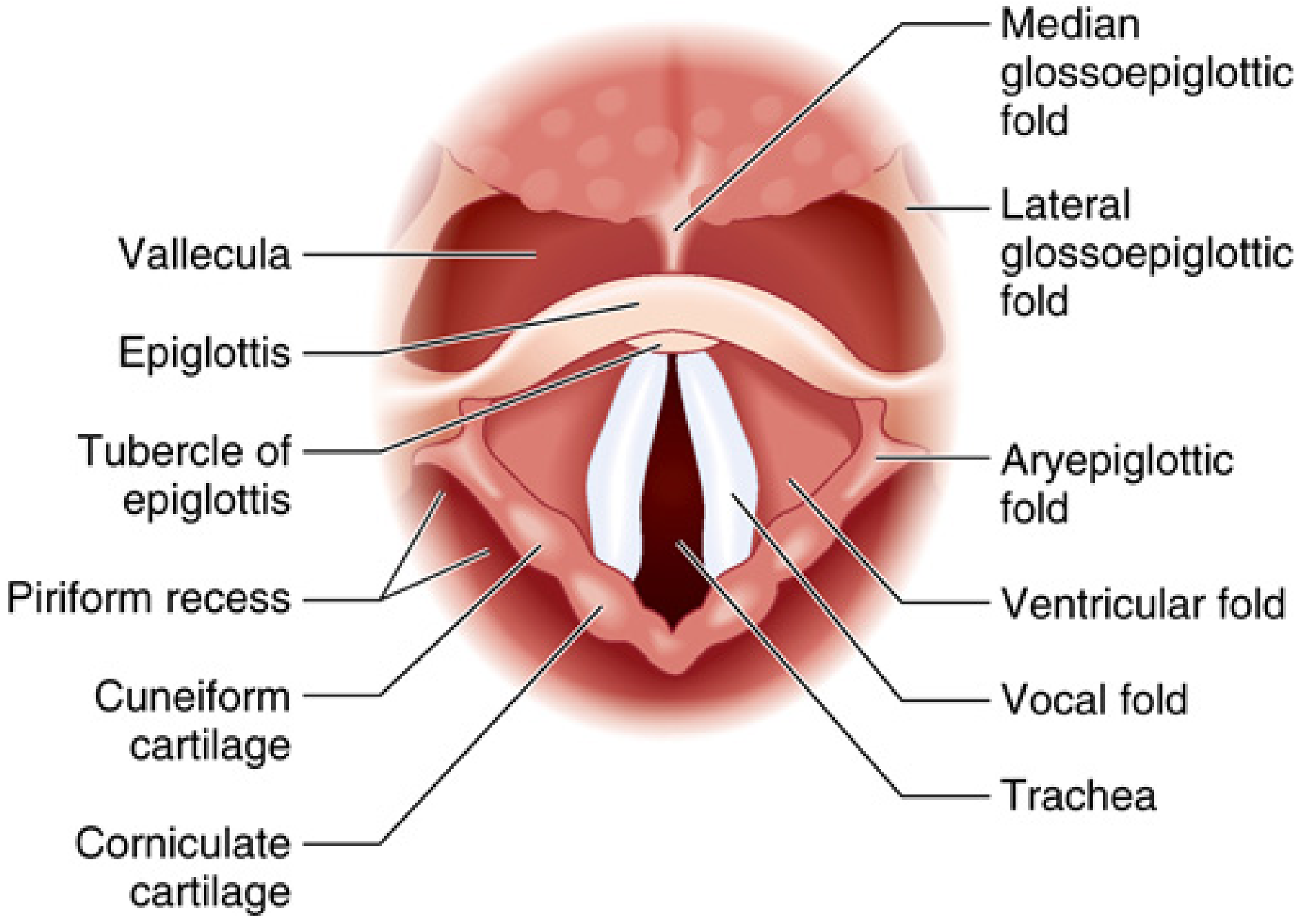
Laryngoscopic view of the larynx (Miller's Anesthesia, 10e)
The laryngeal cavity is divided into three compartments:
-
Vestibule (supraglottis) - from laryngeal inlet to false vocal cords
- Contains the aryepiglottic folds (lateral borders of laryngeal inlet)
- Contains ventricular folds (false cords) - redundant mucosa superior to true cords
-
Glottis - the space between the true vocal cords
- The true vocal cords attach anteriorly to the thyroid cartilage (anterior commissure) and posteriorly to the arytenoid cartilages
- In adults: anterior-posterior diameter ~23 mm (male), ~17 mm (female)
- The narrowest part of the adult airway is the glottis
-
Subglottis - from below the vocal cords to the inferior border of the cricoid
- In children, the subglottis (at the level of the cricoid ring) is the narrowest part of the airway - this is why uncuffed ETTs are used in young children; using an oversized tube causes subglottic edema and post-extubation stridor
E. Laryngeal Muscles
Intrinsic muscles (act on the vocal cords):
| Muscle | Action | Nerve |
|---|---|---|
| Posterior cricoarytenoid (PCA) | Abducts vocal cords (opens glottis) - only abductor | Recurrent laryngeal |
| Lateral cricoarytenoid | Principal adductor | Recurrent laryngeal |
| Transverse and oblique arytenoids | Adduct | Recurrent laryngeal |
| Vocalis (thyroarytenoid) | Tenses/shortens cords | Recurrent laryngeal |
| Cricothyroid | Lengthens/tenses cords (pitch) | External laryngeal (branch of SLN) |
Anesthesia key point: Since the PCA is the only abductor, bilateral RLN palsy results in both cords in the adducted (paramedian) position - causing stridor and respiratory distress.
PART 4: THE TRACHEA AND TRACHEOBRONCHIAL TREE
A. Trachea
- Begins at the inferior border of the cricoid cartilage (C6) and extends to the carina at T4-T5 (sternal angle of Louis)
- Length: 10-15 cm in adults; 4-5 cm in neonates
- Diameter: ~2 cm in adults
- Composed of 16-20 C-shaped cartilaginous rings open posteriorly
- The trachealis muscle forms the posterior (membranous) wall
- The trachea is mobile: it can be palpated and displaced laterally by masses
- In neck flexion the trachea moves cephalad (ETT may enter bronchus); in neck extension it moves caudal
Anesthesia relevance:
- The optimal ETT tip position is mid-trachea, 2-4 cm above the carina
- An ETT inserted too far will enter the right mainstem bronchus (right-sided endobronchial intubation) due to the less acute angle of the right bronchus
- Tracheal rings are palpable during emergency cricothyrotomy/tracheostomy
(Miller's Anesthesia, 10e, p. 5845)
B. Carina
- Located at T4-T5, the level of the sternal angle (Louis)
- The trachea bifurcates here into right and left mainstem bronchi
- Rich in cough receptors - most reactive part of the airway
- Anesthesia relevance: ETT cuff should be above the carina; confirm position with bilateral breath sounds and end-tidal CO2
C. Right and Left Mainstem Bronchi
| Feature | Right Mainstem Bronchus | Left Mainstem Bronchus |
|---|---|---|
| Angle from trachea | ~25° (more vertical) | ~45° (more horizontal) |
| Length | ~2.5 cm | ~5 cm |
| Width | Wider | Narrower |
Anesthesia consequence of right-sided anatomy:
- ETTs inserted too far enter the right bronchus preferentially
- Foreign bodies preferentially enter the right bronchus
- One-lung ventilation: Double-lumen tubes (Robertshaw) are placed with the bronchial limb in the left mainstem bronchus for most thoracic procedures, given the longer length of the left bronchus
D. Bronchial Divisions
The bronchial tree divides progressively:
- Mainstem bronchi → lobar bronchi (3 right, 2 left lobes)
- Lobar → segmental (tertiary) bronchi (10 right, 8-10 left)
- Segmental → subsegmental → bronchioles → terminal bronchioles → respiratory bronchioles → alveolar ducts → alveoli
Cartilage is present down to the bronchiolar level (> 1mm diameter); below this are cartilage-free bronchioles.
PART 5: NERVE SUPPLY OF THE LARYNX
The entire sensory and motor supply of the larynx is via branches of the vagus nerve (CN X).
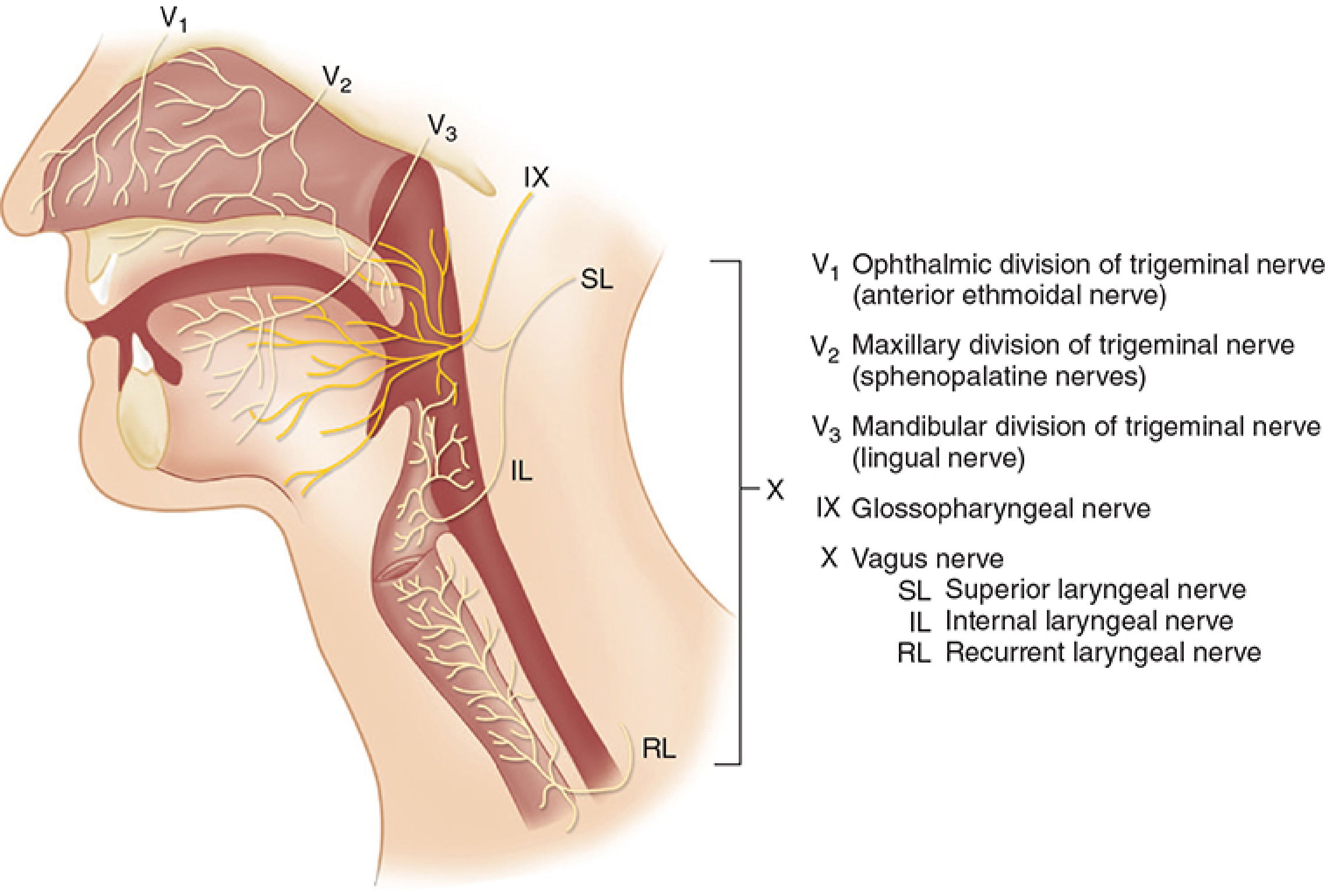
Sensory nerve supply of the airway (Morgan & Mikhail's Clinical Anesthesiology, 7e)
A. Complete Sensory Map of the Airway
| Region | Nerve(s) |
|---|---|
| Anterior nasal mucosa | V1 (ophthalmic) - anterior ethmoidal nerve |
| Posterior nasal mucosa | V2 (maxillary) - sphenopalatine nerves |
| Hard and soft palate | V2 - palatine nerves |
| Anterior 2/3 tongue (general) | V3 (lingual nerve) |
| Posterior 1/3 tongue + tonsils + soft palate undersurface | CN IX (glossopharyngeal) |
| Epiglottis to vocal cords (above cords) | Internal laryngeal nerve (branch of SLN, CN X) |
| Below vocal cords + trachea | Recurrent laryngeal nerve (CN X) |
(Morgan & Mikhail's Clinical Anesthesiology, 7e, p. 567)
B. Branches of the Vagus Relevant to the Larynx
1. Superior Laryngeal Nerve (SLN)
Arises from the inferior (nodose) ganglion of the vagus and divides into:
| Branch | Type | Function |
|---|---|---|
| Internal laryngeal nerve | Sensory | Mucosa from epiglottis down to vocal cords; pierces thyrohyoid membrane |
| External laryngeal nerve | Motor | Cricothyroid muscle only |
Block technique: Injecting local anesthetic at the greater cornu of the hyoid (where the internal laryngeal nerve pierces the thyrohyoid membrane) provides topical anesthesia above the cords - used for awake intubation.
2. Recurrent Laryngeal Nerve (RLN)
- Right RLN: Loops around the right subclavian artery, ascends in the right tracheoesophageal groove
- Left RLN: Loops around the arch of the aorta (ligamentum arteriosum), ascends in the left tracheoesophageal groove - has a longer intrathoracic course, making it more vulnerable to pathology (lung tumors, aortic aneurysm, mediastinal masses)
Supplies:
- Motor: All intrinsic laryngeal muscles except cricothyroid
- Sensory: Mucosa below the vocal cords and upper trachea
Block technique (transtracheal block): Injecting local anesthetic through the CTM into the trachea anesthetizes subglottic/tracheal mucosa. Coughing distributes the anesthetic upward to the vocal cords.
PART 6: VOCAL CORD PALSIES AND SIGNIFICANCE IN ANESTHESIA
A. Pathophysiology
The RLN is the principal motor nerve to the larynx. Injury produces predictable patterns of cord dysfunction. The SLN (external branch) is less critical for cord movement but important for pitch.
Semon's Law: Abductor fibers (PCA muscle) are more susceptible to injury than adductor fibers; thus in partial/incomplete RLN injury, adductor function is preserved while abductor function is lost, resulting in a midline/paramedian cord that cannot abduct. In complete injury, both functions are lost and the cord lies in the paramedian (cadaveric) position.
(Morgan & Mikhail's Clinical Anesthesiology, 7e; Localization in Clinical Neurology, 8e, p. 871)
B. Classification of Vocal Cord Palsies
1. Superior Laryngeal Nerve Palsy
| Type | Effect |
|---|---|
| Unilateral SLN palsy | Minimal - slight voice change, loss of high pitch; cricothyroid unilaterally paralyzed; airway not jeopardized |
| Bilateral SLN palsy | Hoarseness, easy tiring of voice; airway control preserved |
2. Recurrent Laryngeal Nerve Palsy
Causes:
- Thyroid surgery (most common - especially total thyroidectomy)
- Lung carcinoma (left hilar lesion - left RLN)
- Aortic aneurysm
- Mediastinal masses, lymph nodes
- Post-intubation injury (cuff pressure on RLN at anterior cricoarytenoid joint)
- Neck dissection, carotid endarterectomy
- Idiopathic (20-25%)
Unilateral RLN Palsy:
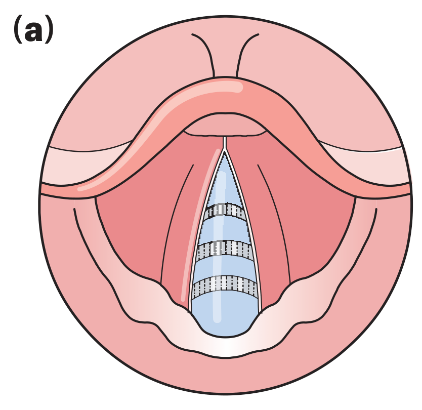
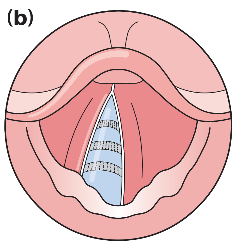
Vocal fold positions - (a) normal; (b) unilateral palsy (Bailey & Love's Surgery, 28e)
- Paralyzed cord lies paramedian (near midline)
- Hoarseness, breathiness, weakened cough, difficulty swallowing liquids
- Diplophonia (two pitch levels) due to unequal cord vibration
- Mild inspiratory stridor possible
- Compensation occurs over weeks as the normal cord crosses the midline to meet the paralyzed one - voice may normalize
- Anesthesia: Generally does not compromise airway; intubation is safe; be aware of aspiration risk (weakened cough reflex, incomplete glottic closure)
Bilateral RLN Palsy:
| Subtype | Cord Position | Voice | Airway | Clinical Significance |
|---|---|---|---|---|
| Acute bilateral | Paramedian (adducted) | Weak but clear | Severely compromised | Stridor, respiratory distress, may need immediate tracheostomy |
| Chronic bilateral | Paramedian | Near normal | Narrowed to 2-3 mm | Dyspnea on exertion, inspiratory stridor |
Critical anesthesia scenario: Post-total thyroidectomy bilateral RLN palsy - on extubation/reversal of anesthesia, both cords in paramedian position cause acute dyspnea and stridor as the glottis is reduced to 2-3 mm and cords are sucked together on inspiration. Positive pressure mask ventilation temporarily relieves it, but immediate reintubation or tracheostomy may be life-saving. Death from asphyxia can occur.
(Bailey & Love's Surgery, 28e, p. 702)
Table: Effects of Laryngeal Nerve Injury (Morgan & Mikhail's, 7e)
| Nerve | Injury Type | Effect |
|---|---|---|
| Superior laryngeal nerve | Unilateral | Minimal |
| Superior laryngeal nerve | Bilateral | Hoarseness, tiring of voice |
| Recurrent laryngeal nerve | Unilateral | Hoarseness |
| Recurrent laryngeal nerve | Bilateral - Acute | Stridor, respiratory distress |
| Recurrent laryngeal nerve | Bilateral - Chronic | Hoarseness, stridor |
| Both SLN + RLN | Unilateral (complete vagotomy) | Moderate hoarseness |
| Both SLN + RLN | Bilateral | Aphonia, risk of aspiration |
C. Anesthesia-Specific Significance of Vocal Cord Palsies
-
Pre-op assessment: Always enquire about voice changes and stridor before thyroid, esophageal, aortic, or mediastinal surgery. A pre-existing unilateral palsy must be documented because inadvertent injury to the contralateral nerve during surgery causes bilateral palsy.
-
Intubation-related RLN injury: Post-intubation vocal cord paralysis can occur due to:
- Cuff pressure compressing the anterior branch of the RLN at the cricoarytenoid joint
- Arytenoid dislocation during laryngoscopy
- Prevention: Use the lowest effective cuff pressure; use appropriate-sized ETT
-
Extubation after thyroid surgery: Always inspect vocal cord mobility (direct laryngoscopy or fiberoptic) before extubation if bilateral RLN injury is suspected. Have a tracheostomy set and reintubation equipment immediately available.
-
Awake intubation planning: Presence of RLN palsy does not contraindicate awake fiberoptic intubation but requires careful topicalization; the superior laryngeal nerve block alone is sufficient above the cords if the RLN territory is already anesthetized by the palsy.
-
Seimon's Law clinical implication: Early partial RLN injury shows adductor function preserved (cord moves to midline on phonation) but abductor function lost. Do not mistake this for a normal cord.
-
Treatment of bilateral palsy:
- Acute: Reintubation/tracheostomy
- Chronic: Endoscopic cordotomy or arytenoidectomy (CO2 laser) to widen posterior glottic airway; thyroplasty or injection medialization for unilateral palsy
PART 7: CLINICALLY IMPORTANT DIMENSIONS FOR INTUBATION
| Parameter | Value |
|---|---|
| Adult tracheal length | 10-15 cm |
| ETT depth (oral, adult male) | ~23 cm at lips |
| ETT depth (oral, adult female) | ~21 cm at lips |
| Carina level | T4-T5 (sternal angle) |
| Cricoid level | C6 |
| Angle - right bronchus from trachea | ~25° |
| Angle - left bronchus from trachea | ~45° |
| Cricothyroid membrane height | ~9 mm |
| Cricothyroid membrane width | ~30 mm |
| Narrowest airway in adults | Glottis |
| Narrowest airway in children | Subglottis (cricoid level) |
Summary: Key Points for Exam
- The cricoid cartilage is the only complete cartilaginous ring - landmark for Sellick's maneuver and emergency airway access.
- The CTM is the site for emergency surgical airway (cricothyrotomy) - avascular, accessible.
- Macintosh blade tip sits in the vallecula; Miller blade tip goes posterior to the epiglottis.
- The right bronchus is more vertical - ETTs and foreign bodies preferentially enter it.
- All intrinsic laryngeal muscles are supplied by the RLN except cricothyroid (external laryngeal nerve).
- Only abductor of the cords = posterior cricoarytenoid (PCA).
- Bilateral RLN palsy = adducted cords → stridor → airway emergency.
- Left RLN has a longer intrathoracic course → more susceptible to mediastinal/thoracic pathology.
- Children's narrowest airway is at the cricoid (subglottis), not the glottis.
- Semon's Law - abductor fibers injured first in partial RLN palsy.
Sources:
- Miller's Anesthesia, 10e - Chapter 40 (Airway Management), pp. 5841-5845
- Morgan & Mikhail's Clinical Anesthesiology, 7e - Chapter 19 (Airway Management), pp. 567-568
- Gray's Anatomy for Students - Chapter 8 (Larynx), p. 1192
- Bailey and Love's Short Practice of Surgery, 28e - Chapter 52 (Vocal Fold Palsy), pp. 702-724
- Localization in Clinical Neurology, 8e - Lesions of the Recurrent Laryngeal Nerve, p. 871
This is a shared conversation. Sign in to Orris to start your own chat.