
To be able to advise this couple appropriately the candidate needs to understand the following facts: Ovulation occurs 1 4 days prior to the period, as the length of the luteal phase is 1 4 days irrespective of the follicular phase length. In this woman her ovulation, therefore, has occurred as early as day 1 2, or as late as day 1 5, of her cycle. Sperm survival has been shown to be much longer than originally thought, in the presence of adequate and normal cervical mucus, with intercourse occurring up to 6 days before the known time of ovulation resulting in pregnancy. In this woman, therefore, intercourse should be ceased six days before the earliest ovulation (i.e. on day six) . The duration over which the ovulated egg can be fertilised is generally accepted to be about 24-36 hours. Intercourse should therefore not be resumed until two days after the latest ovulation. In this woman this would be day 1 7. Abstinence is therefore required between days 6 and 1 7 of the cycle.explain this in easy language also explain the menstrual cycle separately with all the phases and their duration and action.
menstrual cycle phases diagram ovulation follicular luteal
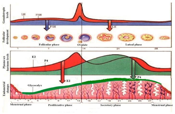
A multi-panel pathophysiology diagram illustrating the synchronous physiological changes during a standard 28-day human menstrual cycle. The top panel graphs gonadotropin levels, showing a sharp surge in Luteinizing Hormone (LH) and a moderate rise in Follicle-Stimulating Hormone (FSH) at day 14, correlating with the follicular phase, ovulation, and luteal phase stages. The corresponding follicular development shows the maturation of a primary follicle into a Graafian follicle, ovulation, and the formation of the corpus luteum and corpus albicans. The middle panel tracks plasma sex hormone levels, highlighting the mid-cycle peak of Estradiol (E2) and the post-ovulatory rise of Progesterone (P4). The bottom panel depicts endometrial morphology, transitioning from the menstrual phase to the proliferative and secretory phases. A green-shaded layer highlights the glycocalyx development, which thickens and becomes most prominent during the secretory phase under the influence of P4, before thinning during menstruation. The diagram demonstrates the hormonal regulation of the ovary and uterine lining for medical educational purposes.
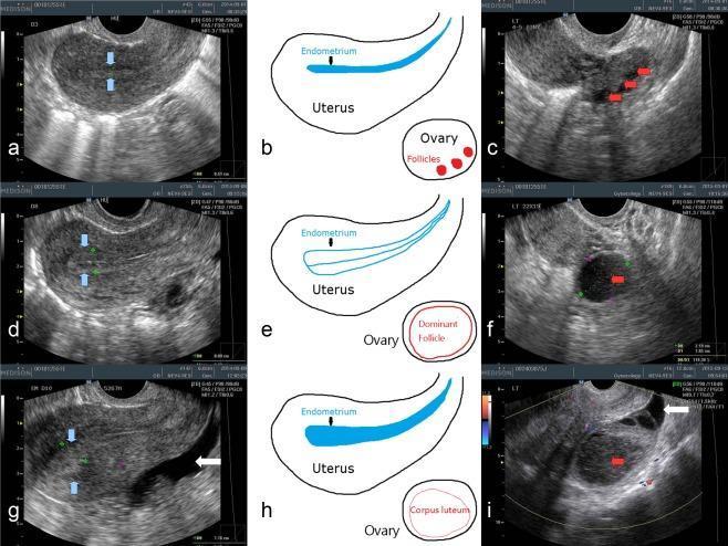
This composite figure illustrates transvaginal ultrasonography (TVUS) findings of the uterus and ovaries throughout the menstrual cycle, organized into three phases: early follicular (a-c), late follicular (d-f), and post-ovulatory luteal (g-i). In the early follicular phase, the endometrium is thin and homogenous (blue arrows), with multiple small antral follicles (red arrows) visible in the ovary. The late follicular phase displays a thickened endometrium with a characteristic 'triple-line' pattern (d) and a large, hypoechoic dominant follicle approximately 2 cm in diameter (f). The luteal phase shows a 'luteinized' endometrium with increased echogenicity (g) and the transformation of the dominant follicle into a corpus luteum (i), characterized by an inhomogenous internal structure and filament-like contents. Anechoic free fluid in the pelvis, indicative of recent ovulation, is highlighted by white arrows in the late follicular and luteal phase images. Schematic diagrams (b, e, h) provide anatomical context for the sonographic changes in endometrial morphology and ovarian follicle development.
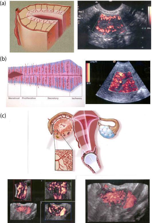
This composite educational resource compares 2D and 3D transvaginal color Doppler sonography (TV-CDS) for evaluating pelvic vascularity. (a) A cross-sectional anatomical diagram and 2D TV-CDS image highlight the uterine vascular hierarchy during the follicular phase, depicting arcuate, radial, and spiral vessels. (b) A timeline diagram illustrates endometrial vascular changes across the menstrual cycle phases: Menstrual, Proliferative, Secretory, and Ischemic. This is paired with a 2D TV-CDS showing the characteristic increase in spiral vessel density during the luteal phase. (c) The bottom section focuses on the corpus luteum, featuring a diagram of the female reproductive system with a magnified view of ovarian vascularization. Below this, multiplanar 3D TV-CDS images (longitudinal, axial, and coronal planes) and a high-definition 3D reconstruction demonstrate the pathognomonic vascular 'wreath'—a dense circumferential ring of blood vessels surrounding the functioning corpus luteum. This content illustrates key gynecological sonography concepts, including the cyclical nature of endometrial perfusion and the advanced spatial assessment afforded by 3D Doppler imaging for ovarian structures.
Part 1: The Menstrual Cycle - All Phases Explained Simply
The Ovarian Cycle (What the Ovary Does)
Phase 1: Follicular Phase (Days 1-14)
- At the start of the period, hormone levels are low.
- The brain's pituitary gland releases FSH (Follicle Stimulating Hormone), which tells several follicles to start growing.
- One follicle "wins" and becomes dominant - the others shrink away.
- The dominant follicle produces estrogen (estradiol), which steadily rises throughout this phase.
- Rising estrogen signals the pituitary to release a large surge of LH (Luteinizing Hormone) near the end of this phase.
- This LH surge triggers ovulation.
Ovulation (Around Day 14 in a 28-day cycle)
| Cycle Length | Ovulation Day |
|---|---|
| 24-day cycle | Day 10 |
| 28-day cycle | Day 14 |
| 35-day cycle | Day 21 |
- Cervical mucus becomes clear, slippery and stretchy (like raw egg white) - this helps sperm travel
- A slight rise in basal body temperature (due to progesterone)
- Some women feel mild one-sided pelvic pain (mittelschmerz)
Phase 2: Luteal Phase (Days 14-28)
- The corpus luteum produces large amounts of progesterone (and some estrogen).
- Progesterone prepares the womb lining to receive a fertilized egg.
- If the egg is fertilized: The pregnancy hormone hCG is produced, keeping the corpus luteum alive, and the period does NOT come.
- If the egg is not fertilized: The corpus luteum dies after 14 days, progesterone and estrogen drop sharply, and the womb lining sheds - this is the period (menstruation).
The Uterine Cycle (What the Womb Does)
| Uterine Phase | Timing | Driven By | What Happens |
|---|---|---|---|
| Menstrual phase | Days 1-5 | Estrogen & progesterone drop | Womb lining sheds - this is the period. Blood loss ~20-60 mL |
| Proliferative phase | Days 6-14 | Estrogen (from growing follicle) | Womb lining thickens and rebuilds itself |
| Secretory phase | Days 14-28 | Progesterone (from corpus luteum) | Womb lining becomes spongy, rich in blood vessels, ready to receive a fertilized egg |
The Hormone Orchestra - A Quick Summary
| Hormone | Made By | Role |
|---|---|---|
| FSH | Pituitary gland | Stimulates follicle growth |
| LH | Pituitary gland | Triggers ovulation; forms corpus luteum |
| Estrogen | Growing follicle | Builds up womb lining; causes LH surge |
| Progesterone | Corpus luteum | Prepares womb for implantation; raises body temperature |
Part 2: The Couple's Case - Explained Simply
The Situation
The Woman's Cycle
Step 1: When Does She Ovulate?
- If her shortest cycle is 26 days: Ovulation = Day 26 - 14 = Day 12
- If her longest cycle is 29 days: Ovulation = Day 29 - 14 = Day 15
Step 2: How Long Do Sperm Live?
Earliest ovulation = Day 12 12 - 6 = Day 6 No intercourse from Day 6 onwards.
Step 3: How Long Does the Egg Survive After Ovulation?
Latest ovulation = Day 15 15 + 2 = Day 17 Intercourse can resume from Day 17 onwards.
The Final Answer: The "Danger Zone"
| Days | Status | Reason |
|---|---|---|
| Days 1-5 | SAFE | Too early - sperm cannot survive long enough to reach a future ovulation |
| Days 6-17 | ABSTAIN | Sperm could fertilize the egg - dangerous zone |
| Days 18 onwards | SAFE | Egg has already degenerated; corpus luteum phase; next ovulation is weeks away |
Why Is the Danger Zone So Wide?
- Uncertainty about when exactly she ovulates - she could ovulate anywhere between Day 12 and Day 15 (a 3-day window of variability in her cycle)
- Sperm survival - sperm can live up to 6 days, extending the danger backwards from Day 12 to Day 6
A Simple Visual Summary
Day:   1  2  3  4  5 | 6  7  8  9 10 11 12 13 14 15 16 17 | 18 19 20 21 ...
       ✅ SAFE        | ❌ ABSTAIN (sperm alive / egg viable) |  ✅ SAFE
- Days 1-5: Period is occurring; safe
- Day 6: Last day of intercourse (sperm from Day 6 could survive to Day 12 ovulation)
- Days 12-15: Her ovulation window
- Day 17: Two days after latest ovulation (Day 15); safe to resume
- Day 18+: Safe - egg long gone, next ovulation weeks away
Key Points to Remember
- The luteal phase is always 14 days - this is the basis for calculating ovulation from the next expected period.
- The follicular phase varies - this is what makes cycles longer or shorter in different women.
- Sperm survival is up to 6 days - much longer than previously thought.
- The egg survives only 24-36 hours after ovulation.
- To be safe: Stop intercourse 6 days before the earliest possible ovulation; resume 2 days after the latest possible ovulation.