
Structure of tesstis with schematic diagram and full functional anatomy
Reading File
Now let me search the medical library for comprehensive testis anatomy content, including images.
Finding Sources
Finding Sources
Excellent - multiple rich sources found. Let me now read from the best anatomy/histology sources in parallel.
Reading File
Reading File
Reading File
Found a key image URL. Let me fetch it and also get more content from the histology textbook, Harrison's, and look for the gross anatomy diagram.
Loading Image
Reading File
Finding Sources
Excellent! Let me fetch the key images and read the THIEME atlas and Junqueira histology content for a comprehensive picture.
Loading Image
Reading File
Reading File
Excellent! I have rich textbook content and two great diagrams. Let me fetch the Junqueira sagittal section diagram and the blood supply diagram, then get the Sertoli/Leydig cell details.
Loading Image
Loading Image
Reading File
I now have all the diagrams and rich content needed. Let me fetch one more key image - the seminiferous tubule schematic diagram showing cell layers.
Loading Image
Reading File
I now have all the key diagrams and comprehensive content. Let me compile the full, detailed answer.
Structure of the Testis - Full Functional Anatomy
Overview
The testis is the male gonad with two primary functions: spermatogenesis (sperm production) and testosterone secretion. Both testes are normally located in the scrotum, maintained at 35-36°C (1-2°C below core body temperature), which is essential for normal spermatogenesis. This temperature regulation is achieved by the countercurrent heat exchange between the coiled testicular artery and the surrounding pampiniform plexus of veins.
1. Gross Anatomy and Coverings
Schematic Diagram - Sagittal Section of the Testis
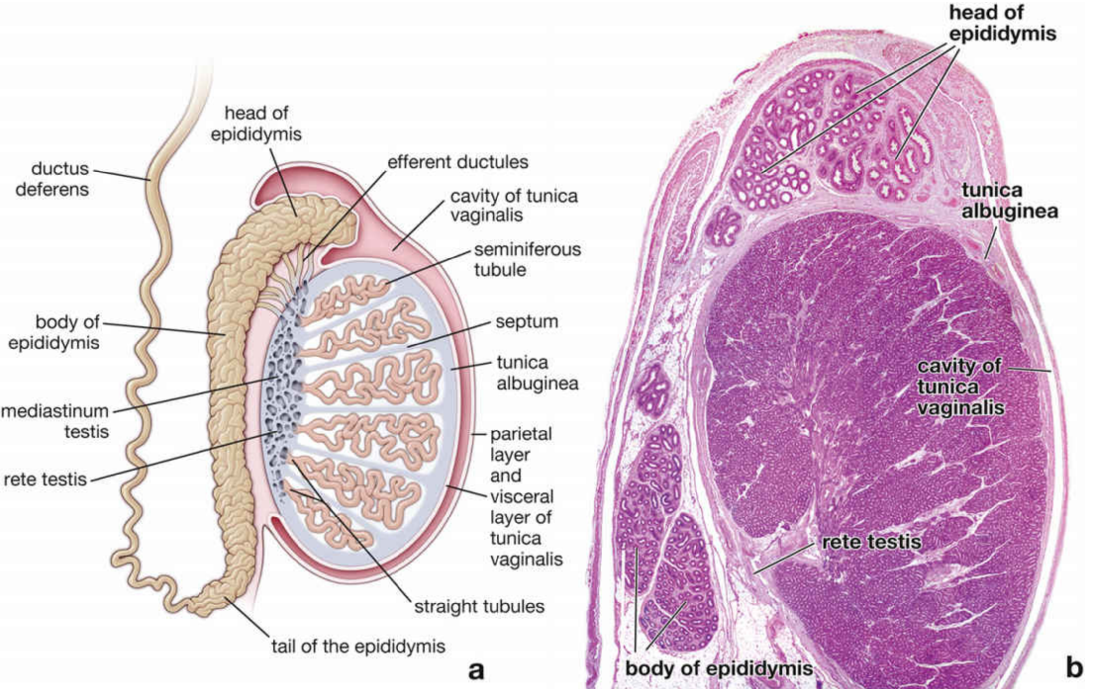
Fig. Sagittal section of the human testis (left: schematic; right: H&E histology). Labeled structures include tunica albuginea, tunica vaginalis, seminiferous tubules, septa, straight tubules, rete testis, efferent ductules, head/body/tail of epididymis, mediastinum testis, and ductus deferens. - Junqueira's Basic Histology, 17e
Coverings (from outermost to innermost)
| Layer | Description |
|---|---|
| Skin of scrotum | Thin, rugose, with dartos smooth muscle |
| Dartos layer | Smooth muscle responsible for scrotal rugae |
| External spermatic fascia | From external oblique aponeurosis |
| Cremaster muscle & fascia | From internal oblique; responsible for cremasteric reflex |
| Internal spermatic fascia | From transversalis fascia |
| Tunica vaginalis | Double-layered peritoneal remnant; parietal and visceral layers with a potential serous cavity between them |
| Tunica albuginea | Dense white fibrous capsule; the true capsule of the testis |
| Tunica vasculosa | Inner loose connective tissue with blood and lymphatic vessels |
Internal Organization
- The tunica albuginea is an unusually thick, dense fibrous capsule that encases the testis
- It sends radial fibrous septa (septula testis) inward, dividing the parenchyma into 250-370 conical lobules
- These septa converge posteriorly at the mediastinum testis - a thickened wedge of the tunica albuginea through which all blood vessels, lymphatics, and ducts enter/exit
2. Microstructure - The Two Compartments
Tubular Compartment (80% of testis volume)
Each lobule contains 1-4 highly convoluted seminiferous tubules. Key figures:
- ~250 m total tubule length in the testis (Campbell-Walsh states up to 600 m)
- Each tubule is 30-80 cm long, 150-300 µm in diameter
- Tubules are closed loops - both ends drain into the tubuli recti (straight tubules)
Interstitial Compartment (20% of testis volume)
- Contains loose connective tissue, blood vessels, lymphatics, and Leydig cells
- The testis contains ~700 million Leydig cells in the young adult
3. Microscopic Structure of the Seminiferous Tubule
Histology - Wall of the Tubule
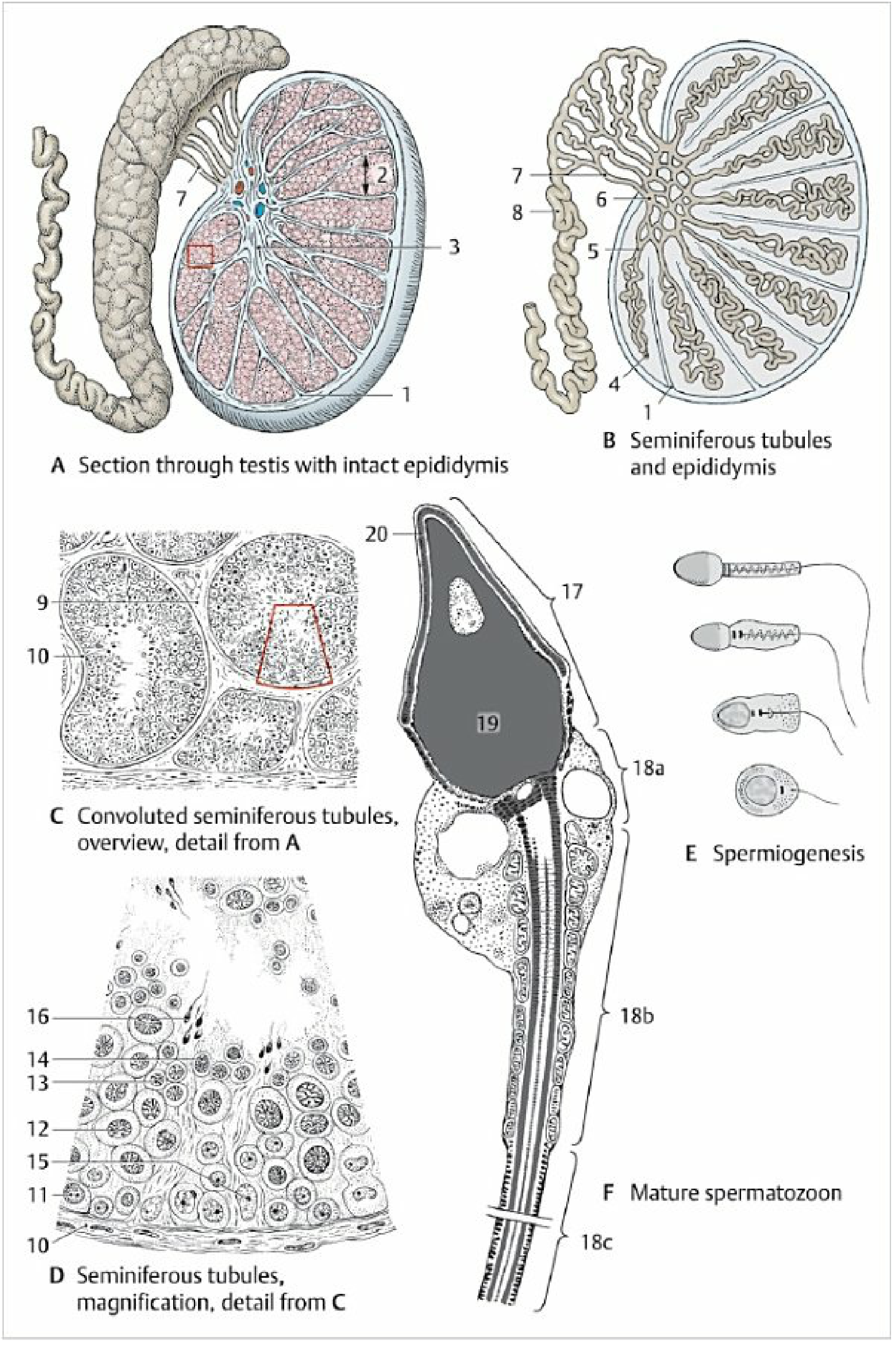
Fig. Microscopic structure of the testis and epididymis. A: Section through testis with intact epididymis. B: Seminiferous tubules and epididymis. C: Convoluted seminiferous tubules (overview). D: High magnification of seminiferous tubules showing cell layers. E: Spermiogenesis stages. F: Mature spermatozoon with labeled head (17), nucleus (19), acrosome (20), neck (18a), middle piece (18b), and principal piece (18c). - Color Atlas of Human Anatomy, Vol. 2
The tubule wall from outside to lumen:
- Basal lamina - the basement membrane on which Sertoli cells and spermatogonia rest
- Tunica propria (peritubular tissue) - 3-5 layers of myoid cells (peritubular contractile cells). These have smooth muscle-like actin filaments; their rhythmic contractions create peristaltic waves that propel sperm and tubular fluid toward the rete testis
- Seminiferous epithelium - the main functional layer
Seminiferous Epithelium - Detailed Cell Schematic
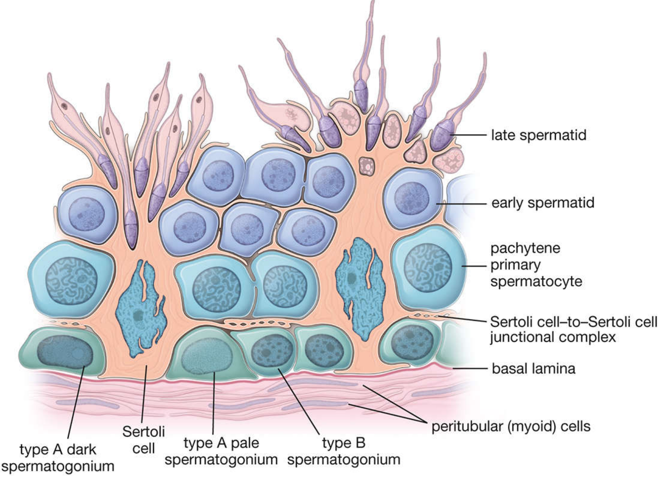
Fig. Schematic drawing of the human seminiferous epithelium showing: (basal compartment) type A dark and type A pale spermatogonia, type B spermatogonia, Sertoli cells, Sertoli-to-Sertoli junctional complex forming the blood-testis barrier; (abluminal compartment) pachytene primary spermatocytes, early spermatids, and late spermatids. Peritubular myoid cells and basal lamina are also labeled. - Junqueira's Basic Histology, 17e
4. Cell Types and Their Functions
A. Sertoli Cells (Supporting / Sustentacular Cells)
- Tall columnar cells that extend from basal lamina to the lumen; do not replicate after puberty
- Form the blood-testis barrier via tight (occluding) junctions between adjacent Sertoli cells - this divides the epithelium into a basal compartment and an adluminal (abluminal) compartment
- The blood-testis barrier protects developing spermatocytes from the immune system (since they express foreign antigens after puberty)
Four key functions (Costanzo Physiology, 7e):
- Nutritional support - provide nutrients to differentiating sperm cells that are isolated from the bloodstream
- Blood-testis barrier - tight junctions that selectively permit testosterone but block noxious substances
- Fluid secretion - secrete an aqueous fluid into the tubule lumen to transport sperm toward the epididymis
- Androgen-binding protein (ABP) secretion - binds testosterone and keeps local concentrations high near developing sperm
Additional functions: secrete inhibin B (FSH feedback), produce anti-Müllerian hormone (AMH) in fetal life, and phagocytose residual bodies shed during spermiogenesis
B. Leydig Cells (Interstitial Cells)
- Large, polygonal, eosinophilic cells in the interstitium between tubules
- Contain lipid droplets, lipofuscin, and characteristic Crystals of Reinke (rod-shaped cytoplasmic crystalloids, ~3 × 20 µm; function unknown, pathognomonic of Leydig cells)
- Function: Synthesize and secrete testosterone in response to LH
- Testosterone has:
- Local (paracrine) effects on Sertoli cells to support spermatogenesis
- Endocrine effects on skeletal muscle, prostate, and other target organs
C. Spermatogenic Cells (from basal to luminal)
| Cell Type | Location | Ploidy | Key Feature |
|---|---|---|---|
| Type A dark spermatogonia | Basal lamina | 2n | Reserve stem cells, rarely divide |
| Type A pale spermatogonia | Basal lamina | 2n | Active stem cells, undergo mitosis |
| Type B spermatogonia | Basal lamina | 2n | Committed precursors, enter meiosis |
| Primary spermatocytes | Basal → adluminal | 4n (after DNA replication) | Undergo meiosis I (lasts up to 22-24 days - longest phase) |
| Secondary spermatocytes | Adluminal | 2n | Short-lived; undergo meiosis II |
| Spermatids | Near lumen | 1n | Undergo spermiogenesis |
| Spermatozoa | Lumen | 1n | Released into lumen (spermiation) |
5. Spermatogenesis - Three Phases
The total cycle takes ~64-74 days in humans. ~128 million sperm are produced daily.
Phase 1 - Spermatogonial Phase (Mitotic)
Type A dark spermatogonia serve as reserve stem cells. Type A pale spermatogonia divide mitotically to renew themselves and produce type B spermatogonia, which are committed to become sperm.
Phase 2 - Spermatocyte Phase (Meiotic)
- Type B spermatogonia → primary spermatocytes (DNA duplicates to 4n)
- Meiosis I (up to 22-24 days, prophase I is the longest stage) → two secondary spermatocytes (2n each)
- Meiosis II (only hours) → four spermatids (1n each, haploid)
- Genetic recombination occurs during prophase I
Phase 3 - Spermiogenesis (Spermatid Remodeling)
Spermatids undergo extensive morphological transformation without further division:
- Nuclear chromatin condensation
- Formation of the acrosomal cap (contains acrosin and hydrolytic enzymes for penetrating the zona pellucida)
- Development of the axoneme (flagellum, 9+2 microtubule arrangement)
- Mitochondria wrap helically around the middle piece
- Shedding of excess cytoplasm as residual bodies (phagocytosed by Sertoli cells)
The resulting spermatozoon is ~60 µm long, comprising:
- Head (flattened nucleus + acrosomal cap)
- Neck (connecting piece)
- Middle piece (mitochondrial sheath for energy)
- Principal piece (longest segment of flagellum)
- End piece (terminal flagellum)
6. Excurrent Duct System
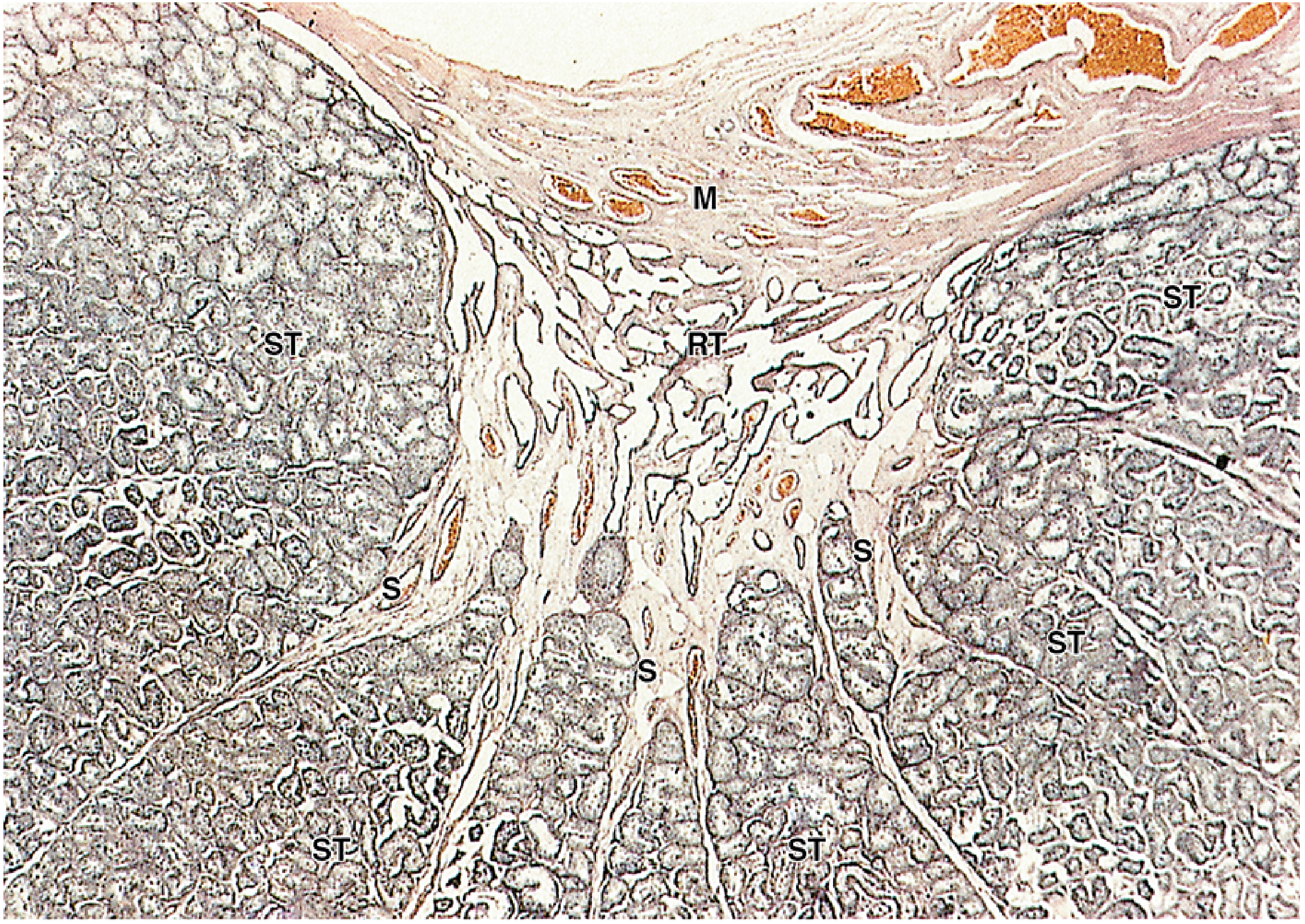
Fig. Low-power H&E section showing seminiferous tubules (ST) in lobules separated by septa (S), converging at the rete testis (RT) within the mediastinum (M). - Junqueira's Basic Histology, 17e
Sperm travel through the following pathway:
Seminiferous tubules → Tubuli recti (straight tubules) → Rete testis → 10-15 efferent ductules → Duct of epididymis (head → body → tail) → Ductus (vas) deferens → Ejaculatory duct → Prostatic urethra
- Rete testis: An anastomosing network of channels within the mediastinum testis lined by simple cuboidal/low columnar epithelium
- Efferent ductules: 10-15 ducts connecting rete testis to epididymis; lined by alternating ciliated and non-ciliated cells (giving a scalloped luminal appearance)
- Epididymis: Pseudostratified columnar epithelium with stereocilia (long microvilli); smooth muscle coat increases from head to tail. Sperm acquire motility and undergo final maturation here; viable for several months
- Ductus (vas) deferens: Thick (1-1.5 mm) muscular coat of smooth muscle; pseudostratified columnar epithelium with stereocilia. During ejaculation, forceful peristaltic contractions propel sperm
7. Blood Supply and Temperature Regulation
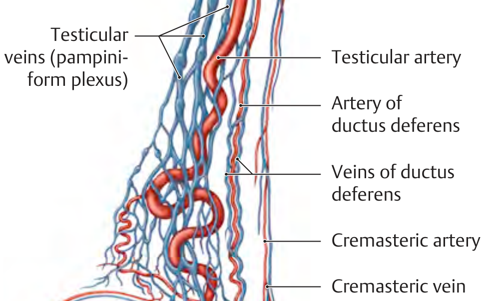
Fig. Blood supply of the testis and pampiniform plexus (THIEME Atlas of Anatomy)
Arterial Supply (3 anastomosing arteries):
- Testicular (gonadal) artery - arises directly from the aorta at L2; primary supply
- Artery of the ductus deferens - from the internal iliac artery
- Cremasteric artery - from the inferior epigastric artery
Venous Drainage:
- Venous blood drains into the pampiniform plexus - a network of veins that surrounds the coiled testicular artery in the spermatic cord
- This arrangement creates a countercurrent heat exchange: warm arterial blood is pre-cooled by the venous blood returning from the cooler scrotum, maintaining testicular temperature 1-2°C below body temperature
- Right testicular vein → inferior vena cava (direct, at an angle)
- Left testicular vein → left renal vein (enters at a right angle - physiologically significant; creates a tendency for venous obstruction → varicocele is more common on the left)
Clinical relevance of varicocele:
When the pampiniform plexus can no longer cool the testicular artery, local temperature rises and can impair spermatogenesis → male factor infertility. The left-right asymmetry of venous drainage explains why left-sided varicoceles are far more common.
Lymphatic Drainage:
- Lymphatics of the testis drain to the para-aortic (lumbar) lymph nodes at L1-L2 (following the gonadal vessels)
- This is clinically distinct from scrotal lymphatics, which drain to the inguinal nodes
- Clinical implication: testicular tumors metastasize to para-aortic nodes, not inguinal nodes
Nerve Supply:
- Sympathetic fibers from T10-T11 travel with the testicular artery
- Responsible for testicular pain radiating to the flank/loin (T10 dermatome)
8. Hormonal Regulation (HPG Axis)
| Hormone | Source | Target | Effect |
|---|---|---|---|
| GnRH | Hypothalamus | Anterior pituitary | Stimulates LH and FSH release |
| LH | Anterior pituitary | Leydig cells | Stimulates testosterone synthesis |
| FSH | Anterior pituitary | Sertoli cells | Stimulates ABP secretion, supports spermatogenesis |
| Testosterone | Leydig cells | Sertoli cells, whole body | Drives spermatogenesis (paracrine); virilization (endocrine) |
| Inhibin B | Sertoli cells | Anterior pituitary | Negative feedback on FSH |
| DHT | Periphery (5α-reductase) | External genitalia, prostate | Development of accessory sex structures |
Summary Table: Key Structures and Functions
| Structure | Cell/Tissue | Primary Function |
|---|---|---|
| Tunica albuginea | Dense fibrous CT | Capsule; provides structural integrity |
| Septula testis | CT septa | Divide testis into lobules |
| Mediastinum testis | Thickened tunica albuginea | Entry/exit point for vessels and ducts |
| Seminiferous tubule | Complex stratified epithelium | Site of spermatogenesis |
| Sertoli cells | Supporting columnar cells | Blood-testis barrier, nurture sperm, secrete ABP and inhibin |
| Spermatogonia | Stem cells | Self-renewal and initiation of spermatogenesis |
| Leydig cells | Interstitial polygonal cells | Testosterone synthesis (LH-stimulated) |
| Myoid cells | Peritubular contractile cells | Peristaltic propulsion of sperm |
| Rete testis | Anastomosing channels | Collect sperm from all tubules |
| Epididymis | Pseudostratified epithelium | Sperm maturation, motility acquisition, and storage |
| Pampiniform plexus | Venous network | Countercurrent cooling of testicular artery |
Sources: Junqueira's Basic Histology - Text and Atlas, 17e; Costanzo Physiology, 7e; Histology: A Text and Atlas (Ross & Pawlina), 9e; Color Atlas of Human Anatomy Vol. 2 (Thieme); THIEME Atlas of Anatomy - General Anatomy and Musculoskeletal System; Campbell-Walsh-Wein Urology, 4th ed.
Can you give me this information but at neet ug lebel instead of mbbs
Here's the complete testis structure at NEET UG level - focused on what's asked in MCQs, with clean diagrams and memory tricks.
Structure of Testis - NEET UG Level
QUICK OVERVIEW
The testis is the male gonad with 2 functions:
- Spermatogenesis (makes sperm)
- Testosterone secretion (male hormone)
Located in the scrotum - kept ~2-3°C BELOW body temperature (essential for sperm production).
GROSS STRUCTURE (What it looks like from outside)
OUTER → INNER layers of coverings:
Skin of scrotum
↓
Dartos muscle (wrinkles skin in cold)
↓
External spermatic fascia
↓
Cremaster muscle (cremasteric reflex)
↓
Internal spermatic fascia
↓
Tunica vaginalis (peritoneum-derived, 2 layers)
↓
TUNICA ALBUGINEA ← actual capsule of testis
↓
Tunica vasculosa (vessels inside)
Inside the Testis:
Tunica albuginea
|
sends septa inward
|
250-370 LOBULES
|
Each lobule has 1-4 seminiferous tubules
|
Tubules → Tubuli recti → RETE TESTIS
(rete = network, located at mediastinum testis)
SCHEMATIC DIAGRAM
Key labels to know:
- Tunica albuginea (white fibrous capsule)
- Septa → divide into lobules
- Mediastinum testis (posterior thickening where vessels enter)
- Seminiferous tubules (site of sperm production)
- Rete testis → Efferent ductules → Epididymis → Vas deferens
SEMINIFEROUS TUBULE - MOST IMPORTANT FOR NEET
80% of testis volume = seminiferous tubules
20% = interstitium (connective tissue + Leydig cells)
Two Cell Types (NEET Favourite):
| Cell | Location | Function | Hormone control |
|---|---|---|---|
| Sertoli cells | Inside tubule (lining) | Nourish sperm, form blood-testis barrier, secrete ABP & Inhibin | FSH acts on these |
| Leydig cells | OUTSIDE tubule (interstitium) | Secrete testosterone | LH acts on these |
Memory trick: Leydig → LH → Located outside tubule Sertoli → Supports sperm → Secrets ABP/Inhibin → FSH acts on it (FSH = Fostering Sertoli)
CELL DIAGRAM OF SEMINIFEROUS TUBULE
Reading from OUTSIDE → INSIDE (base to lumen):
Basal lamina + Peritubular myoid cells (contractile, push sperm forward)
↓
Spermatogonia (stem cells) - 2n, on basement membrane
↓ ← BLOOD-TESTIS BARRIER (Sertoli cell tight junctions)
Primary spermatocytes (4n, undergo Meiosis I)
↓
Secondary spermatocytes (2n, undergo Meiosis II - very brief)
↓
Spermatids (1n = haploid) - undergo spermiogenesis
↓
Spermatozoa → released into lumen
SPERMATOGENESIS - STEP BY STEP (NEET MCQ Focus)
SPERMATOGONIUM (2n)
| Mitosis
↓
PRIMARY SPERMATOCYTE (4n)
| Meiosis I (longest step - takes ~22 days)
↓
SECONDARY SPERMATOCYTE (2n)
| Meiosis II (very fast - few hours)
↓
SPERMATID (1n = haploid)
| Spermiogenesis (NO division - just shape change)
↓
SPERMATOZOON (mature sperm)
Total time = ~64-74 days
Spermiogenesis (shape changes in spermatid):
- Nucleus condenses
- Acrosome forms (cap on head - contains digestive enzymes to penetrate egg)
- Flagellum develops (for movement)
- Mitochondria coil around middle piece (energy supply)
- Excess cytoplasm shed as residual bodies (eaten by Sertoli cells)
STRUCTURE OF A MATURE SPERM
HEAD
├── Flattened nucleus
└── Acrosome cap (enzymes: hyaluronidase, acrosin)
NECK (connecting piece)
MIDDLE PIECE
└── Mitochondria wrapped in a helix
(= powerhouse for sperm motility)
PRINCIPAL PIECE
└── Long flagellum
END PIECE
└── Short terminal flagellum
NEET tip: Acrosome contains hyaluronidase (digests corona radiata) and acrosin (penetrates zona pellucida)
BLOOD-TESTIS BARRIER (BTB)
- Formed by tight junctions between Sertoli cells
- Divides tubule into:
- Basal compartment - spermatogonia + early spermatocytes (accessible to blood)
- Adluminal compartment - later spermatocytes + spermatids (protected from immune system)
- Why important? Post-pubertal sperm carry new antigens - without BTB, immune system would attack them (auto-immune infertility)
HORMONAL CONTROL (HPG Axis) - MUST KNOW FOR NEET
HYPOTHALAMUS
| GnRH (pulse)
↓
ANTERIOR PITUITARY
| |
LH FSH
| |
↓ ↓
LEYDIG SERTOLI
CELLS CELLS
| |
Testosterone ABP + Inhibin
| |
(-)feedback (-) feedback
on pituitary on FSH only
| Hormone | Made by | Acts on | Result |
|---|---|---|---|
| GnRH | Hypothalamus | Pituitary | Releases LH + FSH |
| LH | Pituitary | Leydig cells | Testosterone ↑ |
| FSH | Pituitary | Sertoli cells | Spermatogenesis support + Inhibin ↑ |
| Testosterone | Leydig cells | Body + Sertoli cells | Spermatogenesis + male features |
| Inhibin | Sertoli cells | Pituitary | FSH ↓ (negative feedback) |
| ABP | Sertoli cells | Tubule lumen | Keeps testosterone high locally |
NEET trick: "LH → Leydig → Testosterone" and "FSH → Sertoli → Sperm support"
BLOOD SUPPLY (Important for NEET)
3 arteries supply the testis:
- Testicular artery - from abdominal aorta at L2 (main supply)
- Artery of vas deferens - from internal iliac
- Cremasteric artery - from inferior epigastric
Pampiniform plexus = network of veins that coil around the testicular artery
- Function: countercurrent cooling - keeps testis 2°C cooler than body
- Right testicular vein → IVC (inferior vena cava)
- Left testicular vein → Left renal vein (enters at 90° angle → prone to backup → left varicocele more common)
LYMPHATIC DRAINAGE
- Testis lymphatics → Para-aortic (lumbar) lymph nodes (L1-L2)
- NOT inguinal nodes (common exam trap!)
- Scrotum lymphatics → inguinal nodes
- NEET/USMLE trap: Testicular cancer spreads to para-aortic nodes, not inguinal
SPERM PATHWAY OUT OF TESTIS
Seminiferous tubules
↓
Tubuli recti (straight tubules)
↓
Rete testis (network in mediastinum)
↓
Efferent ductules (10-15)
↓
Epididymis (head → body → tail) ← sperm mature here, stored here
↓
Vas deferens (ductus deferens)
↓
Ejaculatory duct
↓
Prostatic urethra → outside
Memory: "STRE-EVE" → Seminiferous → Tubuli recti → Rete testis → Efferent ductules → Epididymis → Vas deferens → Ejaculatory duct
HIGH-YIELD NEET MCQ FACTS
| Fact | Answer |
|---|---|
| Site of spermatogenesis | Seminiferous tubules |
| Hormone for Leydig cells | LH |
| Hormone for Sertoli cells | FSH |
| Blood-testis barrier formed by | Sertoli cell tight junctions |
| Inhibin is secreted by | Sertoli cells |
| Inhibin inhibits | FSH (not LH) |
| ABP secreted by | Sertoli cells |
| Temperature for spermatogenesis | 35-36°C (2°C below body temp) |
| Countercurrent cooling by | Pampiniform plexus |
| Which varicocele is more common | Left (left testicular vein → left renal vein) |
| Testicular lymphatics drain to | Para-aortic nodes (NOT inguinal) |
| Cryptorchidism causes | Infertility (high temperature) but NOT testosterone loss (Leydig cells still work) |
| Acrosome contains | Hyaluronidase + acrosin |
| Energy for sperm motility from | Mitochondria in middle piece |
| Total spermatogenesis time | ~64-74 days |
| Longest phase of spermatogenesis | Meiosis I (prophase I, ~22 days) |
| Number of lobules in testis | 250-370 |
CRYPTORCHIDISM (UNDESCENDED TESTIS) - NEET FAVOURITE
- Testis fails to descend into scrotum → stays in abdomen (37°C)
- Effect: Spermatogenesis fails (temperature too high) → infertility
- Testosterone production is NOT affected (Leydig cells are more heat-resistant)
- Increased risk: Testicular cancer (seminoma most common)
- Needs surgical correction (orchiopexy) before age 2
That covers everything you need for NEET UG on testis structure. Focus especially on: Sertoli vs Leydig cells, FSH vs LH targets, blood-testis barrier, spermatogenesis sequence, and the lymphatic drainage rule (para-aortic, not inguinal).
Ejaculator duct
Reading File
Finding Sources
Finding Sources
Reading File
Reading File
Loading Image
Loading Image
Reading File
I have everything needed. Now let me write the full NEET UG-level answer on the ejaculatory duct.
Ejaculatory Duct - NEET UG Level
WHAT IS IT?
The ejaculatory duct is a short paired duct formed by the union of:
Ampulla of VAS DEFERENS
+
Duct of SEMINAL VESICLE
↓
= EJACULATORY DUCT
↓
Opens into PROSTATIC URETHRA
(at the VERUMONTANUM / colliculus seminalis)
Think of it as the final common pathway before sperm enter the urethra.
FORMATION (Embryology - NEET Favourite)
- Derived from the Wolffian (mesonephric) duct - same origin as vas deferens, epididymis, and seminal vesicles
- The seminal vesicle develops as an outgrowth of the lower end of the vas deferens
- The duct of seminal vesicle joins the ampulla of vas deferens → forms ejaculatory duct
NEET Trick: All male genital ducts (epididymis, vas deferens, ejaculatory duct, seminal vesicles) = Wolffian duct derivatives Fallopian tubes, uterus, upper vagina = Müllerian duct derivatives
ANATOMY AT A GLANCE
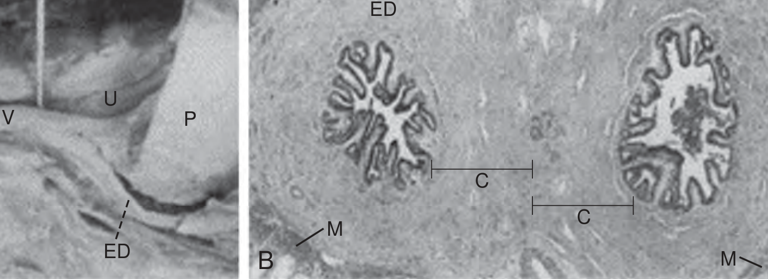
Fig. Human ejaculatory duct anatomy: (Left) Sagittal cadaver section - ED runs through prostate (P) and opens at the verumontanum (V) into urethra (U). (Right) Histology of paired ejaculatory ducts showing folded mucosa, thick collagenous layer (C) and thin muscle (M). - Campbell-Walsh-Wein Urology
| Feature | Detail |
|---|---|
| Number | Paired (one on each side) |
| Length | ~2 cm each |
| Start | Posterior base of prostate (union of vas + seminal vesicle) |
| Course | Passes THROUGH the prostate gland (obliquely, downward and forward) |
| Opening | Into prostatic urethra at the verumontanum (seminal colliculus) |
THREE REGIONS OF EJACULATORY DUCT
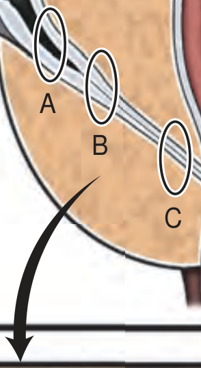
Fig. Three regions of the ejaculatory duct: A = Proximal, B = Intraprostatic (middle - muscle layer is thinnest here), C = Distal opening at verumontanum. - Campbell-Walsh-Wein Urology
- Proximal (A) - where vas ampulla meets seminal vesicle duct
- Intraprostatic / Middle (B) - runs through prostate; muscle layer is thinnest here
- Distal (C) - opens into prostatic urethra at the verumontanum
HISTOLOGY (Wall Structure)
- Mucosa - folded epithelium (pseudostratified columnar, with secretory function)
- Cells contain yellow pigment granules (from lipofuscin)
- NO muscular wall of its own - the fibromuscular tissue of the prostate substitutes for it
- Surrounded by a thick collagenous layer
Key NEET fact: The ejaculatory duct has NO muscularis layer - unlike the vas deferens which has a very thick muscular wall. The prostate's smooth muscle serves this function.
FUNCTION
The ejaculatory duct:
- Conducts sperm from the vas deferens toward the urethra
- Carries seminal vesicle secretions (which make up 60-80% of semen volume)
- Mixes sperm + seminal vesicle fluid just before ejaculation
- During ejaculation - propelled by contraction of the prostate's smooth muscle (since the duct has no muscle of its own)
WHAT IS THE VERUMONTANUM?
- Also called seminal colliculus or colliculus seminalis
- A small midline elevation on the posterior wall of the prostatic urethra
- The two ejaculatory ducts open here (one on each side)
- The prostatic utricle (a small blind pouch, Müllerian remnant) also opens here in the midline
- Landmark used in cystoscopy and TURP surgery
POSTERIOR WALL of PROSTATIC URETHRA
↓
VERUMONTANUM
/ | \
Left ED Utricle Right ED
(ejac duct) (ejac duct)
COMPLETE SPERM PATHWAY (Review)
Seminiferous tubules
↓
Tubuli recti
↓
Rete testis
↓
Efferent ductules (10-15)
↓
Epididymis (head → body → tail)
↓
Vas deferens → Ampulla of vas deferens
+
Duct of seminal vesicle
↓
EJACULATORY DUCT (~2 cm)
(passes THROUGH prostate)
↓
Prostatic urethra (at verumontanum)
↓
Membranous urethra → Penile urethra → Outside
WHAT DOES SEMEN CONTAIN?
| Component | Contributed by | % of semen volume |
|---|---|---|
| Sperm + fluid | Testis + Epididymis | ~5% |
| Fluid rich in fructose, prostaglandins, alkaline | Seminal vesicles | ~60-70% |
| Fluid rich in PSA, citric acid, zinc, acidic | Prostate | ~25-30% |
| Lubricating mucus | Bulbourethral (Cowper's) glands | ~5% |
NEET tip: Seminal vesicle fluid = fructose (energy source for sperm). Ejaculatory duct carries this fluid into the urethra. Total semen pH = mildly alkaline (neutralizes acidic vaginal environment)
CLINICAL RELEVANCE - EJACULATORY DUCT OBSTRUCTION (EDO)
| Feature | Detail |
|---|---|
| Cause | Stone/calculus, cyst (midline mullerian cyst or off-midline ejaculatory duct cyst), infection/scarring, congenital |
| Presentation | Azoospermia or severe oligospermia + low semen volume + acidic semen pH (<7.2) |
| Clue | Low fructose in semen (seminal vesicle secretions blocked) |
| Diagnosis | Transrectal ultrasound (TRUS) - dilated seminal vesicles (>1.5 cm), dilated ejaculatory duct (>2.3 mm) |
| Treatment | Transurethral resection of ejaculatory ducts (TURED) - resect the obstruction endoscopically |
NEET/USMLE high-yield pattern: Azoospermia + LOW semen volume + LOW pH + LOW fructose = Ejaculatory duct obstruction (or seminal vesicle absence)
HIGH-YIELD MCQ FACTS
| Question | Answer |
|---|---|
| Ejaculatory duct formed by | Ampulla of vas deferens + duct of seminal vesicle |
| Length of ejaculatory duct | ~2 cm |
| Ejaculatory duct opens into | Prostatic urethra at verumontanum |
| Embryological origin | Wolffian (mesonephric) duct |
| Ejaculatory duct passes through | Prostate gland |
| Has its own muscular wall? | NO - prostate muscle substitutes |
| Structure at verumontanum | Both ejaculatory ducts + prostatic utricle |
| Prostatic utricle = remnant of | Müllerian duct (Paramesonephric) |
| EDO presents with | Azoospermia + low semen volume + acidic pH + low fructose |
| Treatment of EDO | TURED (Transurethral Resection of Ejaculatory Ducts) |
| Seminal vesicle secretion % | 60-70% of semen volume |
| Energy source in seminal vesicle | Fructose |
MEMORY TRICKS
"VAS + SV = ED → VU" Vas deferens ampulla + Seminal Vesicle = Ejaculatory Duct → opens at Verumontanum in Urethra
Verumontanum = "THE MEETING POINT" Both ejaculatory ducts + prostatic utricle all meet here
EDO Clue = "ALFA" Azoospermia, Low volume, Fructose absent, Acidic pH
Explain urethra in very short but necessary manner for ug
Finding Sources
Finding Sources
Your credits are exhausted. You can purchase a top-up or subscribe to a plan to continue using Orris. If you're already subscribed, your daily credits will reset tomorrow.
This is a shared conversation. Sign in to Orris to start your own chat.