
Supraglottic airways
supraglottic airway devices LMA laryngeal mask airway types
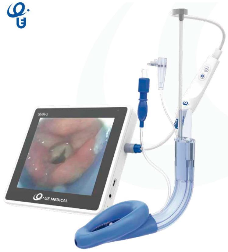
This clinical photograph displays the SaCoVLM video laryngeal mask airway (LMA) system, a specialized supraglottic airway device designed for visualization during airway management. The assembly consists of a curved, semi-transparent breathing tube terminating in a blue, inflatable anatomical mask cuff. Integrated into the device is a video stylus system connected to a handheld control unit with power and capture buttons. Multiple access ports are visible along the tube, including a central channel for gas exchange or endotracheal tube (ETT) insertion, a gastric drainage port, and ports for pressure monitoring and CPAP ventilation. The handheld unit is tethered via a blue connector to a 7-inch portable monitor. The monitor screen displays a high-definition endoscopic view of the glottis, showing the vocal cords and epiglottis, which allows for real-time visual confirmation of device placement and facilitates rescue intubation. This medical technology is utilized by anesthesiologists and emergency physicians to secure the human airway while providing direct visualization of the larynx.
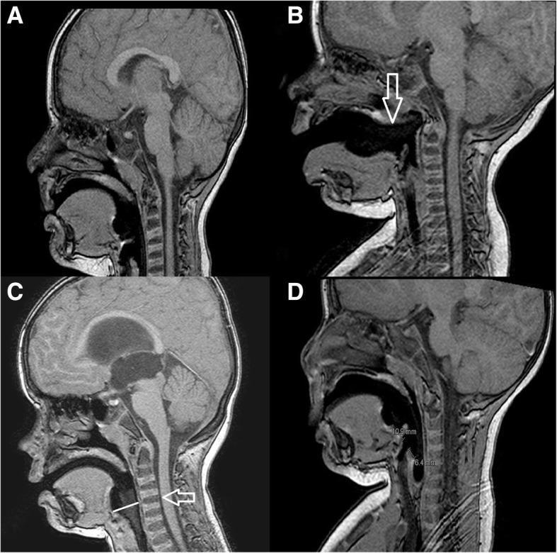
This set of four sagittal T1-weighted MRI images (A, B, C, and D) illustrates the radiological evaluation of laryngeal mask airway (LMA) placement in pediatric patients. Image A displays the radiologically correct position, with the LMA tip seated in the hypopharynx and the proximal cuff aligned with the C1-C2 cervical vertebrae. In contrast, images B, C, and D demonstrate different types of malposition. Image B (Malposition A) shows the mask displaced anteriorly, pointed out by a white arrow, indicating it is outside the hypopharynx. Image C (Malposition B) shows the proximal cuff positioned inferiorly to the C1-C2 level, with a white arrow highlighting the vertebral misalignment. Image D (Malposition C) presents a malposition characterized by spatial measurement discrepancies, where the distance from the proximal cuff to the aditus laryngis (10.9 mm) exceeds the distance from the distal cuff to the aditus laryngis (6.4 mm). These images serve as an educational tool for anesthesiologists to identify anatomical landmarks such as the soft palate, tongue base, and cervical spine relative to supraglottic airway devices.
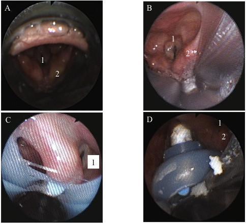
This set of four fiberscopic images (A-D) illustrates the anatomical positioning of various supraglottic airway (SGA) devices relative to the larynx. Panel A shows the i-gel™ device with the glottic inlet (1) and arytenoids (2) clearly visible; the epiglottis is notably resting inside the mask bowl. Panel B displays the LMA-Supreme™, showing similar anatomical landmarks but with visible narrowing of the vocal cords and slightly hyperemic tissue. Panels C and D demonstrate laryngeal views with the Laryngeal Tube Suction-D (LTS-D) in situ. These views illustrate suboptimal or 'awkward' positioning where the device partially obscures the glottic structures, although ventilation remains possible. Across all images, the laryngeal mucosa appears pink and moist, indicating healthy tissue perfusion. This comparative series serves as an educational tool for anesthesiology and emergency medicine to understand how different SGA designs impact glottic visualization and airway alignment during clinical practice.

This diagnostic image series displays four endoscopic clinical photographs categorized as SaCoVLM™ glottic exposure grades, illustrating the airway view obtained via a video laryngeal mask airway (LMA). Grade 1 (Far Left): Minimal visualization showing only the lateral aspect of the right aryepiglottic fold and a small portion of the laryngeal inlet. Grade 2 (Middle Left): Improved exposure revealing bilateral aryepiglottic folds and a wider view of the laryngeal inlet. Grade 3 (Middle Right): Advanced exposure where the entire laryngeal inlet and the posterior portion of the glottis become visible; the mucosa appears more textured with visible vasculature. Grade 4 (Far Right): Optimal exposure showing the whole glottis, including the vocal cords and the dark lumen of the trachea. The sequence demonstrates the progressive visibility of laryngeal landmarks from a tangential epiglottic view to a direct axial glottic view. These grades serve as a clinical tool for anesthesiologists to evaluate the positioning and efficacy of visual-guided supraglottic airway devices during airway management and ventilation.
Supraglottic Airway Devices (SGAs)
Definition
General Structure
- A tube connected to a breathing circuit or bag
- A hypopharyngeal device (cuff, mask, or preshaped form) that seals around or above the glottis
- Directs airflow toward the glottis, trachea, and lungs
- Some occlude the esophagus to varying degrees
- Some include a gastric drainage port to allow passage of an orogastric tube
Classification (Miller's Terminology)
1. Perilaryngeal Sealers (Cuffed)
First-Generation
| Device | Key Features |
|---|---|
| LMA Classic (cLMA) | Silicone, reusable (up to 40×), sizes 1-6 (neonate to large adult); PPV up to 20 cm H₂O |
| LMA Unique | Single-use version of the cLMA |
| LMA Flexible | Kink-resistant tube for head/neck surgery where the tube must be positioned away from the field |
| air-Q, LMA Unique EVO | Designed to facilitate intubation through the device |
Second-Generation (reduced aspiration risk + higher seal pressure)
| Device | Key Features |
|---|---|
| LMA ProSeal (PLMA) | Reusable; posterior cuff for better seal; PPV up to 30 cm H₂O; gastric drainage tube; bite block |
| LMA Supreme (SLMA) | Single-use; gastric drainage tube; fixation tab for securing; integrated bite block |
2. Cuffless Anatomically Preshaped Sealers
| Device | Key Features |
|---|---|
| i-gel (Intersurgical) | Gel-like thermoplastic elastomer; conforms to laryngopharyngeal anatomy; includes a gastric drainage port (classified as 2nd-gen); widely used in emergency/prehospital settings |
| SLIPA (Curveair) | Hollow chamber that can trap regurgitated fluid |
| Baska Mask | Self-sealing membrane; high-pressure capability |
3. Cuffed Pharyngeal Sealers
| Device | Key Features |
|---|---|
| Esophageal-Tracheal Combitube (ETC) | Two lumina + two cuffs (pharyngeal + esophageal); mostly prehospital; >90% esophageal placement; contains latex - avoid in latex-sensitive patients |
| King LT / King LT-D | Single lumen, tapered esophageal tip; ventilation between the two cuffs |
| King LTS / King LTS-D | Same as LT but with open distal tip for gastric suctioning |
| CobraPLA, Tulip Airway | Pharyngeal cuff only (no esophageal cuff) |
LMA Sizing (cLMA / LMA sizes)
| Size | Patient | Weight |
|---|---|---|
| 1 | Neonate/infant | < 5 kg |
| 1.5 | Infant | 5-10 kg |
| 2 | Child | 10-20 kg |
| 2.5 | Child | 20-30 kg |
| 3 | Small adult | 30-50 kg |
| 4 | Adult (female) | 50-70 kg |
| 5 | Adult (male) | 70-100 kg |
| 6 | Large adult | > 100 kg |
Insertion Technique (LMA Classic)
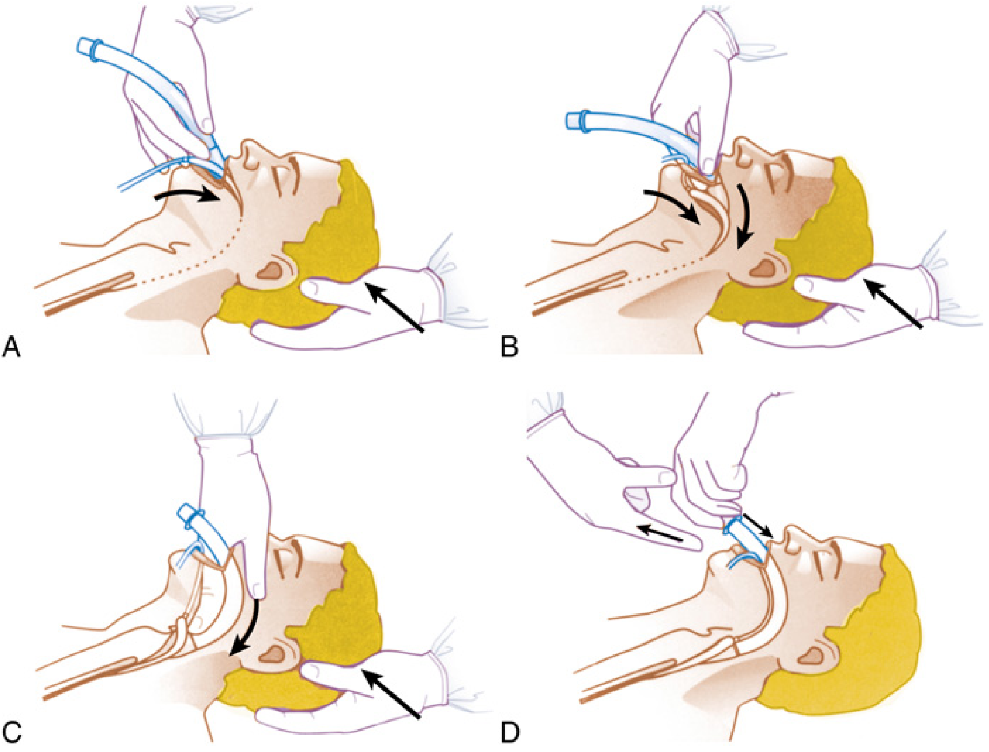
- Choose appropriate size; check for leaks
- Deflate the cuff tightly with the rim facing away from the aperture (no folds near the tip)
- Lubricate only the back of the cuff
- Ensure adequate anesthetic depth (slightly deeper than for an oral airway)
- Place patient in sniffing position (neck flexed, head extended)
- Guide cuff tip along the hard palate and posteriorly into the hypopharynx until resistance is felt
- Inflate with appropriate air volume; the longitudinal black line faces cephalad (upper lip)
- Do not remove until patient is awake (coughing, opening mouth on command)
- Base of tongue - superiorly
- Pyriform sinuses - laterally
- Upper esophageal sphincter - inferiorly
Advantages vs. Disadvantages
| Advantages | Disadvantages |
|---|---|
| Ease and speed of placement | Lower seal pressure than ETT |
| No muscle relaxant required | No protection from laryngospasm |
| Less hemodynamic response | 1st-gen: no aspiration protection |
| Reduced anesthetic requirements | Not a definitive airway |
| Avoids ETT-related trauma (dental, airway, sore throat, bronchospasm) | Ineffective ventilation if high airway pressures needed |
Clinical Uses
- Primary airway - diagnostic and minor surgical procedures (first choice)
- Rescue airway - when BMV and intubation have both failed (part of ASA Difficult Airway Algorithm)
- Conduit for intubation - tracheal tube can be passed through select SGAs under fiberoptic guidance
- Prehospital/CPR - placement during cardiac arrest when intubation is not feasible
- Pediatric trauma/TBI - bridge until definitive airway can be placed - Current Surgical Therapy, 14e
SGA as Intubation Conduit
Confirming Correct Placement
- Adequate tidal volumes with reasonable peak pressures
- Leak pressure > 20 cm H₂O (2nd-gen)
- Normal capnography waveform
- For the ProSeal: gel test - a small layer of gel over the drainage tube orifice moves with suprasternal notch palpation, but not during PPV ventilation
- Easy passage of an orogastric tube through the gastric drainage channel
Key Points to Remember
- SGAs are not definitive airways - in trauma, they must eventually be replaced with an ETT
- Cuff pressure should never exceed 60 cm H₂O (to avoid oropharyngeal nerve injury and tissue ischemia)
- Size 4 for average adult female, size 5 for average adult male (LMA)
- If the LMA is not functioning after repositioning attempts, try one size larger or smaller
- The i-gel is popular in emergency settings because it requires no cuff inflation and has a built-in gastric port
- The ETC/Combitube contains latex - avoid in latex-sensitive patients
- SGAs can be used in lateral and prone positions with second-generation devices